X-ray CT apparatus

An X-ray and equipment technology, applied in the field of X-ray CT equipment, can solve problems such as rough X-ray dose information and excessive X-ray dose

Inactive Publication Date: 2007-03-14
GE MEDICAL SYST GLOBAL TECH CO LLC
View PDF1 Cites 11 Cited by
  • Summary
  • Abstract
  • Description
  • Claims
  • Application Information

AI Technical Summary

Problems solved by technology

There is a high probability that the X-ray dose applied to the object tends to be too large when the operator tries to have a higher image quality thin tomographic image
Thus, only a series of images of helical scans, conventional scans (axial scans) or cine scans are available only based on more accurate x-ray dose information of the subject size or to account for changes in sensitivity to damage caused by x-rays in the subject area Acquired X-ray dose information is too coarse to be used as future X-ray dose information

Method used


Image

  • X-ray CT apparatus
  • X-ray CT apparatus
  • X-ray CT apparatus

Examples


example 1

[0172] When this embodiment is applied to an actual helical scan, the X-ray dose information of the entire image acquisition area, the X-ray dose information of the ROI 1 (heart) and the X-ray dose information of the ROI 2 (liver) are known. Considering the sensitivity of the X-ray exposure amount of each organ, it is considered to reduce the exposure amount of the subject.

[0173] Also in conventional scanning (axial scanning) or cine scanning, similarly, each of the X-ray dose information of the entire image acquisition area and the X-ray dose information of the region of interest 1 is known, as shown in FIG. 28 , so that The X-ray exposure of each organ and the X-ray exposure of the whole area can be considered.

example 2

[0175] In Example 2, the case of variable-pitch helical scanning as shown in FIG. 29 will be described. In variable-pitch helical scanning, as shown in FIG. 29 , the helical pitch and noise index (image noise index value) vary in the z-direction range, for example, within the heart, liver, and lung regions. Thus, it is not easy to immediately know the X-ray dose information at a position in the z direction compared with normal conventional scan (axial scan), cine scan or helical scan, and thus it is more necessary to display the X-ray dose information. Also in this case, by displaying the X-ray dose information on each of the entire region, region of interest 1 (heart), region of interest 2 (lung region), and region of interest 3 (liver), it is possible to or display the information more clearly. Therefore, with respect to the sensitivity of the X-ray exposure amount of each organ, it is possible to consider reducing the exposure amount of the subject.

example 3

[0177] In Example 3, the X-ray profile area Sx obtained from the scout view was used to obtain a correlation with the water substitution model to be referenced. Statistical surveys were performed on height, weight, age, image acquisition area, and gender. As shown in FIG. 30A , the relationship between body weight, height, and a region sectional area is obtained for each of sex, age range, and region, and a regression plane or regression curve is derived from the distribution statistics. Alternatively, as shown in Figure 30B, the relationship between weight, height and cross-sectional area of ​​the water replacement model is obtained and a regression plane or curve is derived from the distribution statistics. Expressions for regression planes or regression curves are also available.

[0178] When gender, age, area, weight and height are input, the cross-sectional area of ​​the area and the cross-sectional area of ​​the water substitution model are obtained through the express...


PUM


Abstract

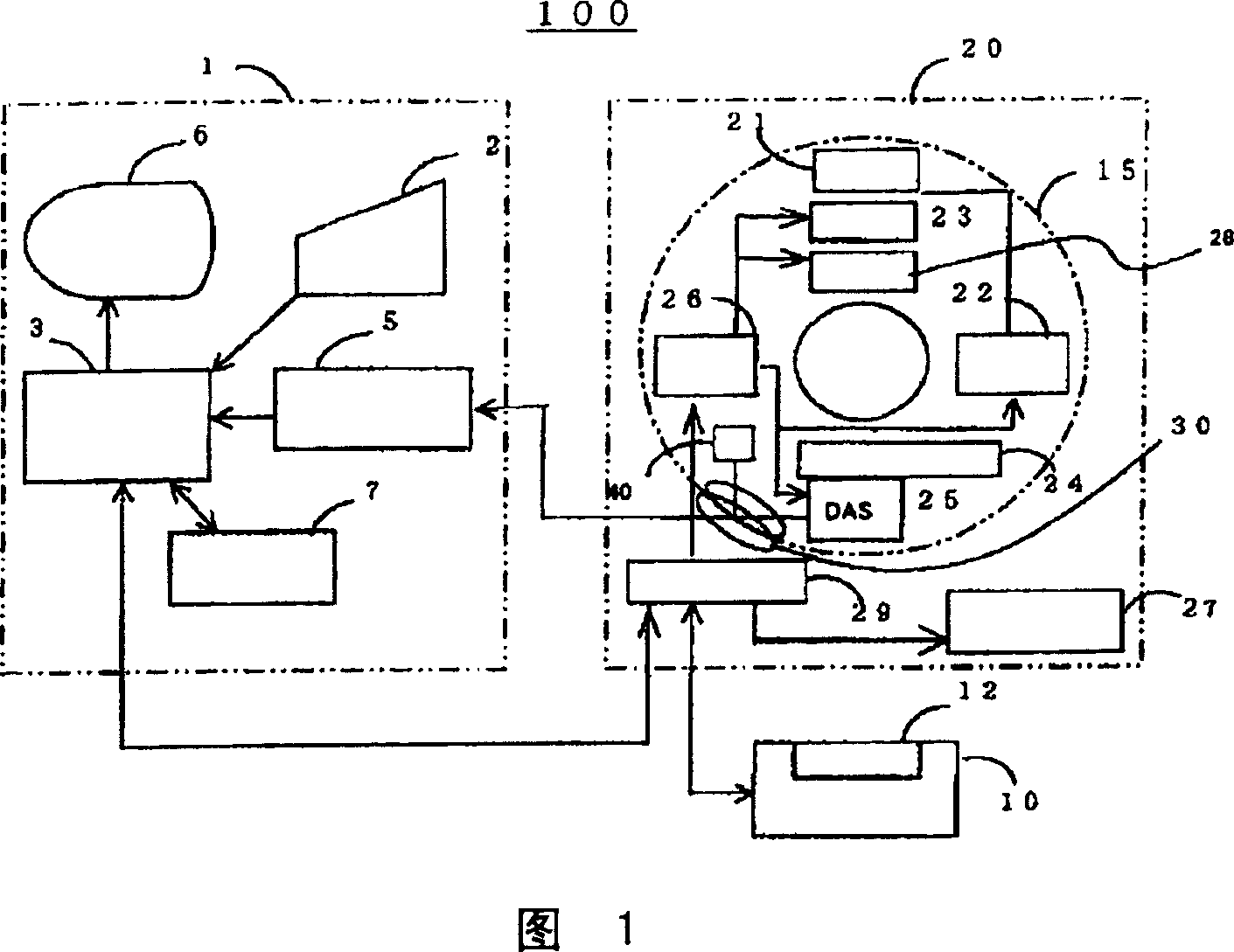
This invention provides an X-ray CT capable of presenting information of exposure to the operator by displaying X-ray dose information of each of regions of interest to be scanned by an X-ray CT apparatus, thereby encouraging reduction in exposure and optimization. X-ray dose information of each region to be scanned by a conventional scan (axial scan), a cine scan, a helical scan, or a variable-pitch helical scan of an X-ray CT apparatus is displayed so that the operator can recognize the X-ray dose information before acquisition of an image of a subject. The X-ray dose information can be predicated with higher precision and displayed by using a dose prediction value obtained by an interpolation value and an extrapolation value of the first or higher order on at least three or more kinds of phantom measurement values, not a simple prediction value such as a zero-th order interpolation value or a zero-th order extrapolation value obtained by using measurement values of two kinds of phantoms like in the present CTDI display.

Description

technical field [0001] The present invention relates to a method for X-ray CT (computed tomography) image acquisition using an X-ray CT device for medical or industrial use, and to an X-ray CT device. More specifically, the present invention relates to a method for displaying to the operator X-ray dose information for each region of interest in a conventional scan (also known as an axial scan), a cinescan, a helical scan, or a variable pitch helical scan. display method, thereby helping to reduce and optimize exposure. Background technique [0002] Conventionally, in an X-ray CT apparatus using a multi-row X-ray detector or an X-ray CT apparatus using a matrix-structured two-dimensional X-ray area detector typified by a flat panel, the helical scanning from the neck to the liver or In the case of an image from the lung area to the liver, as shown in FIG. 12 , when the image acquisition parameters are set by the image acquisition parameter setting device, an X is displayed w...

Claims


Application Information

Patent Timeline
Patent Type & Authority Applications(China)
IPC IPC(8): A61B6/03G01N23/00
CPCA61B6/542A61B6/032A61B6/027A61B6/00G01N23/046G01N2223/419G01N2223/612A61B6/583G01N2223/1016
Inventor 西出明彦堀内哲也今井靖浩
Owner GE MEDICAL SYST GLOBAL TECH CO LLC
Why Eureka
Try Eureka