Elastography measurement system and method
a measurement system and elastography technology, applied in ultrasonic/sonic/infrasonic image/data processing, instruments, applications, etc., can solve the problems of poor placement of roi, poor elastography measurement results, contamination of stiffness reconstruction, etc., to achieve better measurement results, visualize suitability, and good quality
- Summary
- Abstract
- Description
- Claims
- Application Information
AI Technical Summary
Benefits of technology
Problems solved by technology
Method used
Image
Examples
embodiment 80
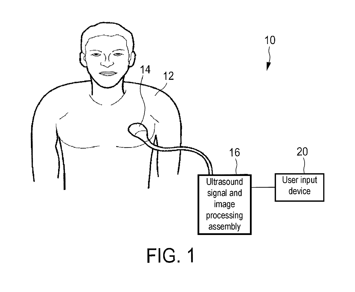
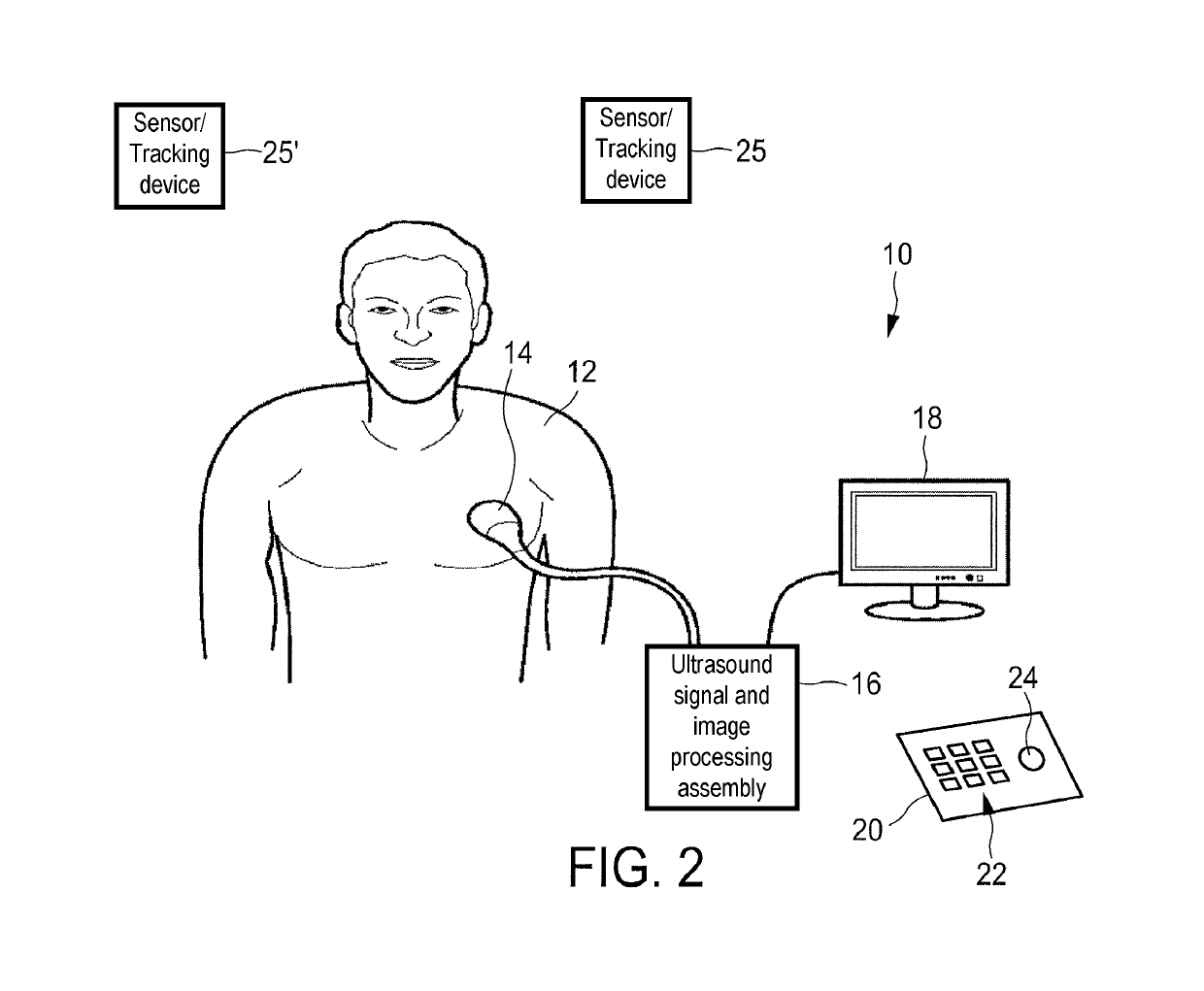
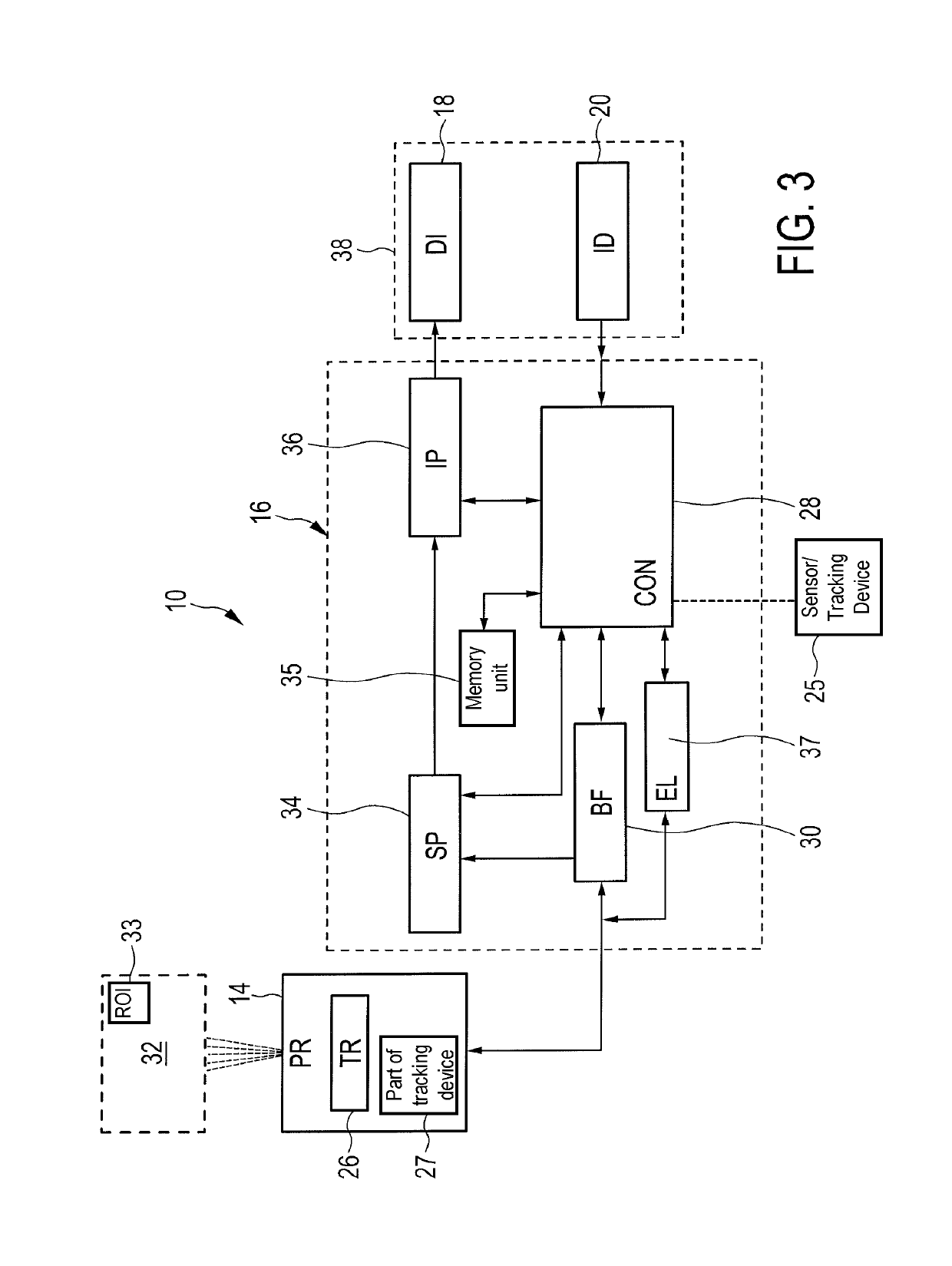
[0062]FIG. 9 shows an embodiment 80 of a method for inspecting an anatomical site 32 with an ultrasound elastography system 10. The method starts at step 82. In step 84, a reference image 86 of the anatomical site 32 is acquired. This may be conducted using a further modality like computer tomography or magnetic resonance tomography. However, it may also be the case that the reference image 86 is acquired via the ultrasound system 10. Further, a reference image 86 acquired via ultrasound may two-dimensional or three-dimensional. Then, in a step 88, at least one recommendation feature 58 representative of a suitability for a shear wave elastography of a region of interest 33 in the reference image 86 is determined. Such a recommendation feature 58 may be for example a position of vessels, homogenous tissue regions or suspicious lesion regions in the anatomical structure.
[0063]Then, at least one of the following two steps is conducted. The recommendation features 58 may either be used...
embodiment 180
[0065]In FIG. 10, a further embodiment 180 of a method is shown.
[0066]In this embodiment, the local and global approaches take the steps 190 to 194 similarly toward vessel delineation, homogeneous area detection, visualizing go / no-go zone on B-mode ultrasound image 52, and toggling the proposed feature on and off. In step 184, the acquisition and integration of a live 2D / 3D ultrasound image to pre-procedure images is conducted. This can be achieved using a variety of different tracking techniques. In one embodiment, the tracking could be electromagnetic tracking wherein the probe and patient are tracked live in an electromagnetic field. The pre-procedures images could be either tracked three-dimensionally to reconstruct three-dimensional ultrasound images / volumes. Alternatively, earlier acquired MR / CT images could be used. These pre-procedure images can be registered to the electromagnetic tracking system using fiducials, for example. Using this integration, a variety of different v...
PUM
 Abstract
Description
Claims
Application Information
 - R&D
- Intellectual Property
- Life Sciences
- Materials
- Tech Scout
- Unparalleled Data Quality
- Higher Quality Content
- 60% Fewer Hallucinations
Browse by: Latest US Patents, China's latest patents, Technical Efficacy Thesaurus, Application Domain, Technology Topic, Popular Technical Reports.
© 2025 PatSnap. All rights reserved.Legal|Privacy policy|Modern Slavery Act Transparency Statement|Sitemap|About US| Contact US: help@patsnap.com


