
Method for culturing myogenic cells, cultures obtained therefrom, screening methods, and cell culture medium

a cell culture and cell technology, applied in the field of cell culturing, can solve the problems of mrna degradation and failure to produce gaa protein, difficult identification, and only partially effective current enzyme replacement therapy (ert), and achieve the effect of high expansion and retention of terminal differentiation ability

Active Publication Date: 2022-06-07
ERASMUS UNIV MEDICAL CENT ROTTERDAM ERASMUS MC
View PDF1 Cites 0 Cited by
  • Summary
  • Abstract
  • Description
  • Claims
  • Application Information

AI Technical Summary

Benefits of technology

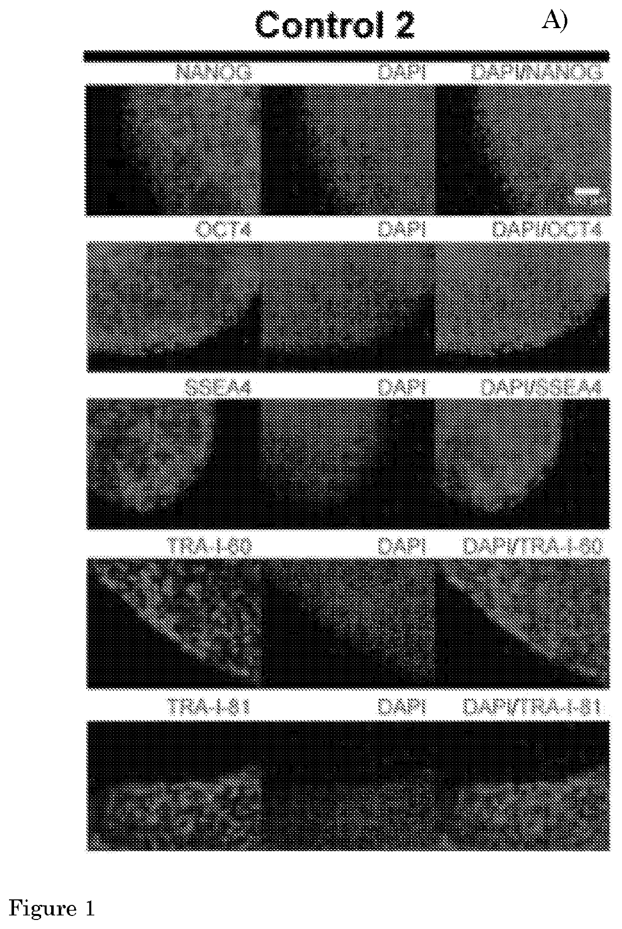
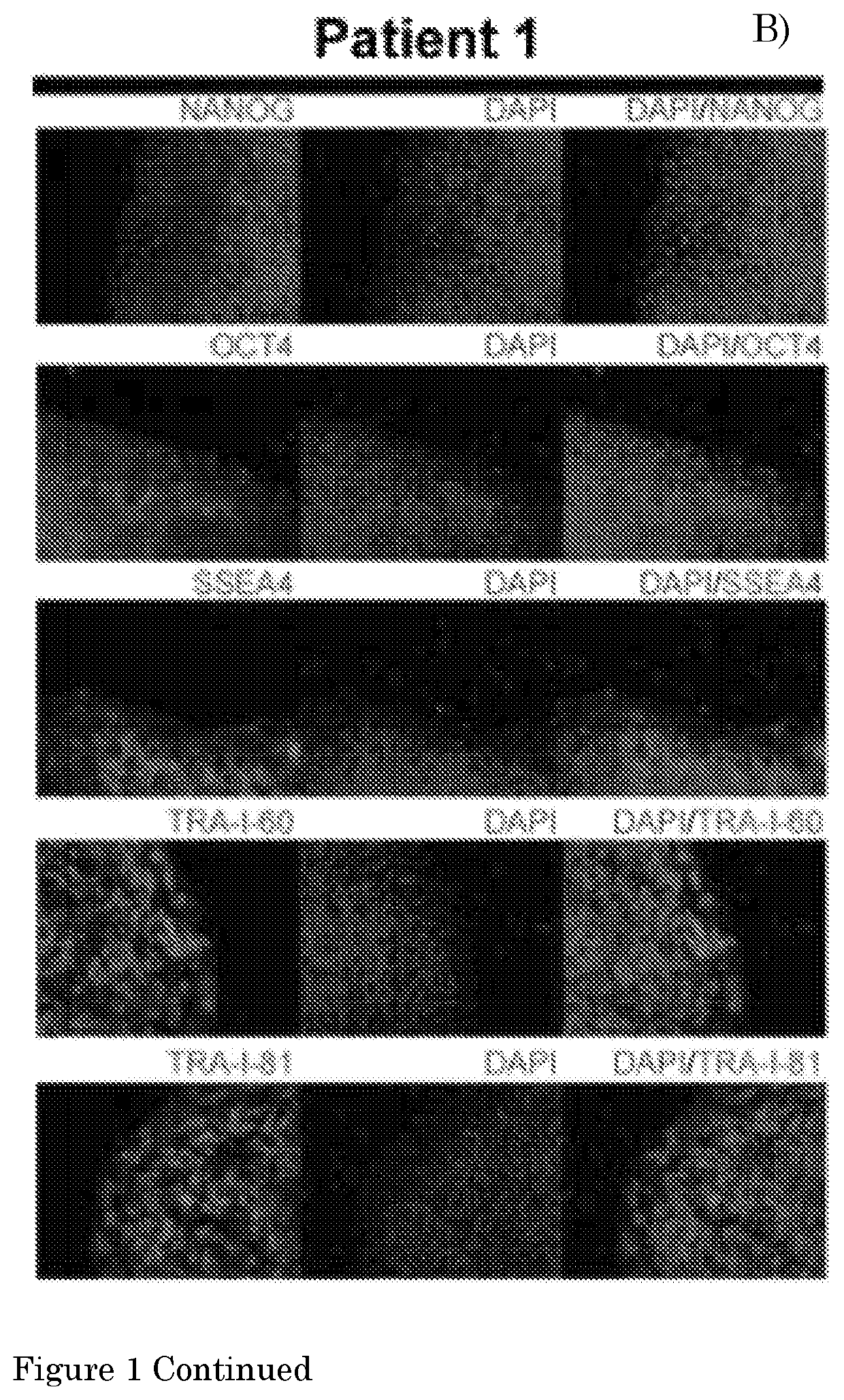
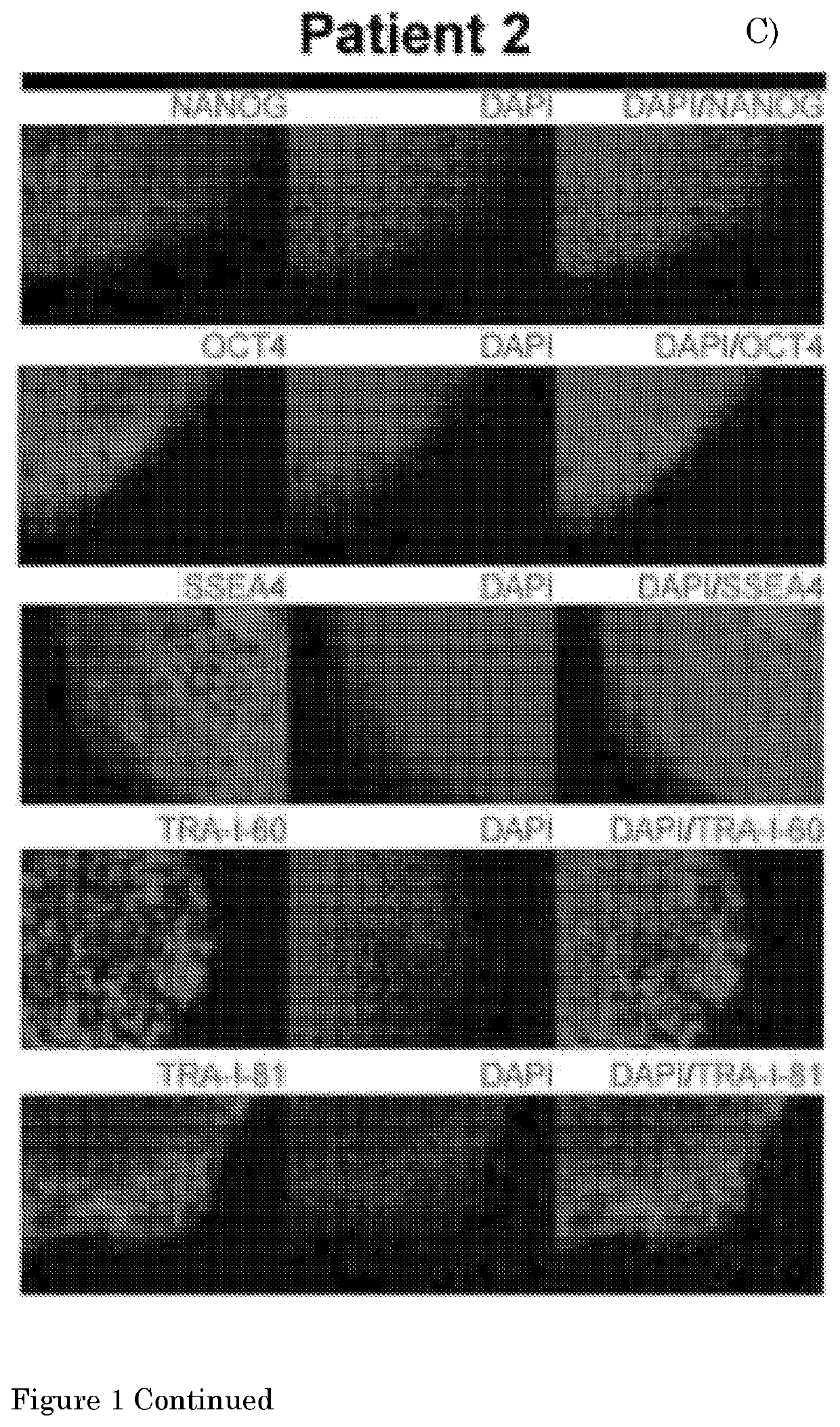
[0007]The present inventors have now surprisingly established pluripotent stem cell culturing conditions allowing expansion of individually isolated myogenic cells up to at least about 7×10⁶ fold following their isolation by, for instance, fluorescence activated cell sorting (FACS). It was further unexpectedly found that an expanded cell culture obtained by a method of the invention constitutes a novel class of myogenic cells, which are homogeneous and can be held in culture for a prolonged period of time. These cells are highly expandable when cultured under the appropriate conditions, and retain their ability for terminal differentiation. Using this novel class of cells, it was possible to generate differentiated myogenic tissue structures such as myotubes which ultimately provided for the possibility of quantitative in vitro testing, and discovery, of compounds or compositions that could potentially be of great benefit in the treatment of muscle disease, including such diseases as Pompe disease and other (neuro)muscular disorders. The differentiation conditions applied, such as the differentiation media, allow for the functional maturation of expanded cells of the invention into myotubes and even in more mature myofibers.
[0008]The culturing methods presented here enable the large-scale generation of myogenic cells, from, inter alia, iPS cells. Assuming that a drug screen will be performed in 96 wells plates containing 20,000 cells / well, the testing of 10,000 compounds in triplicate would require the seeding of 6×10⁸ myogenic progenitors. This quantity of cells can now easily be attained using the methods presented herein, which are capable of yielding at least 1012 cells per line. The myogenic cells prepared by methods of the invention offer important advantages over primary human myoblasts preparations, as these latter preparations can be contaminated with fibroblasts and endothelial cells. Moreover, primary human myoblast preparations show a progressive loss of myogenic potential, in terms of both proliferation potential and / or differentiation potential upon successive passaging. The methods and means of the present invention allow for the generation of expanded myogenic progenitor cells that resemble myoblasts in terms of differentiation status, but remain culturable and expandable for prolonged periods of time, and will differentiate into myogenic end-structures under differentiation conditions, thereby overcoming the problems of culturing myoblasts, and the problem of low yields of viable and expandable myogenic cells after isolating by, for instance, FACS. The methods of the present invention allow for the generation of patient-derived cell lines at high cell densities, and their application in personalized medicine.

Problems solved by technology

Current Enzyme Replacement Therapy (ERT) is only partially effective and very expensive, warranting the search for alternatives.
Exon 2 is the first coding exon, and its skipping results in mRNA degradation and failure to produce GAA protein.
Promotion of exon inclusion may be achieved by blocking of a splicing repressor, which is difficult to identify due to the difficulty of reliable in silico prediction of such elements.
A major obstacle for the modeling of human diseases affecting skeletal muscle and the testing of potential drugs in vitro has been the lack of methods for reproducibly obtaining sufficient amounts of pure, preferably patient-derived, differentiated human skeletal muscle cells.
It has not been possible to generate large amounts of isolated skeletal muscle cells from pluripotent stem cells, and to test therapies quantitatively.
This is one of the major reasons that, as to date, very few therapies have been developed, as these therapies cannot be tested quantitatively in vitro.
Application of induced pluripotent stem (iPS) cells to model human disorders affecting skeletal muscle has been hampered by the difficulty to differentiate pluripotent stem cells into skeletal muscle cells in vitro and to obtain sufficient amounts of pure, patient-derived, differentiated human skeletal muscle cells.
Alternatively, transgene overexpression of MyoD or Pax7, which are markers for certain stages of myogenic differentiation, is difficult.
We have found that the use of small molecules that activate the WNT pathway to drive stem cell differentiation into the myogenic lineage still generally results in low recoveries of myogenic cells after purification by FACS (typically 250,000 cells from 106 iPS cells) and variable viability and differentiation potential.
All this prevents proper quantitative analysis of potential treatments.

Method used


Image

  • Method for culturing myogenic cells, cultures obtained therefrom, screening methods, and cell culture medium
  • Method for culturing myogenic cells, cultures obtained therefrom, screening methods, and cell culture medium
  • Method for culturing myogenic cells, cultures obtained therefrom, screening methods, and cell culture medium

Examples


embodiment 1

[0138]The present invention provides a method for culturing an isolated myogenic cell, comprising the step of

[0139]a) culturing an isolated C-Met+ and Hnk1− myogenic cell in a culture medium comprising an FGF pathway activator; wherein said myogenic cell is isolated from a myogenic cell culture obtained by culturing a pluripotent stem cell (PSC) with (i) a Wnt agonist and / or a glycogen synthase kinase 3 beta (GSK3B) inhibitor, and (ii) an FGF pathway activator.

embodiment 2

[0140]The present invention provides a method according to Embodiment 1, wherein the pluripotent stem cell (PSC) is an embryonic stem cell (ESC) or induced PSC (iPSC), preferably a human PSC (hPSC), more preferably an iPSC derived from a fibroblast of a human subject, preferably a human subject suffering from a (neuro)muscular disorder.

embodiment 3

[0141]The present invention provides a method according to embodiment 1 or embodiment 2, wherein culturing the pluripotent stem cell comprises a first period of culturing with a Wnt agonist and / or a glycogen synthase kinase 3 beta (GSK3B) inhibitor, and a second period of culturing with an FGF pathway activator.


PUM

PropertyMeasurementUnit
concentrationaaaaaaaaaa
timeaaaaaaaaaa

Abstract

The invention is in the field of cell culturing. More specifically, it is in the field of generating and expanding myogenic cells from induced pluripotent stem (i PS) cells. The invention relates inter alia to cells generated and expanded via such a method, a growth medium specifically suited for the purpose of expanding isolated myogenic cells, and methods for screening compounds on cell structures such as myotubes and myofibers.

Description

FIELD OF THE INVENTION[0001]The invention is in the field of cell culturing. More specifically, it is in the field of generating and expanding myogenic cells from induced pluripotent stem (iPS) cells. The invention relates inter alia to cells generated and expanded via such methods, to growth media specifically suited for the purpose of expanding isolated myogenic cells, and methods for in vitro screening and selection of therapeutically active or diagnostic compounds on cell structures such as myotubes and myofibers generated from expanded myogenic cells.BACKGROUND OF THE INVENTION[0002]Although more than 700 human disorders that affect skeletal muscle are known, very few therapies for skeletal muscle disorders have been developed. Notable exceptions are enzyme therapy for Pompe disease and exon skipping for Duchenne Muscular Dystrophy (DMD), which is based on blocking of canonical splice sites of DMD pre-mRNA using antisense oligonucleotides (AONs). AON-mediated therapy has also b...

Claims


Application Information

Patent Timeline
Patent Type & Authority Patents(United States)
IPC IPC(8): C12N5/077C12N5/074C12N5/00C12Q1/02
CPCC12N5/0658C12N5/0018C12N5/0659C12N5/0696C12Q1/025C12N2501/115C12N2501/15C12N2501/602C12N2501/603C12N2501/604C12N2501/606C12N2501/727C12N2506/1307C12N2506/45C12N2533/54C12N2533/90
Inventor PIJNAPPEL, WILHELMUS WENCESLAUS MATTHIASVAN DER PLOEG, ANTJE TJITSKEVAN DER WAL, ERIK
Owner ERASMUS UNIV MEDICAL CENT ROTTERDAM ERASMUS MC
Learn More