Diagnostic ultrasound monitoring system and method

a monitoring system and ultrasound technology, applied in the field of ultrasound diagnostic imaging, can solve the problems of comparing periodic examinations of the same organ with difficulty, reproducing previous scanning, and affecting the accuracy of the results

Active Publication Date: 2022-10-25
URSUS MEDICAL DESIGNS LLC
View PDF17 Cites 0 Cited by
  • Summary
  • Abstract
  • Description
  • Claims
  • Application Information

AI Technical Summary

Benefits of technology

[0012]The task of long-term remote ultrasound monitoring, among other things, requires solving the problem of miniaturization of the diagnostic ultrasound system, scaling weight, power consumption and geometrical dimensions down to the smartphone form factor level and minimization of operator's involvement into the monitoring process, preferably limited to the initial placement and securing the device on the selected location on patient's body.
[0014]One application of the present invention provides a portable ultrasound monitor system configured for use with existing cardiopulmonary resuscitation procedures (CPR) with the portable ultrasound monitor being configured to provide a rescuer with information relating to the efficacy of chest compressions, return of spontaneous circulation, and underlying cardiac rhythm all without cessation of external cardiac compression during CPR.

Problems solved by technology

However, ultrasound systems known in the art are ill suited for any above mentioned tasks since they rely on manual positioning and manipulation of the probe, requiring a constant presence of a trained, experienced sonographer, that essentially preclude any long term observations or receiving a long term data input from a stable monitoring position.
Comparing periodic examinations of the same organ is also met with difficulty, since it is extremely hard to replicate the previous scanning conditions (probe position, orientation, etc.) exactly.

Method used


Image

  • Diagnostic ultrasound monitoring system and method
  • Diagnostic ultrasound monitoring system and method
  • Diagnostic ultrasound monitoring system and method

Examples


Embodiment Construction

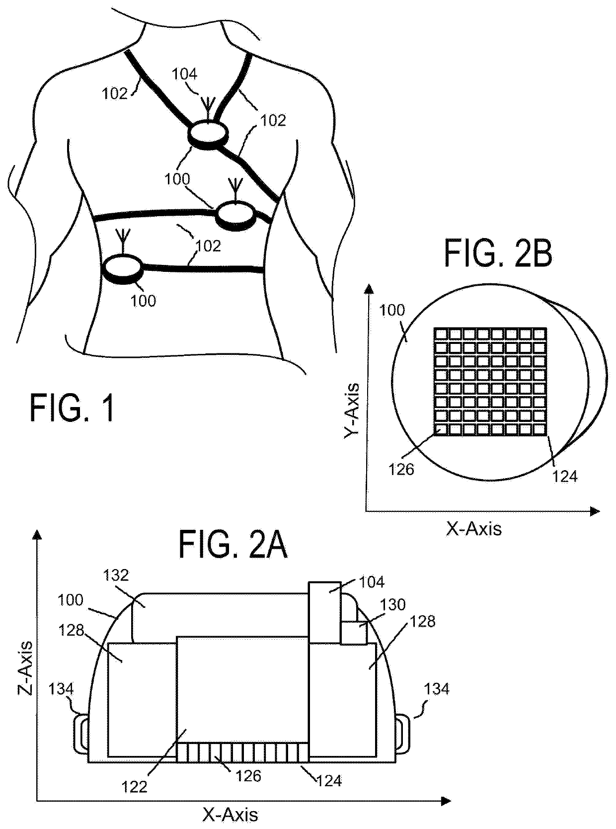
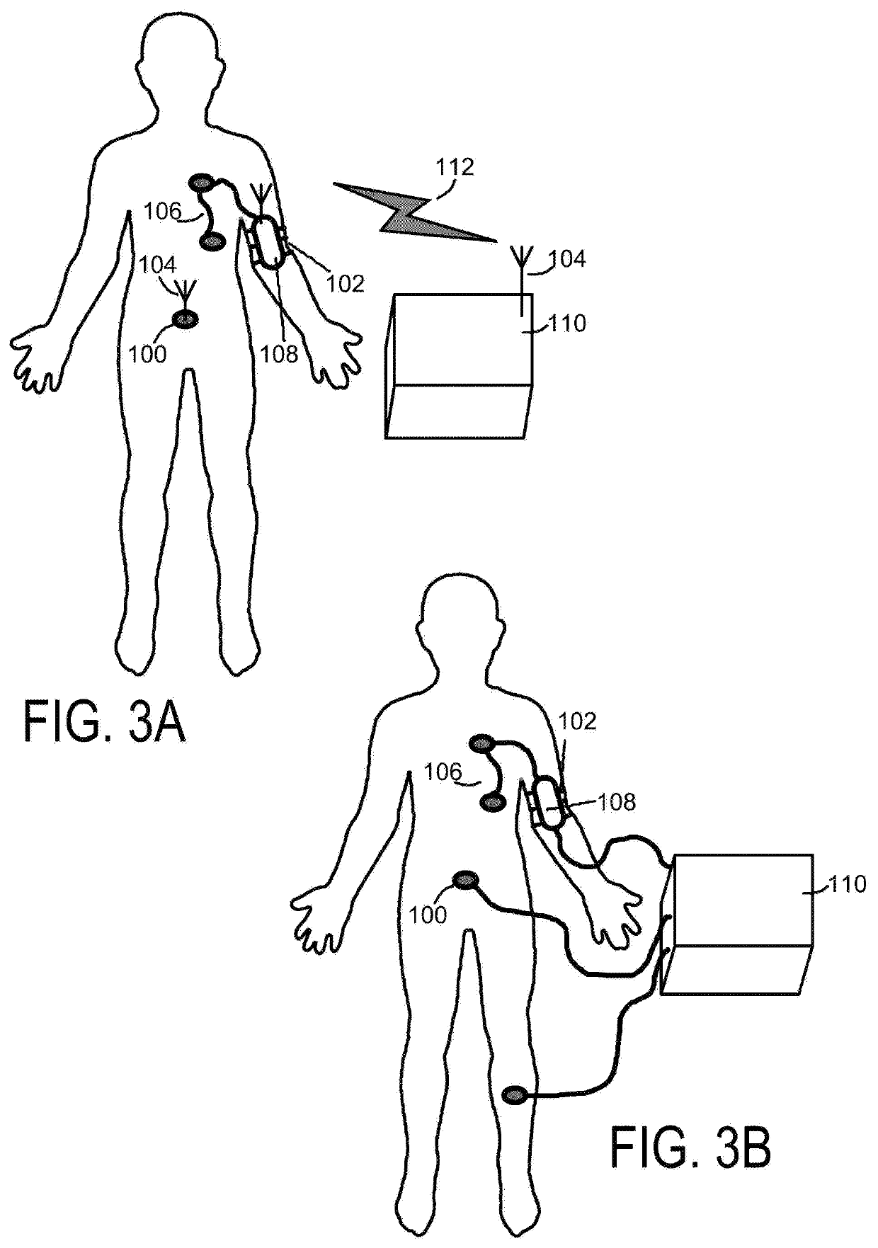
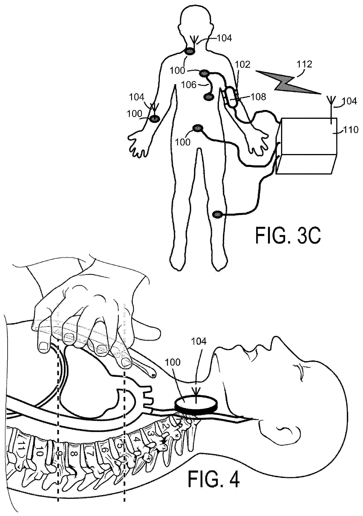
[0043]The present invention relates to ultrasound diagnostic systems, such as used in medical diagnostic systems for medical human and animal applications. Some aspects of the present invention, such as the construction and operation of an analog store digital read beamforming system and method are understood in connection with WO / 2014 / 125371, corresponding to PCT / IB2014 / 000281 and WO / 2016 / 077822 corresponding to PCT / US15 / 60861 which are incorporated herein by reference. The analog store digital read beamforming system and method used in the present system allows for each channel to use less than 40 milliwats in operation and generally less than 25 milliwatts in operation. Outside of human and animal applications, the system and method of the present invention is also applicable to non-destructive testing / evaluation (e.g., pipeline testing, airframe testing, turbine blades testing, bridge and structural testing, and manufacturing testing), particularly where there is a need for task...


PUM


Abstract

A distributed patient monitoring system comprises at least one standalone portable ultrasound imaging unit configured to be fixed to a stable position against the skin on a patient's body and capable of prolonged ultrasound data acquisition, including an ultrasound imaging array, transmit-receive circuitry, a beamformer, backend signal and image processing subsystem, power and communication subsystems, and a monitoring workstation connected to each standalone portable ultrasound imaging unit configured to request and receive ultrasound imaging information from each standalone portable ultrasound imaging unit, and configured to analyze and display acquired ultrasound information.

Description

RELATED APPLICATIONS[0001]The present application claims priority to U.S. Provisional Patent Application Ser. No. 62 / 420,677 entitled DIAGNOSTIC ULTRASOUND MONITORING SYSTEM AND METHOD and filed Nov. 11, 2016 which is incorporated herein by reference.BACKGROUND INFORMATION1. Field of the Invention[0002]The present invention relates to ultrasonic diagnostic imaging. More specifically, the present invention relates to diagnostic and health monitoring applications in the field of medical ultrasonography.2. Background of the Invention[0003]Ultrasound imaging is a universally accepted diagnostic modality used in a range of medical applications. Present-day diagnostic ultrasound imaging systems, as a rule, operate in clinical setting as stand-alone instruments providing static intermittent 2D or 3D snapshots or short video loops of the patient's organ or locality. While this mode of operation meets the great majority of diagnostic needs, there are significant number of clinical tasks that...

Claims


Application Information

Patent Timeline
Patent Type & Authority Patents(United States)
IPC IPC(8): A61B8/06A61B8/08A61B8/00A61N5/10A61H31/00A61B17/00
CPCA61B8/56A61B8/06A61B8/4427A61B8/4472A61B8/4477A61B8/4488A61B8/462A61B8/485A61B8/488A61B8/5246A61H31/005A61N5/1049A61B8/4227A61B8/4281A61B2017/00106A61N2005/1058A61H2230/255A61B8/0883A61B8/4245A61B8/4416A61B8/461A61B5/0024A61B5/01A61B5/026A61H31/00A61B5/14551A61B2505/01A61B2562/046A61H2201/0157A61H2201/1207A61H2201/50A61H2201/5043A61H2201/5058A61H2201/5097
Inventor KOPTENKO, SERGEIHAYES, THOMASLYON, MATTHEW
Owner URSUS MEDICAL DESIGNS LLC
Why Eureka
Try Eureka