Pluripotent stem cells derived without the use of embryos or fetal tissue

a stem cell and embryonic technology, applied in the field of embryonic stem cells, can solve the problems of only obtaining pluripotent es cells, slowed the study of such cells and their application, and substantial risks of immune rejection

Inactive Publication Date: 2003-06-19
STEMA
View PDF6 Cites 159 Cited by
  • Summary
  • Abstract
  • Description
  • Claims
  • Application Information

AI Technical Summary

Benefits of technology

The objective of this patent is to create cells called PNES cells which can develop into different types of body tissue such as those found in the stomach, heart, lungs, etc. These cells have the ability to become any type of cell within the human body.

Problems solved by technology

The technical problem addressed in this patent text needs to be identified by a senior R&D person who can read through it without adding any new information.

Method used


Image

  • Pluripotent stem cells derived without the use of embryos or fetal tissue
  • Pluripotent stem cells derived without the use of embryos or fetal tissue
  • Pluripotent stem cells derived without the use of embryos or fetal tissue

Examples


example ii

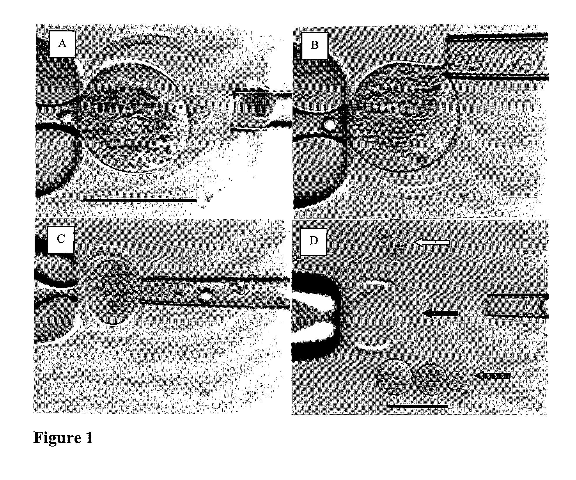
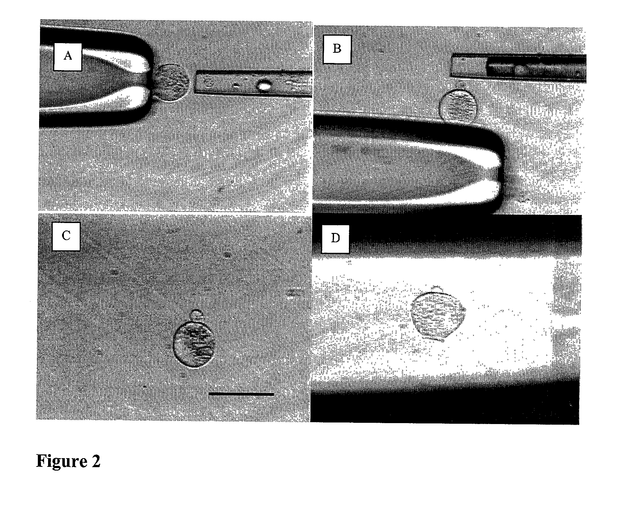
[0387] Micromanipulation and Enucleation of Bovine Oocytes

[0388] Micromanipulation and enucleation of bovine oocytes was performed as follows. Micromanipulation was performed on a inverted microscope (Nikon, Japan) using micromanipulators (Narashige, Japan). The mature metaphase II oocytes were introduced to HECM containing 10% Plasmanate and 7.5-15.0 .mu.g / ml cytochalasin B (Sigma C6762). Next, a holding micropipette (Humagen, Charlottesville, Va.) was used to grasp the oocytes. While holding the oocyte, the zona pellucida of each oocyte was partially dissected (dissolved) by application of an acidic tyrodes solution (Sigma T1788). The acidic tyrodes solution was applied using a 20-30 .mu.m diameter micropipette (Humagen, Charlottesville, Va.). The zona was dissolved adjacent to the polar body of the mature oocyte. Following breach of the zona, a 20-50 .mu.m micrometer polished micropipette (Humagen, Charlottesville, Va.) was used to gently aspirate the polar body and underlying cy...

example iii

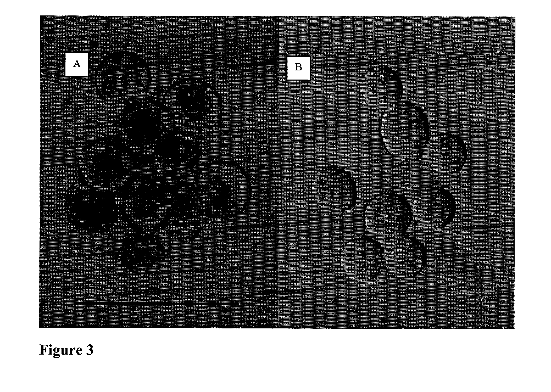
[0389] Ooplastoid Generation from Bovine Oocytes

[0390] Ooplastoid generation for bovine oocytes was performed as follows. Enucleated oocytes were introduced to HECM containing 10% Plasmanate and 7.5-15.0 .mu.g / ml cytochalasin B. A micromanipulator (Narashige, Japan) was used to manipulate the enucleated oocytes. A holding micropipette (Humagen 10MPH-120, Charlottesville, Va.) was used to grasp and orient the enucleated oocytes. A 20-50 .mu.m polished micropipette (Humagen custom, Charlottesville, Va.) was used to gently aspirate and pinch off a portion of the enucleated oocyte. This process was repeated until each enucleated oocyte was partitioned into 3-5 zona pellucida free ooplastoids having from 20 to 33% of the volume of the original oocyte. This procedure was repeated until each enucleated oocyte was appropriately partitioned into ooplastoids. Ooplastoids were washed in HECM with 10% Plasmanate to remove Cytochalasin B for further micromanipulation.

example iv

[0391] Preparation of Bovine Somatic Cells for Nuclear Transfer

[0392] The source of bovine somatic cell nucleus for experiments described here has been granulosa cells. Granulosa cells were obtained from bovine oocyte / granulosa masses. The granulosa masses were subjected to chemical treatment with 0.5-1.0 mg / ml hyaluronidase (Sigma H3757) followed by mechanical removal of granulosa through repeated pipetting of the cells using fine bore Pasteur pipettes. Subsequently, the isolated granulosa cells were washed with HECM with 10% Plasmanate to remove hyaluronidase. Next, granulosa were cultured in ECM or HECM supplemented with 10% FCS or Plasmanate in preparation for further micromanipulation. Alternatively, granulosa or any other type of somatic cell may be cultured in ECM supplemented with 0.5% fetal calf serum or Plasmanate for 24 to 72 h to induce quiescence prior to nuclear transfer.


PUM

PropertyMeasurementUnit
Fractionaaaaaaaaaa
Fractionaaaaaaaaaa
Fractionaaaaaaaaaa

Abstract

This invention provides a method for deriving precursors to pluripotent non-embryonic stem (P-PNES) and pluripotent non-embryonic stem (PNES) cell lines. The present invention involves nuclear transfer of genetic material from a somatic cell into an enucleated, zona pellucida free human ooplastoid having a reduced amount of total cytoplasm. The present invention provides a new source for obtaining human and other animal pluripotent stem cells. The source utilizes as starting materials an oocyte and a somatic cell as the starting materials but does not require the use, creation and/or destruction of embryos or fetal tissue and does not in any way involve creating a cloned being. The oocyte never becomes fertilized and never develops into an embryo. Rather, portions of the oocyte cytoplasm are extracted and combined with the nuclear material of individual mature somatic cells in a manner that precludes embryo formation. Murine, bovine, and human examples of the procedure are demonstrated. Subsequently, the newly constructed P-PNES cells are cultured in vitro and give rise to PNES cells and cell colonies. Methods are described for culturing the P-PNES cells to yield purified PNES cells which have the ability to differentiate into cells derived from mesoderm, endoderm, and ectoderm germ layers. Methods are described for maintaining and proliferating PNES cells in culture in an undifferentiated state. Methods and results are described for analysis and validation of pluripotency of PNES cells including cell morphology, cell surface markers, pluripotent tumor development in SCID mouse, karyotyping, immortality in in vitro culture.

Description


Claims


Application Information

Patent Timeline
Owner STEMA
Why Eureka
Try Eureka