Method and nucleic acids for the analysis of astrocytomas

a technology of astrocytoma and nucleic acids, applied in the field of methods and nucleic acids for the analysis of astrocytoma, can solve the problems of slow growth and generally noninvasive, diagnosis by such methodologies does not utilise the molecular basis of the progression, and no single marker has been shown to be sufficient for the distinction between the two tumors

Inactive Publication Date: 2004-06-17
EPIGENOMICS AG
View PDF2 Cites 36 Cited by
  • Summary
  • Abstract
  • Description
  • Claims
  • Application Information

AI Technical Summary

Benefits of technology

[0024] The object of the present invention is further achieved by an oligonucleotide or oligomer for the analysis of chemically pretreated DNA, for detecting the genomic cytosine methylation state, said oligonucleotide containing at least one base sequence having a length of at least 13 nucleotides which hybridizes to a chemically pretreated genomic DNA according to Seq. ID No.1 through Seq. ID No.120. The oligomer probes according to the present invention constitute important and effective tools which, for the first time, make it possible to ascertain specific genetic and epigenetic parameters of brain tumors, in particular, for use in characterisation, classification, differentiation, grading, staging, treatment and / or diagnosis of astrocytomas. The base sequence of the oligomers preferably contains at least one CpG dinucleotide. The probes may also exist in the form of a PNA (peptide nucleic acid) which has particularly preferred pairing properties. Particularly preferred are oligonucleotides according to the present invention in which the cytosine of the CpG dinucleotide is the 5.sup.th-9.sup.th nucleotide from the 5'-end of the 13-mer; in the case of PNA-oligomers, it is preferred for the cytosine of the CpG dinucleotide to be the 4.sup.th-6.sup.th nucleotide from the 5'-end of the 9-mer.
[0045] According to the present invention, it is preferred that the labels of the amplificates are fluorescence labels, radionuclides, or detachable molecule fragments having a typical mass which can be detected in a mass spectrometer. The mass spectrometer is preferred for the detection of the amplificates, fragments of the amplificates or of probes which are complementary to the amplificates, it being possible for the detection to be carried out and visualized by means of matrix assisted laser desorption / ionization mass spectrometry (MALDI) or using electron spray mass spectrometry (ESI). The produced fragments may have a single positive or negative net charge for better detectability in the mass spectrometer.

Problems solved by technology

They occasionally form cysts, or are enclosed within cysts, and are slow growing and generally noninvasive.
Diagnosis by such methodologies does not utilise the molecular basis of the progression to malignancy.
However, no single marker has been shown to be sufficient for the distinction between the two tumors.
Application as a routine diagnostic tool in a clinical environment is however impeded by the extreme instability of mRNA, the rapidly occuring expression changes following certain triggers (e.g. sample collection), and, most importantly, the large amount of mRNA needed for analysis (Lipshutz, R. J. et al., Nature Genetics 21:20-24, 1999; Bowtell, D. D. L. Nature genetics suppl.
However, the techniques used in such studies (restriction landmark genomic scanning, imprinting analysis) are: limited to research, they are unsuitable for use in a clinical or diagnostic setting, and do not provide the basis for the development of a medium or high throughput method for the analysis of gliomas.
However, 5-methylcytosine positions cannot be identified by sequencing since 5-methylcytosine has the same base pairing behavior as cytosine.
Moreover, the epigenetic information carried by 5-methylcytosine is completely lost during PCR amplification.
However, 5-methylcytosine remains unmodified under these conditions.
However, currently only individual regions of a length of up to approximately 3000 base pairs are analyzed, a global analysis of cells for thousands of possible methylation events is not possible.
However, this method cannot reliably analyze very small fragments from small sample quantities either.
These are lost through the matrix in spite of the diffusion protection.
For nucleic acids having a multiply negatively charged backbone, the ionization process via the matrix is considerably less efficient.

Method used


Image

  • Method and nucleic acids for the analysis of astrocytomas
  • Method and nucleic acids for the analysis of astrocytomas
  • Method and nucleic acids for the analysis of astrocytomas

Examples


example 2

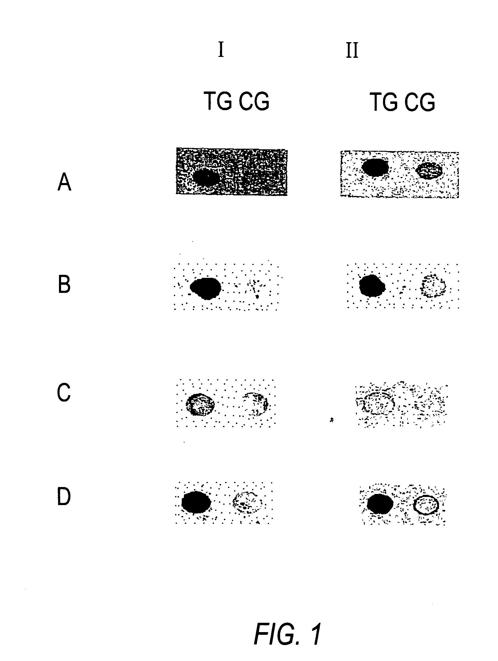
[0075] Methylation Analysis of the Gene NF1.

[0076] The following example relates to a fragment of the gene NF1 in which a specific CG-position is to be analyzed for methylation.

[0077] In the first step, a genomic sequence is treated using bisulfite (hydrogen sulfite, disulfite) in such a manner that all cytosines which are not methylated at the 5-position of the base are modified in such a manner that a different base is substituted with regard to the base pairing behavior while the cytosines methylated at the 5-position remain unchanged.

[0078] If bisulfite solution is used for the reaction, then an addition takes place at the non-methylated cytosine bases. Moreover, a denaturating reagent or solvent as well as a radical interceptor must be present. A subsequent alkaline hydrolysis then gives rise to the conversion of non-methylated cytosine nucleobases to uracil. The chemically converted DNA is then used for the detection of methylated cytosines. In the second method step, the trea...

example 3

[0081] Methylation Analysis of the Gene MLH1.

[0082] The following example relates to a fragment of the gene MLH1 in which a specific CG-position is to be analyzed for methylation.

[0083] In the first step, a genomic sequence is treated using bisulfite (hydrogen sulfite, disulfite) in such a manner that all cytosines which are not methylated at the 5-position of the base are modified in such a manner that a different base is substituted with regard to the base pairing behavior while the cytosines methylated at the 5-position remain unchanged.

[0084] If bisulfite solution is used for the reaction, then an addition takes place at the non-methylated cytosine bases. Moreover, a denaturating reagent or solvent as well as a radical interceptor must be present. A subsequent alkaline hydrolysis then gives rise to the conversion of non-methylated cytosine nucleobases to uracil. The chemically converted DNA is then used for the detection of methylated cytosines. In the second method step, the tr...

example 4

[0087] Methylation Analysis of the Gene CSNK2B.

[0088] The following example relates to a fragment of the gene CSNK2B in which a specific CG-position is to be analyzed for methylation.

[0089] In the first step, a genomic sequence is treated using bisulfite (hydrogen sulfite, disulfite) in such a manner that all cytosines which are not methylated at the 5-position of the base are modified in such a manner that a different base is substituted with regard to the base pairing behavior while the cytosines methylated at the 5-position remain unchanged.

[0090] If bisulfite solution is used for the reaction, then an addition takes place at the non-methylated cytosine bases. Moreover, a denaturating reagent or solvent as well as a radical interceptor must be present. A subsequent alkaline hydrolysis then gives rise to the conversion of non-methylated cytosine nucleobases to uracil. The chemically converted DNA is then used for the detection of methylated cytosines. In the second method step, th...


PUM

PropertyMeasurementUnit
lengthaaaaaaaaaa
heat-resistantaaaaaaaaaa
fluorescenceaaaaaaaaaa

Abstract

The present invention relates to chemically modified genomic sequences, to oligonucleotides and / or PNA-oligomers for detecting the cytosine methylation state of genomic DNA, as well as to a method for ascertaining genetic and / or epigenetic parameters of genes for use in the characterisation, classification, differentiation, grading, staging, treatment and / or diagnosis of astrocytomas, or the predisposition to astrocytomas.

Description

[0001] The levels of observation that have been studied by the methodological developments of recent years in molecular biology, are the genes themselves, the translation of these genes into RNA, and the resulting proteins. The question of which gene is switched on at which point in the course of the development of an individual, and how the activation and inhibition of specific genes in specific cells and tissues are controlled is correlatable to the degree and character of the methylation of the genes or of the genome. In this respect, pathogenic conditions may manifest themselves in a changed methylation pattern of individual genes or of the genome.[0002] The present invention relates to nucleic acids, oligonucleotides, PNA-oligomers, and to a method for the characterisation, classification, differentiation, grading, staging, treatment and / or diagnosis of astrocytomas, or the predisposition to astrocytomas, by analysis of the genetic and / or epigenetic parameters of genomic DNA an...

Claims


Application Information

Patent Timeline
Patent Type & Authority Applications(United States)
IPC IPC(8): C07K14/47C07K14/82C12M1/00G01N27/62C12M1/34C12N15/09C12Q1/68G01N33/53G01N33/58G01N37/00
CPCC07K14/4703C07K14/82C12Q1/6883C12Q2600/154C12Q2523/125C12Q2600/156C12Q1/6886
Inventor OLEK, ALEXANDERPIEPENBROCK, CHRISTIANBERLIN, KURT
Owner EPIGENOMICS AG
Why Eureka
Try Eureka