Method and nucleic acids for the differentiation of prostate tumors

a prostate cancer and nucleic acid technology, applied in combinational chemistry, biochemistry apparatus, chemical libraries, etc., can solve the problems of insufficient or small biopsy samples affecting routine analysis, difficult to distinguish early stages of prostate carcinoma from benign hyperplasia of the prostate, and no single marker has been shown to be sufficient for the distinction between the two lesions

Inactive Publication Date: 2008-06-03
EPIGENOMICS AG
View PDF28 Cites 6 Cited by
  • Summary
  • Abstract
  • Description
  • Claims
  • Application Information

AI Technical Summary

Benefits of technology

[0020]The object of the present invention is further achieved by an oligonucleotide or oligomer for detecting the cytosine methylation state in chemically pretreated DNA, containing at least one base sequence having a length of at least 9 to 13 nucleotides which hybridizes to a chemically pretreated genomic DNA according to Seq. ID No.1 through Seq. ID No.112. The oligomer probes according to the present invention constitute important and effective tools which, for the first time, make it possible to ascertain specific genetic and epigenetic parameters of carcinomas, in particular, for use in differential diagnosis of prostate tumors and for diagnosis of predisposition to prostate carcinomas. The base sequence of the oligomers preferably contains at least one CpG dinucleotide. The probes may also exist in the form of a PNA (peptide nucleic acid) which has particularly preferred pairing properties. Particularly preferred are oligonucleotides according to the present invention in which the cytosine of the CpG dinucleotide is the 5th-9th nucleotide from the 5′-end of the 13-mer; in the case of PNA-oligomers, it is preferred for the cytosine of the CpG dinucleotide to be the 4th-6th nucleotide from the 5′-end of the 9-mer.
[0042]According to the present invention, it is preferred that the labels of the amplificates are fluorescence labels, radionuclides, or detachable molecule fragments having a typical mass which can be detected in a mass spectrometer. The mass spectrometer is preferred for the detection of the amplificates, fragments of the amplificates or of probes which are complementary to the amplificates, it being possible for the detection to be carried out and visualized by means of matrix assisted laser desorption / ionization mass spectrometry (MALDI) or using electron spray mass spectrometry (ESI). The produced fragments may have a single positive or negative net charge for better detectability in the mass spectrometer.

Problems solved by technology

Particularly early stages of prostate carcinoma are often difficult to distinguish from benign hyperplasia of the prostate by routine histological examination even if an adequate biopsy is obtained (McNeal J E et al., Hum Pathol 2001, 32:441-6).
Furthermore, small or otherwise insufficient biopsy samples sometimes impede routine analysis.
However, no single marker has been shown to be sufficient for the distinction between the two lesions so far.
Application as a routine diagnostic tool in a clinical environment is however impeded by the extreme instability of mRNA, the rapidly occurring expression changes following certain triggers (e.g. sample collection), and, most importantly, the large amount of mRNA needed for analysis (Lipshutz, R. J. et al., Nature Genetics 21:20-24, 1999; Bowtell, D. D. L. Nature genetics suppl.
However, 5-methylcytosine positions cannot be identified by sequencing since 5-methylcytosine has the same base pairing behavior as cytosine.
Moreover, the epigenetic information carried by 5-methylcytosine is completely lost during PCR amplification.
However, 5-methylcytosine remains unmodified under these conditions.
However, currently only individual regions of a length of up to approximately 3000 base pairs are analyzed, a global analysis of cells for thousands of possible methylation events is not possible.
However, this method cannot reliably analyze very small fragments from small sample quantities either.
These are lost through the matrix in spite of the diffusion protection.
For nucleic acids having a multiply negatively charged backbone, the ionization process via the matrix is considerably less efficient.

Method used


Image

  • Method and nucleic acids for the differentiation of prostate tumors

Examples


example 2

Differentiation of Prostate Tumors

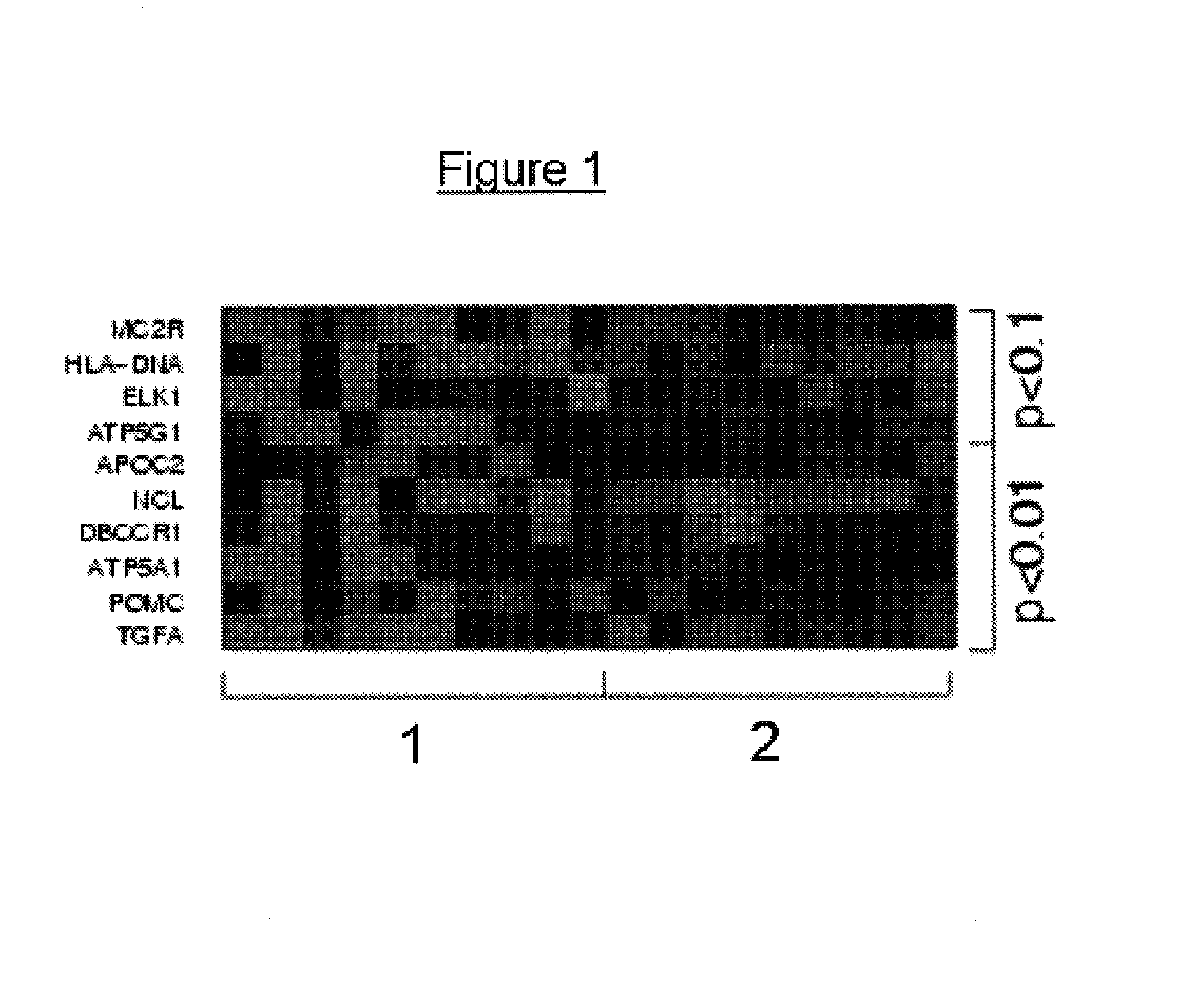
[0065]In order to relate the methylation patterns to a specific tumour type, it is initially required to analyze the DNA methylation patterns of two groups of patients with alternative forms of a tumor, in this case one group of benign prostate hyperplasia and another group of prostate carcinoma. These analyses were carried out, analogously to Example 1. The results obtained in this manner are stored in a database and the CpG dinucleotides which are methylated differently between the two groups are identified. This can be carried out by determining individual CpG methylation rates as can be done, for example, by sequencing, which is a relatively imprecise method of quantifying methylation at a specific CpG, or else, in a very precise manner, by a methylation-sensitive “primer extension reaction”. In a particularly preferred variant the methylation status of hundreds or thousands of CpGs may be analysed on an oligomer array. It is also possible for t...


PUM

PropertyMeasurementUnit
nucleic acidaaaaaaaaaa
peptide nucleic acidaaaaaaaaaa
sizesaaaaaaaaaa

Abstract

The present invention relates to chemically modified genomic sequences, to oligonucleotides and / or PNA-oligomers for detecting the cytosine methylation state of genomic DNA, as well as to a method for ascertaining genetic and / or epigenetic parameters of genes for use in characterisation, classification, differentiation, diagnosis and therapy of prostate lesions.

Description

FIELD OF THE INVENTION[0001]The levels of observation that have been studied by the methodological developments of recent years in molecular biology, are the genes themselves, the translation of these genes into RNA, and the resulting proteins. The question of which gene is switched on at which point in the course of the development of an individual, and how the activation and inhibition of specific genes in specific cells and tissues are controlled is correlatable to the degree and character of the methylation of the genes or of the genome. In this respect, pathogenic conditions may manifest themselves in a changed methylation pattern of individual genes or of the genome.[0002]The present invention relates to nucleic acids, oligonucleotides, PNA-oligomers, and to a method for characterisation, classification, differentiation, and / or diagnosis of prostate lesions, or the predisposition to prostate cancer, by analysis of the genetic and / or epigenetic parameters of genomic DNA and, in...

Claims


Application Information

Patent Timeline
Patent Type & Authority Patents(United States)
IPC IPC(8): C12Q1/68C07H21/04G01N27/62A61K31/7088A61P13/08A61P35/00C12M1/00C12M1/34C12N15/09C12Q1/6827C12Q1/6886C40B40/06G01N33/53G01N37/00
CPCC12Q1/6827C12Q1/6886B01J2219/00497B01J2219/00527B01J2219/00596B01J2219/00605B01J2219/0061B01J2219/00612B01J2219/00659B01J2219/00702B01J2219/00722C12Q2600/154C40B40/06C12Q2565/627C12Q2523/125A61P13/08A61P35/00
Inventor DISTLER, JUERGENMODEL, FABIANADORJAN, PETER
Owner EPIGENOMICS AG
Why Eureka
Try Eureka