Medical-use image data analyzing apparatus and method of analysis using the same

a technology of image data and analysis method, applied in image enhancement, diagnostic recording/measuring, instruments, etc., can solve the problems of not being able to easily compare the timing of expansion and contraction among several local portions

Active Publication Date: 2006-04-20
TOSHIBA MEDICAL SYST CORP
View PDF4 Cites 12 Cited by
  • Summary
  • Abstract
  • Description
  • Claims
  • Application Information

AI Technical Summary

Problems solved by technology

However, conventionally there has been no method of easily comparing timings of expansion and constriction among several local portions.

Method used


Image

  • Medical-use image data analyzing apparatus and method of analysis using the same
  • Medical-use image data analyzing apparatus and method of analysis using the same
  • Medical-use image data analyzing apparatus and method of analysis using the same

Examples


first embodiment

[0017] Referring now to FIG. 1 to FIG. 4, a first embodiment of the present invention will be described.

(1) Structure of Medical Use Image Data Analyzing Apparatus

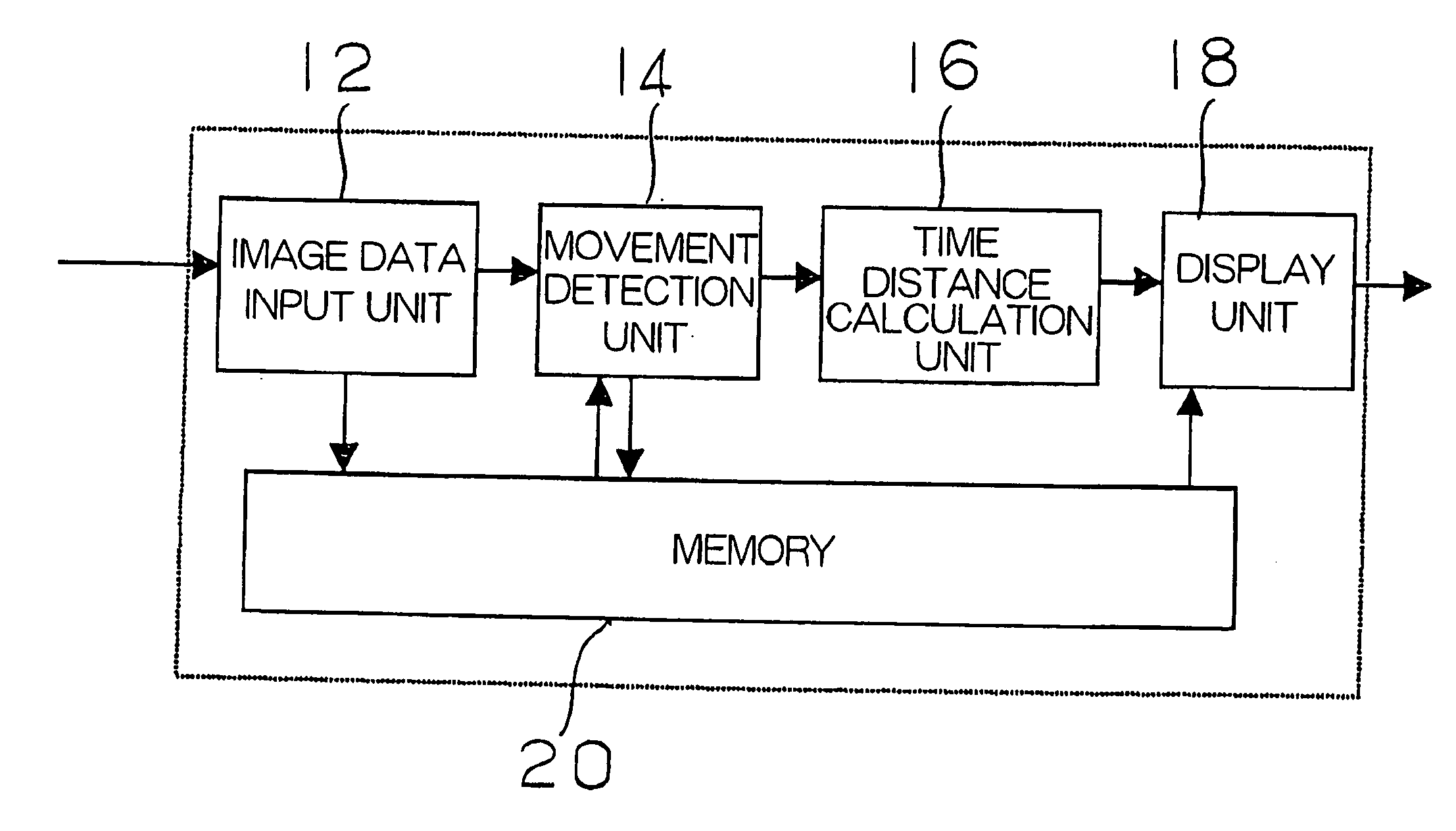
[0018]FIG. 1 is a block diagram of a medical use image data analyzing apparatus 10 according to the first embodiment.

[0019] The medical use image data analyzing apparatus 10 according to the first embodiment is inputted with data of images of a test subject, which is acquired by a separate device, and displays differences in timing of movement of respective local portions of the test subject, as an output data.

[0020] The medical use image data analyzing apparatus 10 according to the first embodiment includes; an image data input unit 12 for inputting an image data; a movement detection unit 14 for detecting movement of a test subject using the image data; a time distance calculation unit 16 for calculating a time distance up to a time point when a movement parameter calculated from the detected movement reaches the ma...

second embodiment

[0029] Subsequently, referring to FIG. 5 to FIG. 8, a medical use image data analyzing apparatus 10 according to a second embodiment will be described.

[0030] The medical use image data analyzing apparatus 10 according to the second embodiment is an example in which the temporal transitions or history of the moving states of the respective local portions are displayed more quantitatively.

(1) Structure of the Medical Use Image Data Analyzing Apparatus

[0031] The medical use image data analyzing apparatus 10 includes, as shown in FIG. 5; the image data input unit 12 for inputting the image data; the movement detection unit 14 for detecting the movement of the test subject using the image data; a movement parameter variation calculation unit 17 for calculating the movement parameters from the detected movement for the respective local portions at each time instance; the display unit 18 for listing and displaying variations of the movement parameters for a plurality of the local porti...


PUM


Abstract

A medical-use image data analyzing apparatus, aiming for detecting with a simple operation, presence or extent of difference in timing of movements among local portions as to facilitate diagnosis of diseases such as cardiac infarction, includes: an image input unit configured to input an image data of a test subject; a movement detector configured to detect movement of the test subject, based on the image data; a time distance calculator which calculates a time distance up to a time point when movement parameter, which is calculated from the movement, reaches local maximum or local minimum; a display device configured to display the time distances for the respective local portions of the test subject; and a memory.

Description

CROSS-REFERENCE TO RELATED APPLICATIONS [0001] This application is based upon and claims the benefit of priority from the prior Japanese Patent Application No. 2004-302123, filed on Oct. 15, 2004; the entire contents of which are incorporated herein by reference. TECHNICAL FIELD [0002] The present invention relates to a medical-use image data analyzing apparatus for analyzing movement of organs or the like. BACKGROUND OF THE INVENTION [0003] It is important to measure movement of organs accurately as exemplified by a diagnosis of cardiac disease. A pumping function of a heart can be obtained by expansion and constriction of the heart at adequate timings. For example, in the case of cardiac infarction, which is one of the cardiac diseases in which part of cardiac muscle does not function normally and hence its pumping function is lowered, it is considered that timings of expansion and constriction are important indicator or “barometer” of symptoms in diagnosis. [0004] Hitherto disclo...

Claims


Application Information

Patent Timeline
Patent Type & Authority Applications(United States)
IPC IPC(8): G06K9/00
CPCG06T7/0012G06T7/2033G06T2207/30048G06T7/0016G06T7/246
Inventor NISHIURA, MASAHIDE
Owner TOSHIBA MEDICAL SYST CORP
Why Eureka
Try Eureka