Segmentation of lesions in ultrasound images

a technology of segmentation and ultrasound, applied in the field of digital image processing of ultrasound images, can solve the problems of common cause of cancer death, misclassification of lesions in everyday clinical practice, and segmentation of lesions from surrounding tissues

Inactive Publication Date: 2006-11-02
CARESTREAM HEALTH INC
View PDF6 Cites 50 Cited by
  • Summary
  • Abstract
  • Description
  • Claims
  • Application Information

AI Technical Summary

Benefits of technology

[0032] According to one aspect of the present invention, there is provided a method for determining a candidate lesion region in a digital ultrasound medical image of anatomical tissue. The method includes the steps of: accessing the digital ultrasound medical image of anatomical tissue; applying an anisotropic diffusion filter to the ultrasound image to generate a filtered ultrasound image; performing a normalized cut operation on the filtered ultrasound image to partition the filtered ultrasound image into a plurality of regions; and selecting, from the plurality of regions, at least one region as a candidate lesion region.
[0033] According to another aspect of the present invention, there is provided a method for determining a candidate lesion region in a digital ultrasound medical image of anatomical tissue. The method includes the steps of: (1) accessing the digital u

Problems solved by technology

Breast cancer is a common cancer of women and a common cause of cancer deaths.
Despite improved radiographic criteria for differentiating malignant from benign lesions of the breast, misclassification of lesions can occur in everyday clinical practice.
A difficulty which has been associated with a computerized system for detecting and diagnosing breast lesions is the segmentation of the lesion regions from the surrounding tissues.
This manual procedure is labor-intensive, can disrupt full automation, and can be prone to human error, inconsistency, and subjectivity.
Given that ultrasound images comprise speckle noise and tissue related textures, accurate segmentation ta

Method used


Image

  • Segmentation of lesions in ultrasound images
  • Segmentation of lesions in ultrasound images
  • Segmentation of lesions in ultrasound images

Examples


Embodiment Construction

[0041] The following is a detailed description of the preferred embodiments of the invention, reference being made to the drawings in which the same reference numerals identify the same elements of structure in each of the several figures.

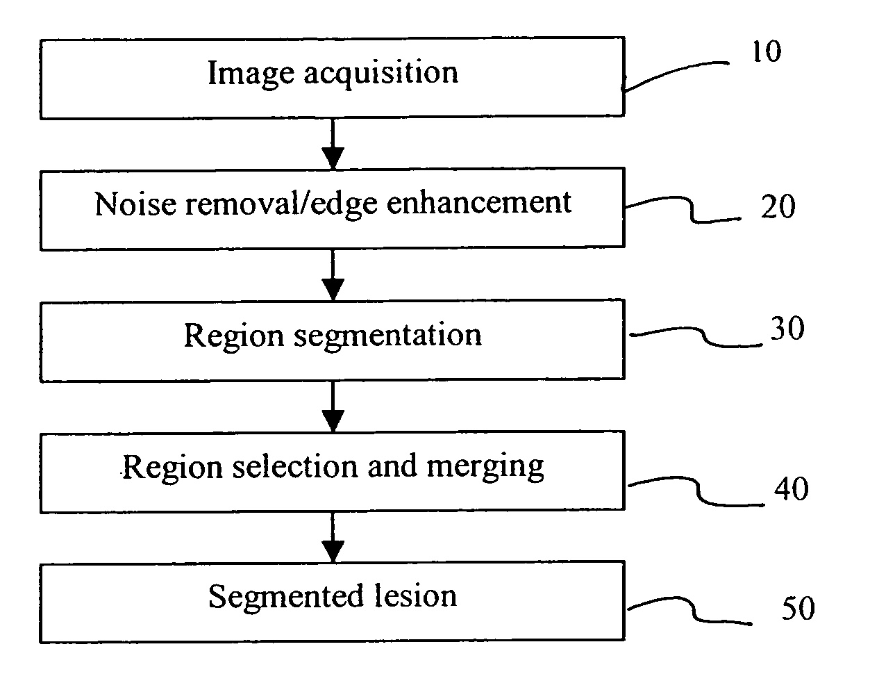
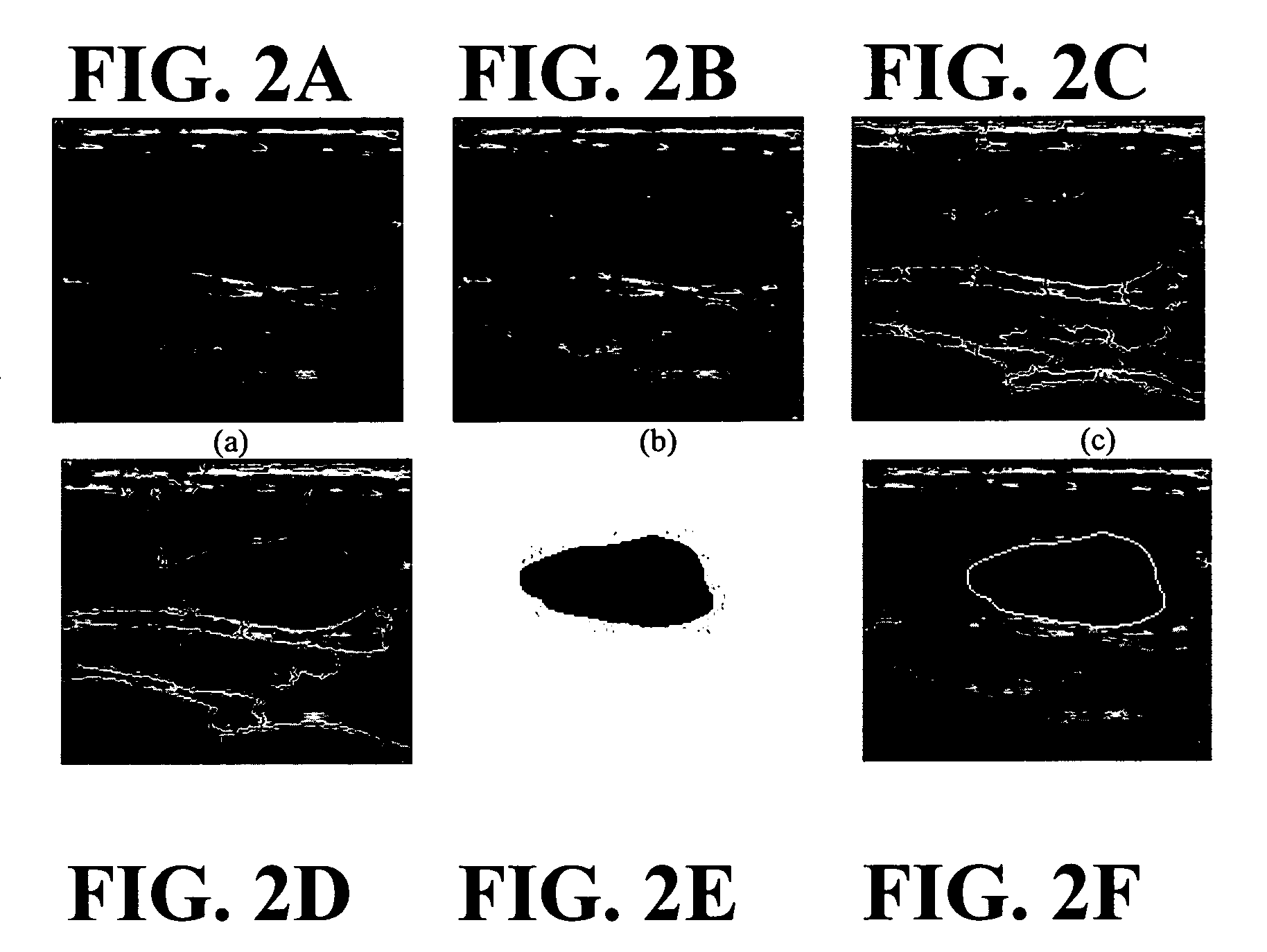
[0042]FIG. 1 shows a flowchart generally illustrating the method in accordance with the present invention. At step 10, an input ultrasound image is accessed / acquired / provided for analysis. A nonlinear filtering is applied on the input ultrasound image to remove the noise while preserving the edge (step 20). At step 30, the region is segmented. Region selection and merging is accomplished at step 40, and the lesion is segmented at step 50.

[0043] More particularly, once the digital ultrasound image of anatomical tissue is accessed, an anisotropic diffusion filter is used to remove noise from the image. Then, spatially contiguous pixels of the digital ultrasound image are segmented into a plurality of regions in accordance with substantially similar...


PUM


Abstract

A method for determining a candidate lesion region in a digital ultrasound medical image of anatomical tissue. The method includes the steps of: accessing the digital ultrasound medical image of anatomical tissue; applying an anisotropic diffusion filter to the ultrasound image to generate a filtered ultrasound image; performing a normalized cut operation on the filtered ultrasound image to partition the filtered ultrasound image into a plurality of regions; and selecting, from the plurality of regions, at least one region as a candidate lesion region.

Description

CROSS REFERENCE TO RELATED APPLICATIONS [0001] Reference is made to, and priority is claimed from, commonly assigned Provisional Patent Application U.S. Ser. No. 60 / 675,629, entitled “SYSTEMS AND METHODS FOR AUTOMATED ANALYSIS OF LESIONS ON ULTRASOUND IMAGES”, filed on Apr. 28, 2005 in the names of Huo et al., and which is assigned to the assignee of this application, and incorporated herein by reference.FIELD OF THE INVENTION [0002] The invention relates to digital image processing of ultrasound images, and more particularly to detecting lesions within such images. BACKGROUND OF THE INVENTION [0003] Breast cancer is a common cancer of women and a common cause of cancer deaths. An effective way to improve prognosis and survival rate is early detection and treatment of breast cancer. Mammography is an imaging modality which has provided some effectiveness in the early detection of clinically occult breast cancer, and is viewed by some to be a primary imaging modality for breast cance...

Claims


Application Information

Patent Timeline
IPC IPC(8): A61B8/00
CPCG06T7/0012G06T7/0081G06T2207/30068G06T2207/10132G06T2207/20036G06T7/0093G06T7/11G06T7/162
Inventor HUO, ZHIMINLIU, XU
Owner CARESTREAM HEALTH INC
Why Eureka
Try Eureka