

Marker device for X-ray, ultrasound and MR imaging
- Summary
- Abstract
- Description
- Claims
- Application Information
AI Technical Summary
Benefits of technology
Problems solved by technology
Method used
Image

Examples
Embodiment Construction
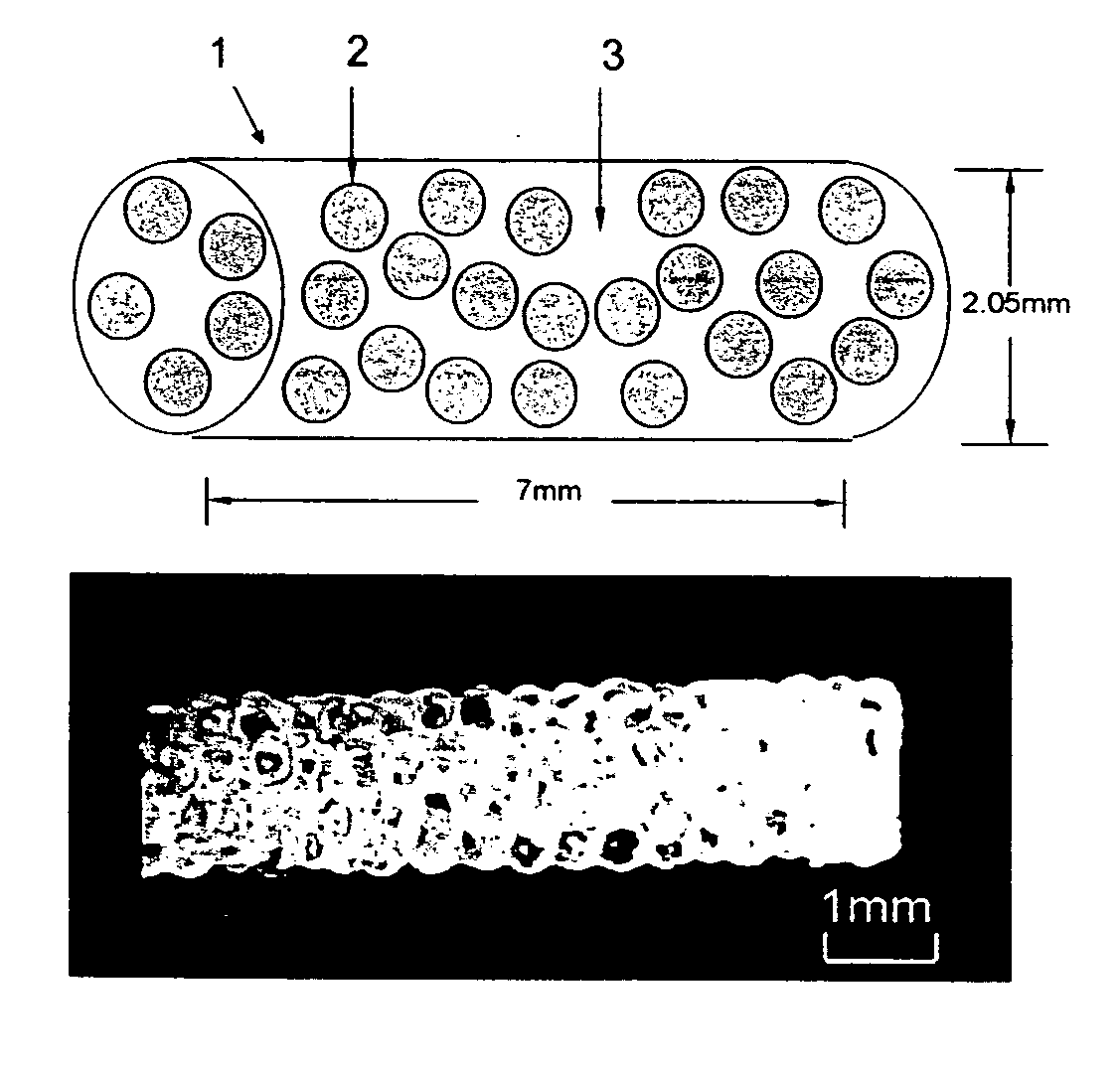
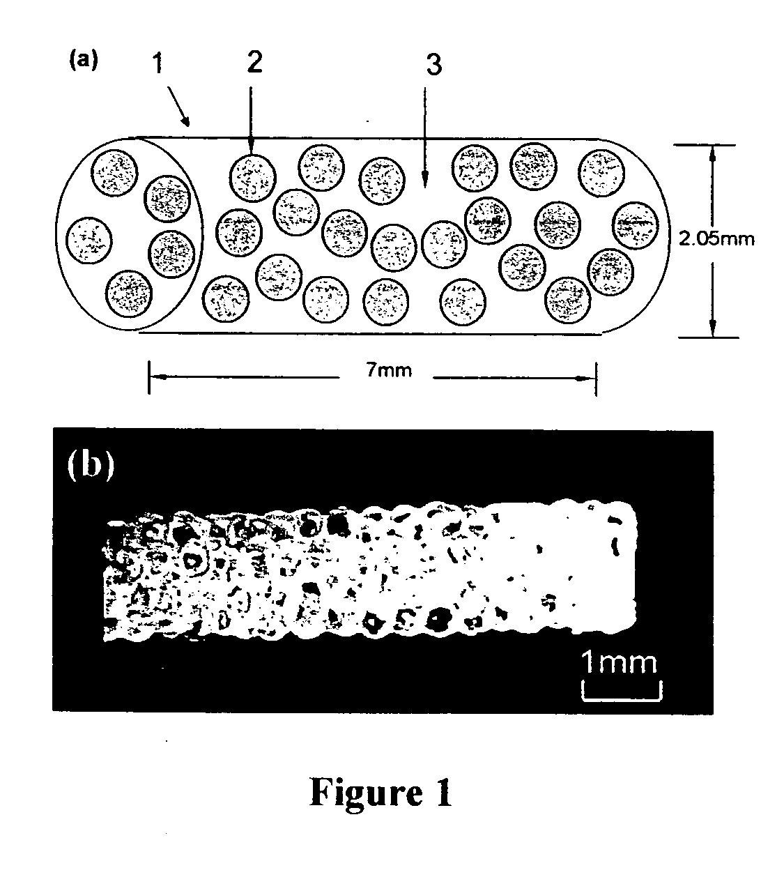
[0031] X-ray mammography remains the primary screening and initial detection method for breast cancer. The distinction between benign and malignant masses is generally made by analysis of the margins, shape, density,15 analysis of the margins, shape, density, and size of any detected lesion. A benign lesion, such as a cyst or fibroadenoma, typically has a sharply circumscribed margin and oval or round shape, whereas malignant masses often exhibit speculated contours due to the infiltrative nature of breast cancer. However, mammography has significant limitations in terms of imaging sensitivity and specificity.
[0032] MR imaging has become a viable adjunct to X-ray mammography for detecting breast lesions. Some reports indicate that MRI can yield 100% sensitivity in the detection of malignant breast lesions. Using contrast enhanced MR imaging methods, malignamt and benign tumors that cannot be seen with mammography are visible on MR images. Furthermore, by incorporating a number of m...
PUM
 Abstract
Description
Claims
Application Information