Method and Apparatus Including Use of Metalloporphyrins for Subsequent Optimization of Radiosurgery and Radiotherapy

a radiotherapy and metalloporphyrin technology, applied in the field of cancer screening and cancer treatment, can solve the problems of inability to repeat interventional procedures to treat neoplastic tissue, and inability to effectively treat many types of diseased tissue, so as to reduce the size of tumors, enhance the ability to provide radiosurgical beams, and eliminate the exposure of healthy tissue

Inactive Publication Date: 2007-02-22
ADAIR
View PDF3 Cites 41 Cited by
  • Summary
  • Abstract
  • Description
  • Claims
  • Application Information

AI Technical Summary

Benefits of technology

[0020] The present invention may make use of porphyrin compounds complexed with various metals such as silver (Ag), aluminum (Al), cadmium (Cd), cobalt (Co), chromium (Cr), copper (Cu), iron (Fe), gadolinium (Gd), indium (In), lutetium (Lu), magnesium (Mg), manganese (Mn), nickel (Ni), palladium (Pd), platinum (Pt), rhodium (Rh), ruthenium (Ru), scandium (Sc), silicon (Si), tin (Sn), titanium oxide (TiO), vanadium oxide (VO), ytterbium (Yb) and zinc (Zn). These complexes are generally categorized as metalloporphyrins meaning a porphyrin moiety having a chelated radioactive isotope of a metal atom. These metalloporphyrins are further processed so that the metal is in the form of a radioactive isotope. The resulting radioactive metalloporphyrins thereby constitute radiopharmaceuticals that can be intravenously introduced to the patient. The affinity of neoplastic tissue for porphyrins results in selective uptake of the radioactive metalloporphyrin, thereby effecting targeted delivery of therapeutic radiopharmaceuticals. For example, in the instance of elemental copper chelated by the porphyrin, the copper can be transformed to radioactive 67Cu. In this way, introduction of the metalloporphyrin radiopharmacuetical to the patient is an effective means to deliver measured radiation therapy to targeted tissue. More specifically, 67Cu provides a source of beta radiation for selective destruction of neoplastic sites. Additionally, metalloporphyrin complexes still provide the ability to simultaneously conduct fluorescence detection and phototherapy if desired. Also, the metalloporphyrins provide the ability for observation of the targeted areas through PET (for example, through the use of 64Cu) or SPECT (for example, through use the of 67Cu).
[0026] One clear advantage to the above method is that in many instances, administration of the metalloporphyrin will greatly shrink a tumor size; therefore, the beam of radiation can be better focused onto a specific targeted area thereby further eliminating exposure of healthy tissue to the irradiating beam.
[0027] Thus, with the present invention, radiosurgery / radiotherapy can be optimized in a manner which enhances the ability to provide a radiosurgical beam to targeted areas in the body and to limit the adverse effects of radiation exposure of healthy tissue.

Problems solved by technology

In all uses of photodynamic therapy, it is well known that there are limitations in such therapy because of the poor penetration of the visible light required to activate the administered porophyrin so as to render it toxic to the targeted tissue.
Particularly for tumors which are found deep within the body of a patient, repeated interventional procedures to treat the neoplastic tissue become infeasible.
Accordingly, many types of diseased tissue cannot be effectively treated through photodynamic therapy.
Although an MRI or CT scan may be adequate under many circumstances, the disadvantages of CT scanning or MRI scanning is that these types of scans image the physical structure of tissue, and do not provide information regarding the body's chemistry, or cell function.

Method used


Image

  • Method and Apparatus Including Use of Metalloporphyrins for Subsequent Optimization of Radiosurgery and Radiotherapy
  • Method and Apparatus Including Use of Metalloporphyrins for Subsequent Optimization of Radiosurgery and Radiotherapy

Examples


Embodiment Construction

[0029] In order to execute the method and apparatus of the present invention, a system is provided for delivering a collimated ionizing beam of radiation to a targeted area of tissue. Examples of such systems to support the present invention are disclosed in the U.S. Pat. Nos. 5,427,097 and 5,207,223.

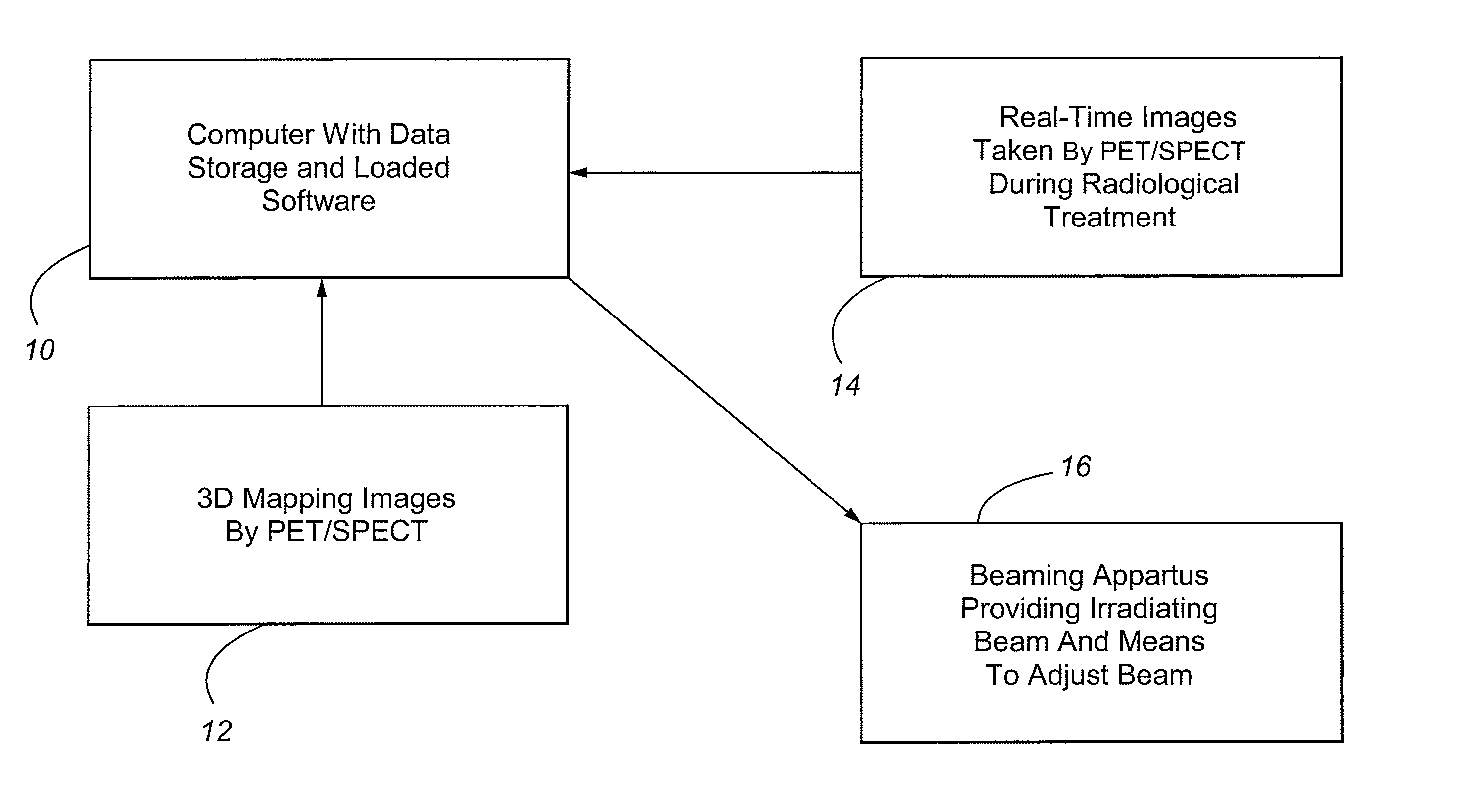
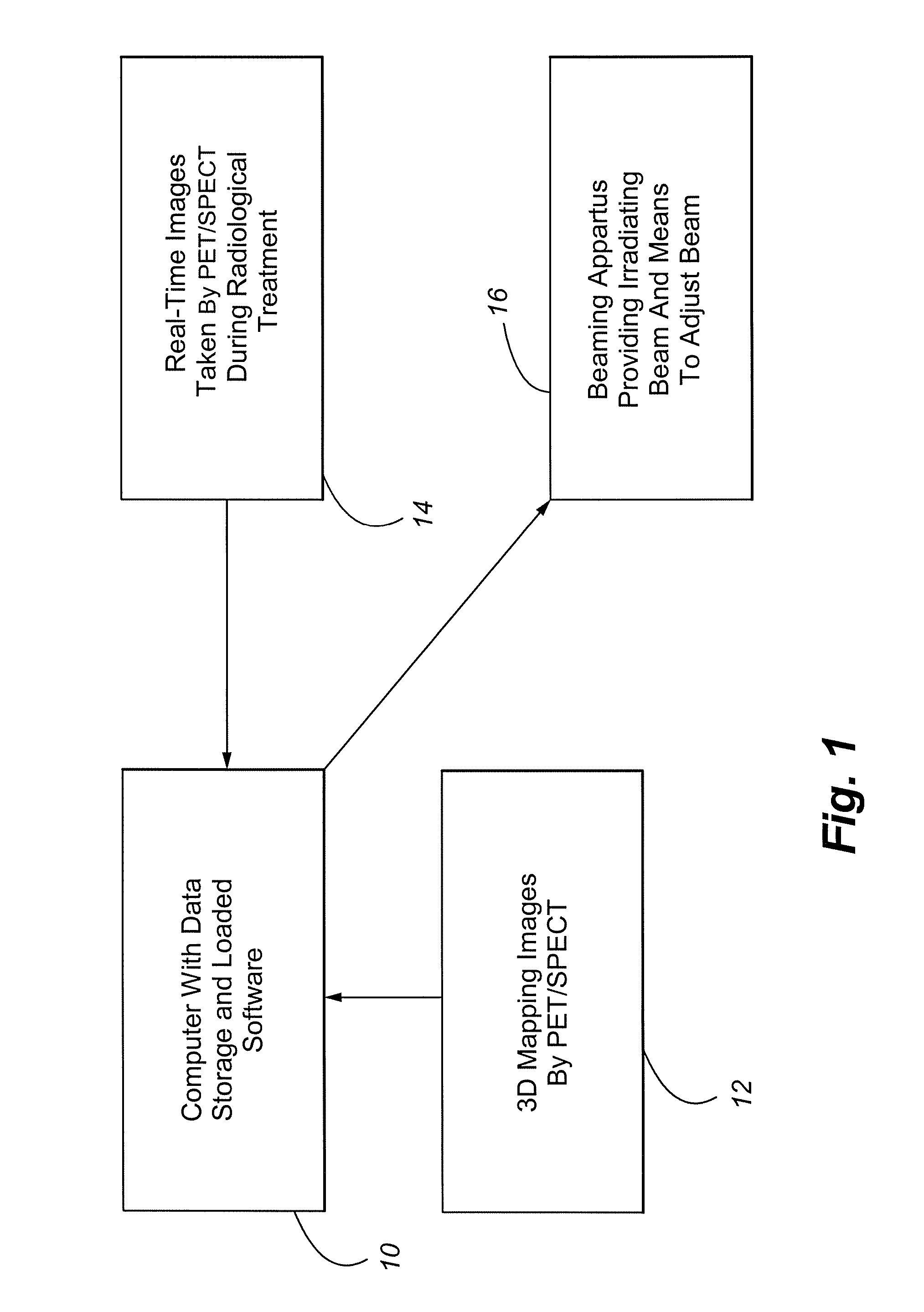
[0030] Referring to FIG. 1, a representative system to achieve the present invention includes a computer 10 with data storage capability that is capable of executing instructions from software loaded within the computer. The computer can store and manipulate 3-dimensional mapping data images 12 of a patient being treated. The 3-dimensional mapping is typically stored in digital form, and is loaded in the computer 10 for later comparison purposes. As mentioned above, the 3-dimensional mapping is preferably achieved by SPECT or PET scanning following administration of a metaloporphyrin to a patient. A beaming apparatus 16 is provided which, when activated, emits a collimated surgical ion...


PUM

PropertyMeasurementUnit
wave lengthsaaaaaaaaaa
half lifeaaaaaaaaaa
half lifeaaaaaaaaaa

Abstract

An apparatus and method for subsequent optimization of radiosurgery and radiotherapy is provided. The invention includes administering a metalloporphyrin to the patient, and then creating a 3-dimensional mapping of tissue through use of PET or SPECT. Malignant and pre-malignant tissue has an affinity for the metalloporphyrin. During treatment, real-time images are also provided which are compared to the previous 3-dimensional mapping. Creation of the real-time images is also achieved through PET or SPECT wherein a metalloporphyrin is administered to the patient. Total administration of radiation is calculated by summing radiation from the inetalloporphyrins and from the radiosurgery / radiotherapy. The amount of radiation delivered by the metalloporphyrins and by the radiosurgery / radiotherapy are adjustable based on a patient's response to the dual delivery.

Description

CROSS REFERENCE TO RELATED APPLICATIONS [0001] This application is a continuation-in-part of co-application Ser. No. 10 / 176,558, filed on Jun. 21, 2002, entitled “METHOD OF CANCER SCREENING PRIMARILY UTILIZING NON-INVASIVE CELL COLLECTION, FLUORESCENCE DETECTION TECHNIQUES, AND RADIO TRACING DETECTION TECHNIQUES”, the disclosure of which is hereby incorporated by reference herein.FIELD OF THE INVENTION [0002] This invention relates to cancer screening and cancer treatment, and more particularly, to the use of metalloporphyrins for subsequent optimization of radiosurgery and / or radiotherapy. BACKGROUND OF THE INVENTION [0003] There are a number of prior art methods and apparatuses which are used in the detection and treatment of cancer. Fluorescent markers have been used to help identify cancerous tissue within a patient. Radio tracers or markers have also been used in the detection and treatment of cancer. [0004] U.S. Pat. No. 5,391,547 discloses a method of using porphyrins to dete...

Claims


Application Information

Patent Timeline
Patent Type & Authority Applications(United States)
IPC IPC(8): A61B6/00A61N5/10C07D487/22G01T1/164A61K49/00G01N21/64A61K51/04C12Q1/02G01N33/48G01N33/483G01N33/50G01N33/574H04N3/15H04N5/225
CPCA61B6/12A61K49/0036A61K51/0485A61N5/1049A61N5/1067A61N2005/1061G01N33/5091G01N33/574H04N5/2253H04N2005/2255Y10S435/968H04N23/555H04N23/54
Inventor ADAIR
Owner ADAIR
Why Eureka
Try Eureka