Method for display of at least one medical finding

a medical finding and display technology, applied in the field of display of at least one medical finding, can solve the problems of increasing the effort of the doctor, unwieldy type of finding review,

Active Publication Date: 2007-04-05
SIEMENS HEALTHCARE GMBH
View PDF10 Cites 11 Cited by
  • Summary
  • Abstract
  • Description
  • Claims
  • Application Information

AI Technical Summary

Benefits of technology

[0007] An object of the present invention to provide a method that clea

Problems solved by technology

This type of finding review is, however, is unwieldy for the treating doctor or radiologist since he or she does not have direct access to the associated image data from the text protocol.
It ma

Method used


Image

  • Method for display of at least one medical finding
  • Method for display of at least one medical finding
  • Method for display of at least one medical finding

Examples


Embodiment Construction

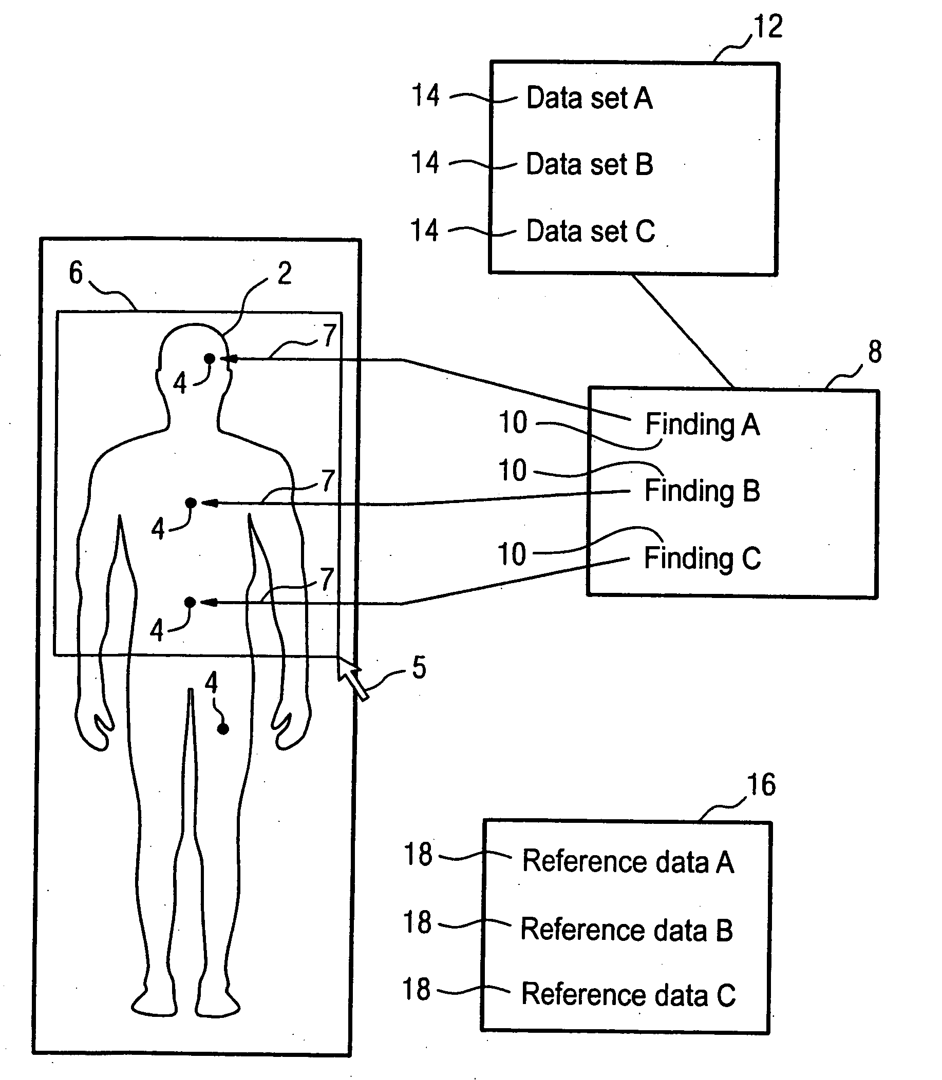
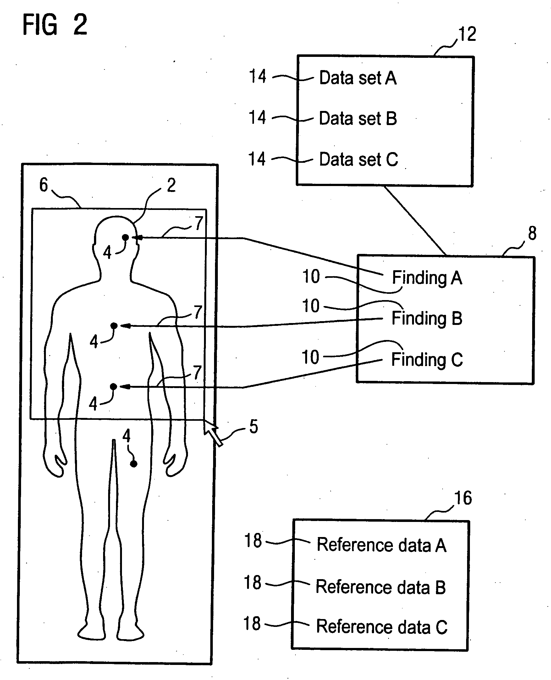
[0018] In the following exemplary embodiment, findings are shown marked on a whole-body view of a cancer patient. The described method, however, is universally usable for all types of medical findings.

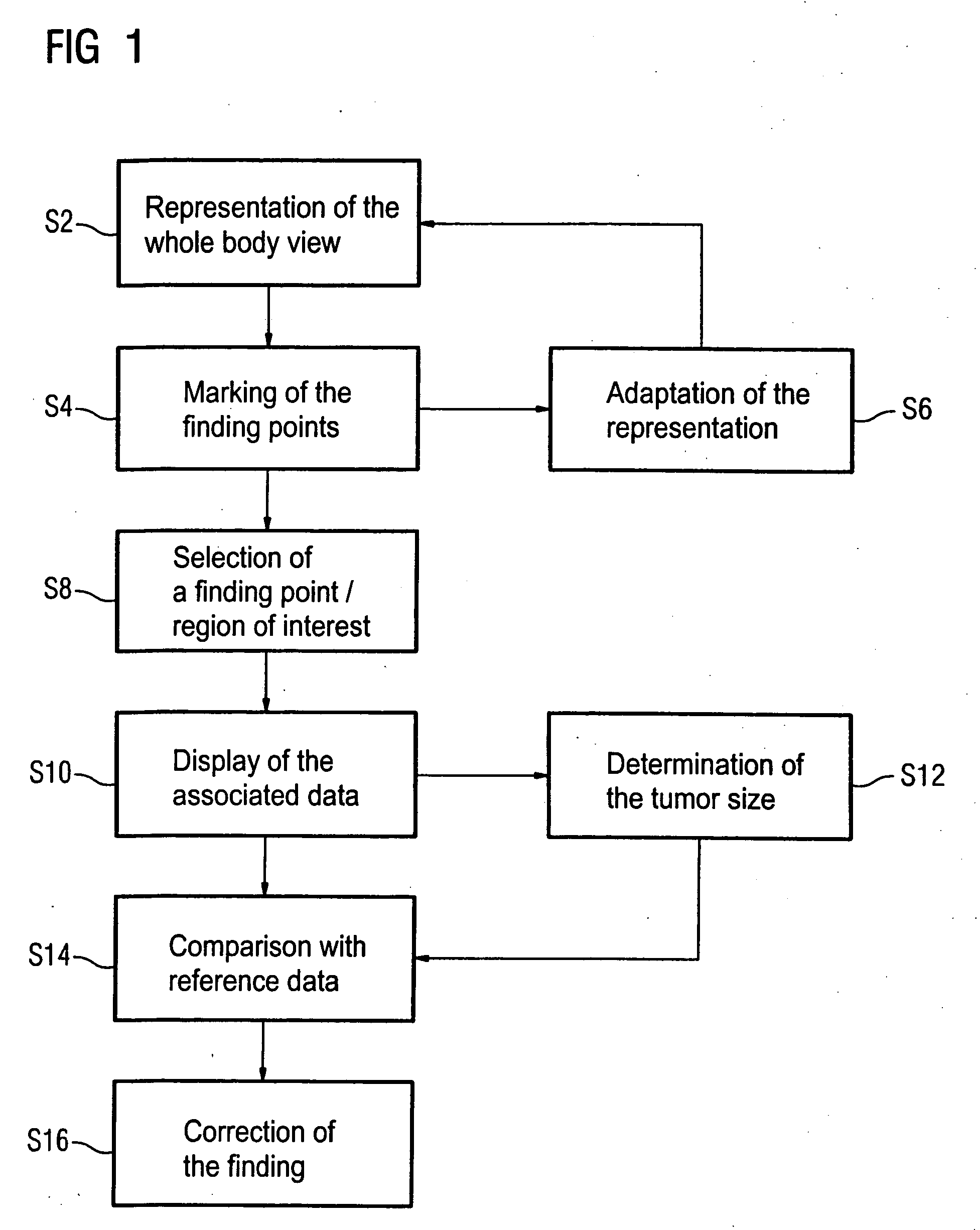
[0019] According to FIG. 1, a whole-body view of a whole-body examination (previously implemented by means of magnetic resonance tomography) is shown on a display medium in a first method step S2. In a second method step S4, the points in the body at which a tumor was determined are marked by a doctor. In a third step S6, the doctor now has the possibility to adapt the viewed representation according to his or her desires. For example, the doctor can modify the contrast, the marking of the finding points and the later display of associated finding data. The method hereupon continues with the first method step. S2 of the representation of the whole-body view. In a fourth method step S8, the doctor alternatively has the possibility to select a finding point or a region of interest by me...


PUM


Abstract

In a method for display of at least one medical finding, at least one part of a body of a patient is shown on a display medium in a body view. A localization in the body of the patient is associated with each finding, and the localizations are marked at a point in the body view corresponding to the localization. The display medium is interactively operable, and a data set associated with the corresponding finding is displayed on the display medium by interaction with a marked localization.

Description

BACKGROUND OF THE INVENTION [0001] 1. Field of the Invention [0002] The present invention concerns a method for display of at least one medical finding. [0003] 2. Description of the Prior Art [0004] In medical diagnostics, in particular imaging diagnostics, it is frequently necessary to process and to evaluate a large number of image data to create a finding. For example, it is possible to map the majority of the body of a patient by means of magnetic resonance tomography. For this purpose, generally several thousands of slices, from the head to the feet of the patient, are acquired by means of a magnetic resonance apparatus. Alternatively, within a partial body examination an overview can initially be created, followed by detailed examinations of specific regions (for example the brain, the upper body or the abdomen). For example, for cancer patients this technique allows a determination as to whether metastases have formed in the body. For this purpose, it is necessary for a docto...

Claims


Application Information

Patent Timeline
IPC IPC(8): G06K9/00
CPCG06F19/3406G16H40/63
Inventor HAIDER, SULTANKREISLER, PETERKUPER, KLAUSALLGAIER, HORSTLICHY, MATTHIAS PHILIPPSCHLEMMER, HEINZ-PETER
Owner SIEMENS HEALTHCARE GMBH
Why Eureka
Try Eureka