Detection of artifacts in bioelectric signals

a bioelectric signal and artifact technology, applied in the field of detection of artifacts in bioelectric signals, can solve the problems of affecting the detection and analysis of events of interest, distorted relevant brain signals, and methods used in brain research that are inappropriate for such clinical applications

Inactive Publication Date: 2007-06-14
GENERAL ELECTRIC CO
View PDF15 Cites 102 Cited by
  • Summary
  • Abstract
  • Description
  • Claims
  • Application Information

AI Technical Summary

Benefits of technology

[0015] The present invention seeks to provide a novel mechanism for detecting artifacts in a bioelectric signal, especially in a frontal EEG signal, thereby to eliminate or suppress the artifacts appearing in the bioelectric signal to be analyzed or the effect of artifacts on an analysis performed based on the bioelectric signal. The present invention further seeks to provide a mechanism, which is suitable for use in a clinical environment, where the number of signal channels is normally low.

Problems solved by technology

During EEG review, these types of artifact may interfere with the detection and analysis of the events of interest.
However, as the EOG is not only sensitive to eye artifacts but also contains brain activity, this method may render the relevant brain signals distorted.
However, the great number of channels / electrodes needed render the methods used in brain research inappropriate for such clinical applications, in which the number of EEG signals / channels is to be kept, due to practical reasons, much lower, typically in one or two.
A drawback of the EOG-based clinical methods is that efficient detection of the contaminated EEG epochs requires separate electrodes for recording the EOG signal(s).
If no separate EOG electrodes are used in clinical applications, the artifact removal remains inefficient, since the omission of the EOG electrodes makes the knowledge about the presence of artifact unreliable.
However, attaching the electrodes adds to the work of the nursing staff and causes inconvenience for the patient.
Movement of the electrodes relative to the skin is another potential source of artifacts.
Apart from the measurement of eye movements or blinks, other measurements of skin surface potential do not provide independent information about the existence of movement artifacts.
First, it is not clear how the head movements and the EEG artifacts are related, because the amplitude of the possible EEG deflections depend on multiple variables, such as the quality of the electrode contact, the direction of the head movement, possible tension in the electrode lead wires, etc.
Second, the method requires a dedicated acceleration transducer component either attached separately on the skin of the patient or integrated as part of one of the electrodes.
This translates to additional cost and increased complexity of the system and its use.
However, predicting low frequency EEG artifacts based on the high frequency signal content is not reliable, because muscle activity does not necessarily imply electrode movement and thus EEG artifact.

Method used


Image

  • Detection of artifacts in bioelectric signals
  • Detection of artifacts in bioelectric signals
  • Detection of artifacts in bioelectric signals

Examples


Embodiment Construction

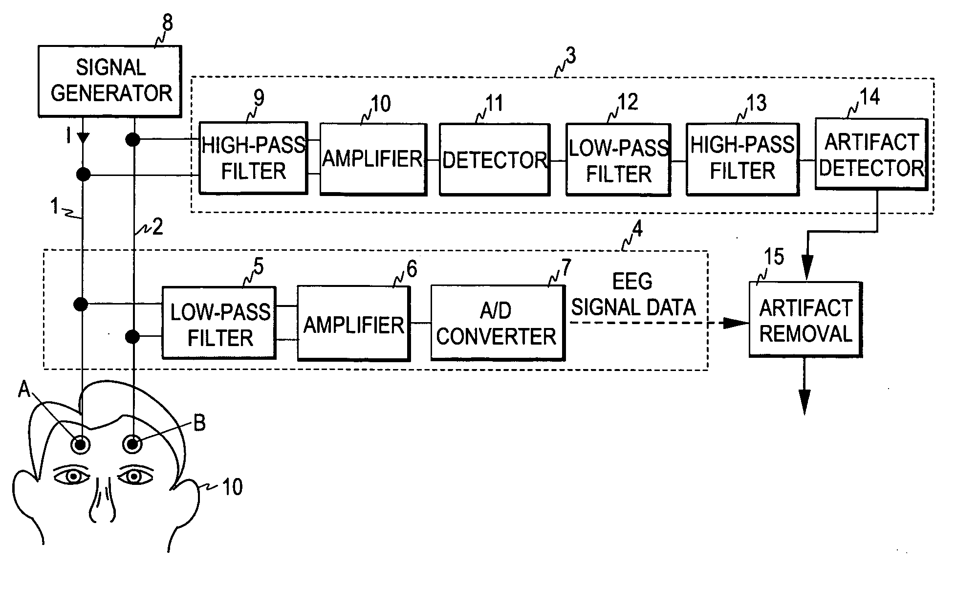
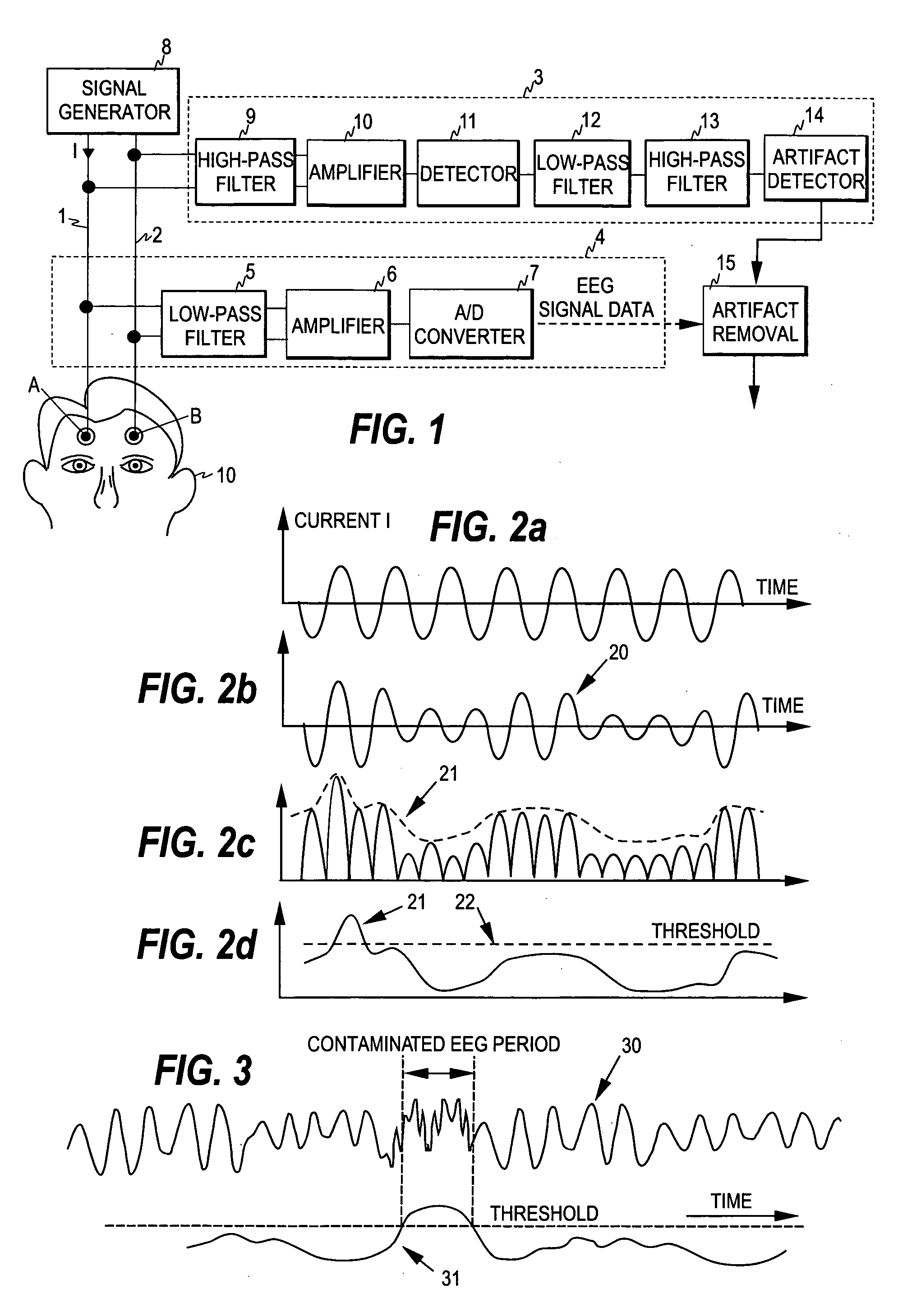
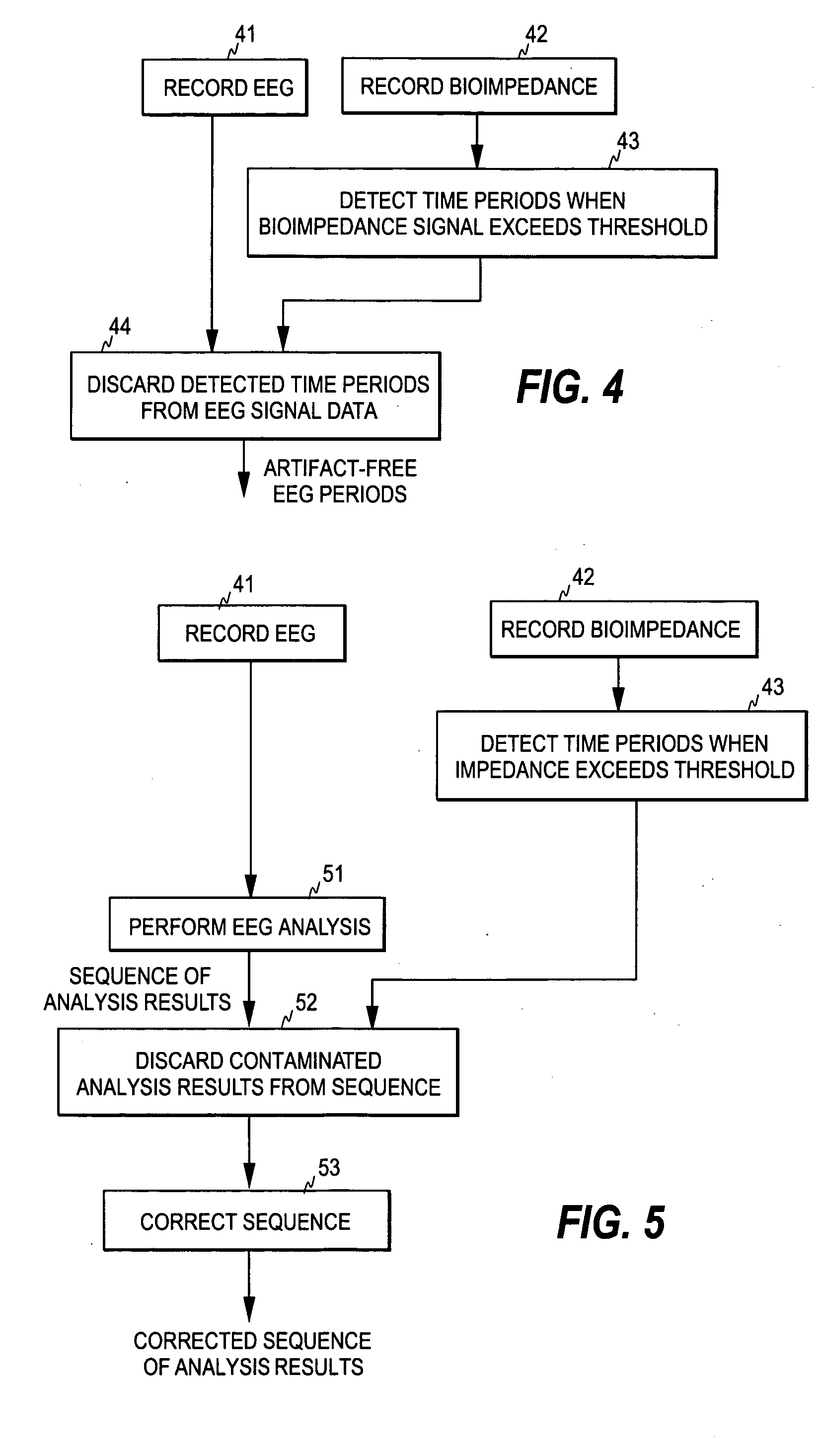
[0032] As discussed above, the present invention rests on the discovery that the major low-frequency interference sources hampering the analysis of an EEG signal measured from the forehead of the patient are such that their presence may be identified from a bioimpedance signal measured from the forehead of the patient. Therefore, a simultaneous bioimpedance measurement indicates when an EEG signal is likely to be distorted by one or more of the said interference sources.

[0033] Bio-impedance measurement combined with biopotential measurement is applied in monitoring of the respiration of a patient, for example. U.S. Pat. No. 5,879,308 discloses a method for measuring bioimpedance in connection with an ECG measurement for monitoring the respiration and / or the blood circulation of the patient. In the bioimpedance measurement, an excitation signal is supplied from a signal generator to the active electrodes of the ECG measurement, whereby an impedance signal indicative of the impedance...


PUM


Abstract

The invention relates to a method and apparatus for detecting artifacts in a bioelectric signal, especially in a frontal EEG signal. In order to accomplish an uncomplicated mechanism for detecting artifacts in clinical applications, an impedance signal is measured through a first electrode set attached to the skin surface in a measurement area of a patient's body, the impedance signal being indicative of the impedance of the signal path formed between individual electrodes of the set. Simultaneously with the impedance measurement, a bioelectric signal is acquired through a second electrode set also attached to the skin surface of the measurement area, and the time periods are determined during which the impedance signal fulfills at least one predetermined criterion indicative of the presence of artifact in the bioelectric signal. In one embodiment, the first and second electrode sets are formed by a common set of two electrodes.

Description

FIELD OF THE INVENTION [0001] The present invention relates generally to the detection of artifacts in bioelectric signals, especially in frontal EEG signals. BACKGROUND OF THE INVENTION [0002] Neuromonitoring is a subfield of clinical patient monitoring focused on measuring various aspects of brain function and on changes therein caused by neurological diseases, accidents, and drugs commonly used to induce and maintain anesthesia in an operation room or sedation in patients under critical or intensive care. [0003] Electroencephalography (EEG) is a well-established method for assessing brain activity. When measurement electrodes are attached on the skin of the skull surface, the weak biopotential signals generated in brain cortex may be recorded and analyzed. The EEG has been in wide use for decades in basic research of the neural systems of the brain as well as in the clinical diagnosis of various central nervous system diseases and disorders. [0004] The EEG signal represents the s...

Claims


Application Information

Patent Timeline
Patent Type & Authority Applications(United States)
IPC IPC(8): A61B5/04
CPCA61B5/04004A61B5/0476A61B5/0531A61B5/30A61B5/369
Inventor VIRTANEN, JUHARANTALA, BORJE
Owner GENERAL ELECTRIC CO
Why Eureka
Try Eureka