Systems and methods for identifying organ transplant risk

a technology of organ transplant and risk, applied in the field of organ transplant rejection diagnosis and prediction, can solve the problems of insufficient use of symptoms alone to detect rejection, graft rejection remains the main complication, and the amount of labeled bounds is reduced

Inactive Publication Date: 2007-12-13
RENOVAR
View PDF6 Cites 9 Cited by
  • Summary
  • Abstract
  • Description
  • Claims
  • Application Information

AI Technical Summary

Problems solved by technology

Despite the availability of potent immunosuppressive agents, graft rejection remains the main complication of renal transplantation.
Statistics indicate that graft rejection is often not detected early enough in the rejection episode to allow initiation of countervailing treatment in time to prevent the organ rejection with immunosuppressive agents at a time when the rejection process could be effectively halted and / or prevented altogether.
However, the use of these symptoms alone to detect rejection is not adequate.
However, this method has major limitations.
For example, since the biopsy procedure itself has complications, and since a portion of the renal transplant is removed during each biopsy, transplant biopsy cannot be performed on a routine or even frequent basis to monitor renal allograft rejection.
In addition, the invasive nature of a renal biopsy is both uncomfortable and inconvenient for patient subjects.
Hence, renal biopsies are reserved for those patients that demonstrate other clinical and / or laboratory evidence of renal allograft rejection, thus limiting its use or potential use in detecting early graft rejection.

Method used


Image

  • Systems and methods for identifying organ transplant risk
  • Systems and methods for identifying organ transplant risk

Examples


example 1

Correlation of Urine IP-10 with Graft Rejection

[0096] This example describes the correlation of urine levels of IP-10 with kidney graft rejection in human subjects. Forty-five human subjects that had undergone kidney transplant were utilized. Urine IP-10 levels were measured repeatedly following organ transplant. IP-10 levels were measured using a quantitative colorimetric sandwich ELISA assay (R & D Systems, Minneapolis, Minn.). Subjects were divided into two groups, rejecters and non-rejecters, based on kidney biopsies. Biopsies were classified using Banff criteria (Solez et al., Kidney Int., 44:411 [1993]). All of the subjects were receiving anti-rejection therapy at the time of the study. Urine from ten normal (non-transplant) subjects was also tested.

[0097] In a majority of the non-rejecters, IP-10 levels remained at a constant, low level or decreased over time. In the rejecters, IP-10 levels remained at a constant, high level or increased over time. A urine level of IP-10 of...

example 2

Correlation of Urinary Chemokines with Graft Rejection and Treatment

[0099] The example describes the correlation of urinary chemokines levels with graft rejection and treatment. Urinary samples were collected from healthy individuals, kidney transplant recipients with stable graft function, and recipients with acute rejection. All patients with acute rejection were hospitalized and received anti-rejection therapy. Urinary samples were centrifuged, and supernatant was aliquoted and stored at −80° C. These samples, after thawing, were evaluated by ELISA for the expression of MCP-1, IP-10, and I-TAC.

Elevated Expression of Chemokines in Urinary Samples from Patients with Acute Rejection

[0100] As shown in Table 1, chemokines IP-10 and I-TAC were significantly increased in the urinary samples of patients with acute graft rejection and acute tubular injury, compared to healthy controls and kidney transplant patients with other pathologic changes. As presented in Table 2, if 100 pg / ml w...

example 3

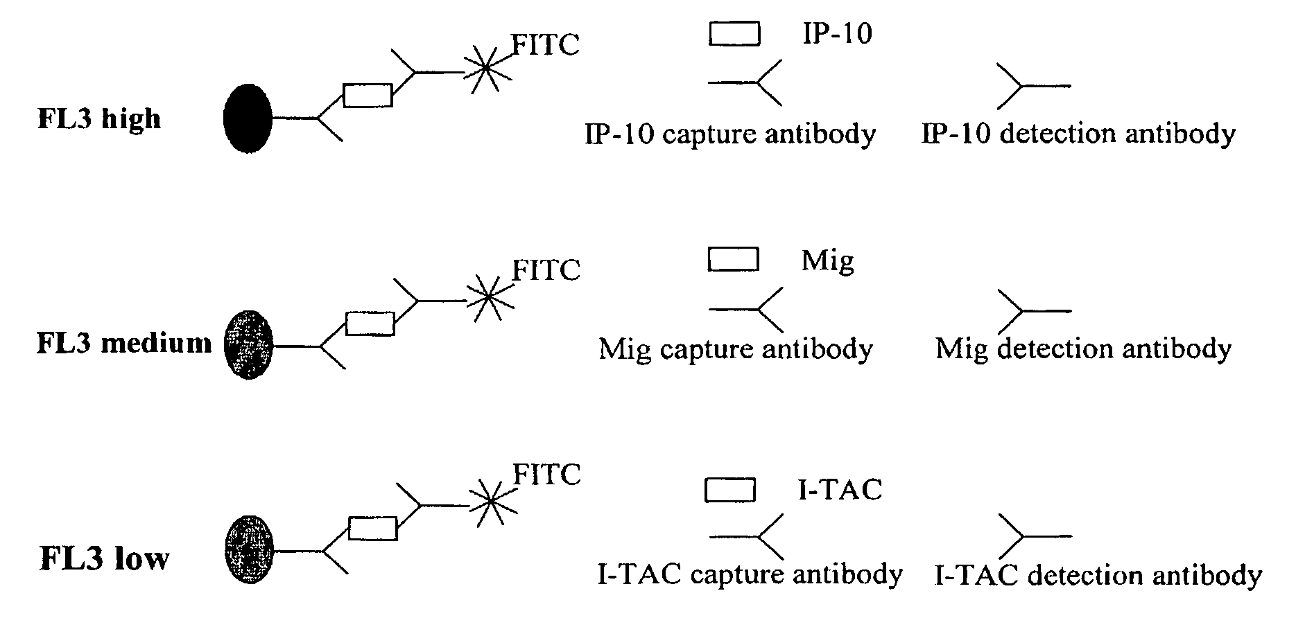
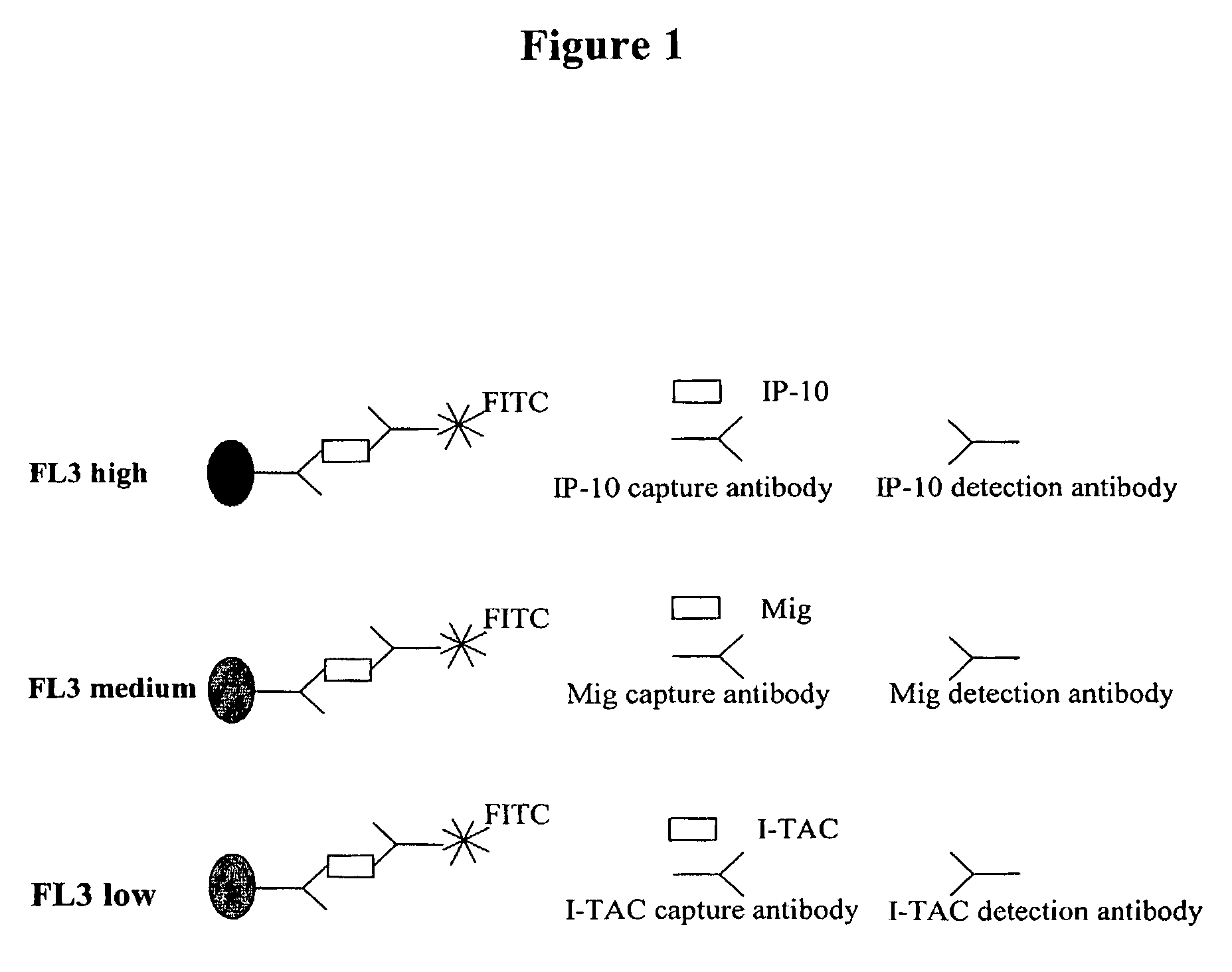
Flow Cytometry Based Technique for Quantification of Chemokines

[0105] This example describes a FACS method for the simultaneous detection of multiple chemokines. The fluorescence activated cell sorting (FACS) method uses fluorescence dye labeled beads that can detect 3 chemokines in one assay. In this example, IP-10, I-TAC and Mig were detected. Detection of these three chemokines was conducted in the same test tube simultaneously as depicted in FIG. 1. As the chemokine concentration increases, the mean fluorescence intensity for each group of beads increases. This correlation between the chemokine concentration and the mean fluorescence establishes the basis for this FACS quantitative method. A standard curve for each chemokine was constructed. This example demonstrates that EP-10, Mig and I-TAC can be simultaneously detected in a urinary sample.


PUM

PropertyMeasurementUnit
concentrationsaaaaaaaaaa
lengthaaaaaaaaaa
body temperatureaaaaaaaaaa

Abstract

The present invention relates to methods of diagnosing and predicting organ transplant rejection. In particular, the present invention relates to the detection and prediction of kidney transplant rejection by detection of CXCR3 and CCL chemokines in urine. The present invention provides improved methods of diagnosing organ rejection and determining the efficacy of anti-rejection drugs.

Description

[0001] This application claims priority to U.S. provisional patent application Ser. No. 60 / 380,569 filed on May 14, 2002.FIELD OF THE INVENTION [0002] The present invention relates to methods of diagnosing and predicting organ transplant rejection. In particular, the present invention relates to the detection and prediction of kidney transplant rejection by detection of CXCR3 and CCL chemokines in urine. The present invention further relates to methods and compositions for assessing the efficacy of anti-rejection agents. BACKGROUND OF THE INVENTION [0003] Approximately 12,000 kidney transplants are performed annually in the United States. Despite the availability of potent immunosuppressive agents, graft rejection remains the main complication of renal transplantation. For example, approximately 50% of all renal allograft recipients are thought to suffer at least one episode of graft rejection. The likelihood of kidney loss due to rejection is highest during the first year post tran...

Claims


Application Information

Patent Timeline
Patent Type & Authority Applications(United States)
IPC IPC(8): G01N33/53G01N33/68
CPCG01N33/6863G01N2800/245G01N2333/715
Inventor HU, HUAIZHONGPUCHALAKI, ALICE
Owner RENOVAR
Why Eureka
Try Eureka