Considerations when colon segmentation differs between CAD processing and visualization

Inactive Publication Date: 2008-01-10
ICAD INC
View PDF15 Cites 44 Cited by
  • Summary
  • Abstract
  • Description
  • Claims
  • Application Information

AI Technical Summary

Problems solved by technology

The resulting colon segmentations may not be identical, and may in fact have large differences if the colon is poorly distended or if significant sections of the small bowel are improperly include

Method used


Image

  • Considerations when colon segmentation differs between CAD processing and visualization
  • Considerations when colon segmentation differs between CAD processing and visualization
  • Considerations when colon segmentation differs between CAD processing and visualization

Examples


Embodiment Construction

[0016]In the following detailed description of the embodiments, reference is made to the accompanying drawings that form a part hereof, and in which are shown by way of illustration, and not by way of limitation, specific embodiments in which the invention may be practiced. It is to be understood that other embodiments may be utilized and that logical, mechanical and electrical changes may be made without departing from the spirit and scope of the present invention.

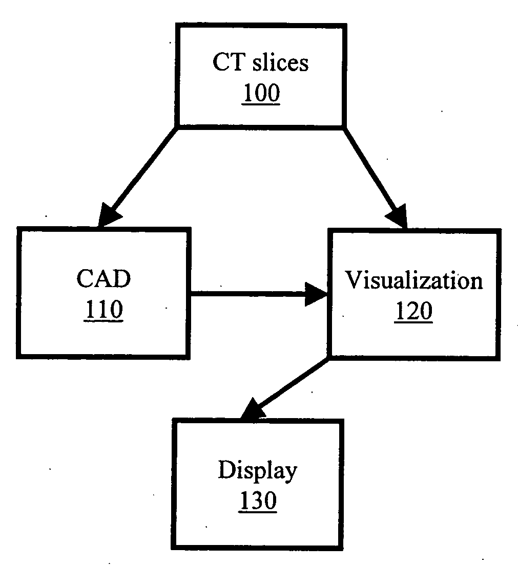
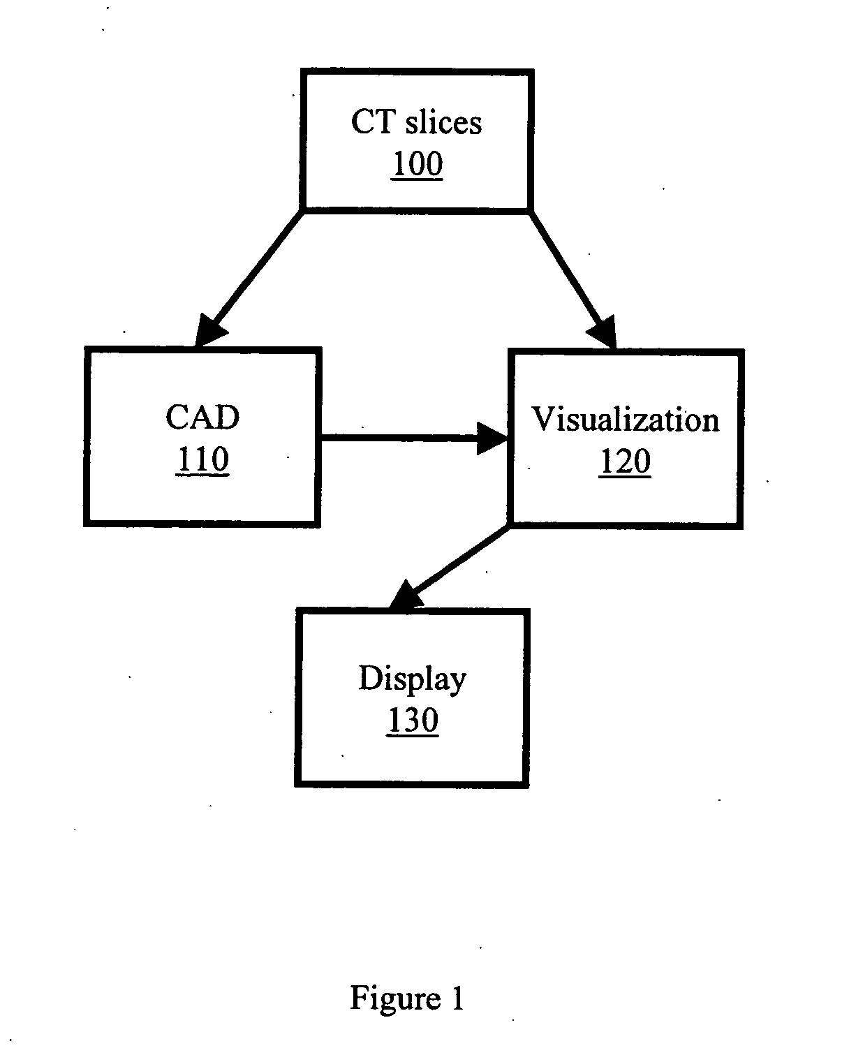
[0017]Referring initially to FIG. 1, when a CT medical image case is processed in step 100, both the visualization workstation 120 and the CAD algorithm 110 segment the medical images. The CT medical images can be taken from a colon or any other anatomical region that may benefit from CAD processing. The CAD algorithm 110 further examines the medical images for suspicious regions and reports the results to the visualization workstation 120 for display 130. On the workstation 120, the suspicious regions from the CAD algori...


PUM


Abstract

A system and method for preserving CAD marks falling outside the three-dimensional rendering on a visualization display following segmentation of CT medical image slices for review by a user is disclosed. The CT medical images are processed by both a CAD algorithm and a visualization workstation. The CAD algorithm reports detected suspicious regions to the visualization workstation for display on the three-dimensional rendering of the medical images as well as creates a list of the detections to notify the user. If the detected CAD marks are not located on the three-dimensional rendering of the medical images, the user is notified. The user can the view the detected CAD marks on the two-dimensional CT medical images.

Description

BACKGROUND OF THE INVENTION[0001]The present invention generally relates to a method and system for processing medical image data and, in particular, relates to a method and system for processing medical image data in which different applications share intermediate processing results.[0002]Many different applications for processing medical image data exist and are used today. For example, there is visualization software in which the medical image is presented for review by a medical professional on a workstation display, or computer monitor. In addition, there is computer-aided detection (CAD) software in which abnormalities are automatically detected in the medical image data by computer and these CAD results are presented to the medical professional. Many times these image processing applications come from different vendors.[0003]One such area of medical image processing is virtual colonography (VC). VC uses x rays and computers to produce two- and three-dimensional images of the ...

Claims


Application Information

Patent Timeline
IPC IPC(8): G06K9/00
CPCG06T2207/30028G06T7/0012
Inventor WOODS, KEVINCOLLINS, MICHAEL J.MCGINNIS, RYAN EDWARD
Owner ICAD INC
Why Eureka
Try Eureka