Probes, systems, and methods for examining tissue according to the dielectric properties thereof

a technology of dielectric properties and probes, applied in the field of probes, systems and methods for examining and characterizing tissue by its dielectric properties, can solve the problems of poor radiological placement, failure to successfully remove the appropriate lesion, and failure to always succeed in traditional methods of biopsy

Inactive Publication Date: 2008-01-24
DUNE MEDICAL DEVICES
View PDF98 Cites 45 Cited by
  • Summary
  • Abstract
  • Description
  • Claims
  • Application Information

AI Technical Summary

Benefits of technology

[0053] In accordance with embodiments of the present invention, the generation of electrical fringe fields in the tissue, substantially to the depth d, as substantially determined by the feature size, with negligible radiation penetrating into and reflected from the tissue beyond the depth d, eliminates almost completely the propagating wave.

Problems solved by technology

The traditional method of biopsy, however, is not always successful and fails to successfully remove the appropriate lesion in about 0.5-17% of the cases.
Some of the reasons given for unsuccessful biopsies include: 1) poor radiological placement of the localization wire; 2) preoperative and intraoperative dislodgment of the wire; 3) surgical inaccuracy and inadequacy in excising the appropriate tissue; 4) failure to obtain a specimen radiograph; and 5) failure by the pathologist to locate the focus of the disease when searching through a larger tissue sample provided by the surgeon.
All of the above reasons stem from a fundamental problem that during the surgery, the surgeon does not have a real time indication or delineation of the tumor.
This trade-off between complete removal of the tumor, and conservation of the breast, is usually difficult to optimize because the surgeon generally does not know the actual margins of the tumor.
Although this method is approved by the FDA, it lacks the necessary accuracy for a screening device mainly because of the inherent limitations of long wavelengths and noise from the contact electrodes.
However, the above devices use a set of electrodes that must be electrically contacted with the tissue or body, and therefore the contact is usually a source of noise and also limits maneuverability of the probe over the organ.
However, these methods lack accuracy and spatial resolution since they are limited by the diffraction limit.
Although non-contact, the device actually measures average values inside the organ, and its ability to detect small tumor is doubtful.
However, such detections have been limited clinically to pre-operative scans, or demand large scanning multi-million-dollar machines, like the MRI, CT, and Mammography.
Furthermore, real-time attempts to use these detecting methods are very sensitive to movement of the body, and cannot really be used to position the cutting knife or the biopsy needle.
Any movement of the organ or the localization device results in an error in biopsy localization.
Also, no information about the tissue being cut by the needle or knife is provided.
However, there is currently no reliable real-time bioimpedance measuring device of sufficiently high accuracy for local tissue characterization and of a spatial resolution comparable to that provided by mammography.

Method used


Image

  • Probes, systems, and methods for examining tissue according to the dielectric properties thereof
  • Probes, systems, and methods for examining tissue according to the dielectric properties thereof
  • Probes, systems, and methods for examining tissue according to the dielectric properties thereof

Examples


Embodiment Construction

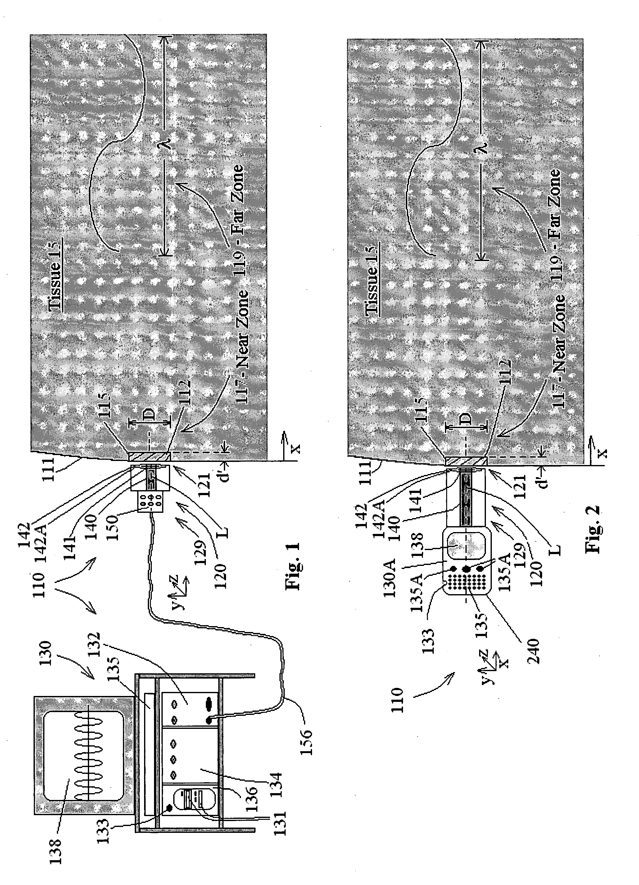
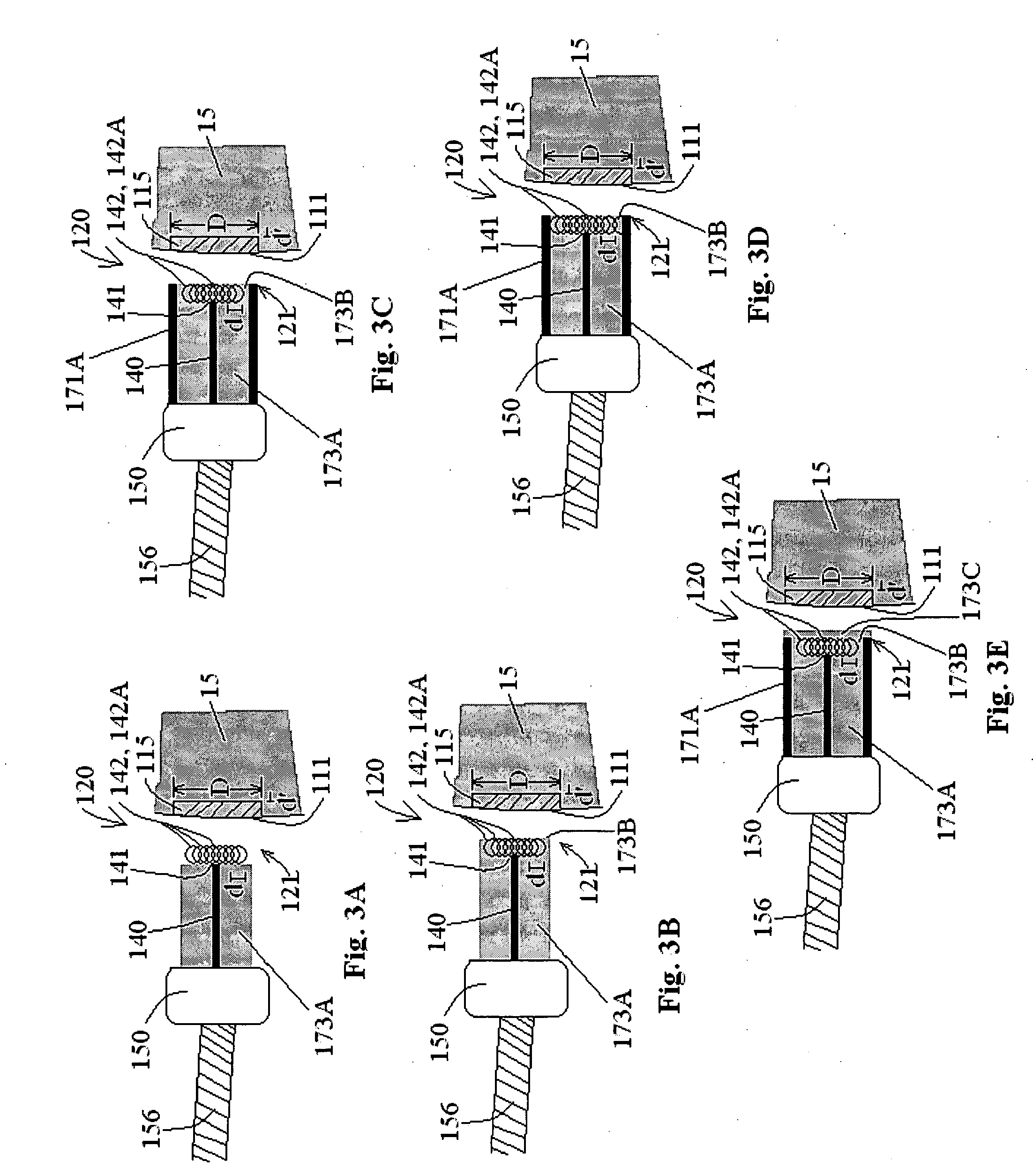
[0107] The present invention relates to probes, systems, and methods for tissue characterization by its dielectric properties, wherein a physical feature of the probe is designed to define and delimit a tissue volume, at the tissue edge, where characterization takes place. Preferably, tissue characterization occurs substantially in real time.

[0108] Before explaining at least one embodiment of the present invention in detail, it is to be understood that the present invention is not limited in its application to the details of construction and the arrangement of the components set forth in the following description or illustrated in the drawings. The invention is capable of other embodiments or of being practiced or carried out in various ways. Also, it is to be understood that the phraseology and terminology employed herein is for the purpose of description and should not be regarded as limiting.

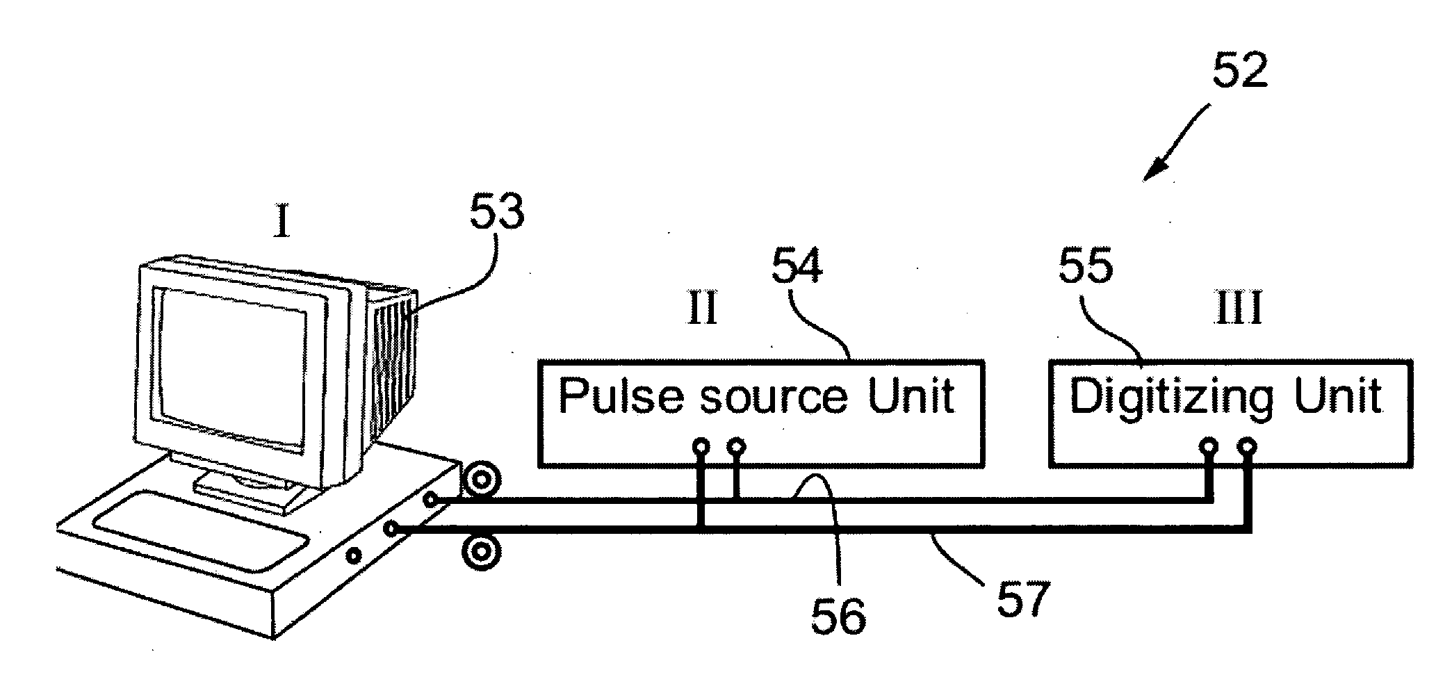
[0109] Referring now to the drawings, FIG. 1 schematically illustrates an overall syste...


PUM


Abstract

The present invention relates to probes, systems, and methods for tissue characterization by its dielectric properties, wherein a physical feature of the probe is designed to define and delimit a tissue volume, at a tissue edge, where characterization takes place. Thus, the probe for tissue-edge characterization comprises: a first inner conductor, which comprises: proximal and distal ends, with respect to a tissue edge, along an x-axis; a first sharp edge, inherently associated with the proximal end; at least one feature, issuing from the first inner conductor, substantially at the proximal end, for forming at least one additional sharp edge, operative to enhance localized electrical fringe fields in the tissue, within a generally predefined tissue volume, at the tissue edge, the tissue volume being generally defined by physical parameters associated with the at least one feature; and a dielectric material, which encloses the conductor, in the y-z planes.

Description

CROSS REFERENCE TO RELATED APPLICATION [0001] This Application is being co-filed with a sister U.S. Patent Application, entitled “Ergonomic Probes” (Attorney Docket No. 38282). [0002] This Application is also a continuation-in-part of pending U.S. patent application Ser. No. 11 / 705,143 filed on Feb. 12, 2007, which is a continuation-in-part of U.S. patent application Ser. No. 10 / 965,752 filed on Oct. 18, 2004, now U.S. Pat. No. 7,184,824 issued on Feb. 27, 2007 and a continuation-in-part of U.S. patent application Ser. No. 10 / 035,428 filed on Jan. 4, 2002, now U.S. Pat. No. 6,813,515 issued on Nov. 2, 2004. [0003] Additionally, this Application is a continuation-in-part of PCT Patent Application No. PCT / IL2006 / 000392 filed on Mar. 29, 2006, which claims the benefit of U.S. Provisional Patent Application No. 60 / 665,842 filed on Mar. 29, 2005, now expired. [0004] Additionally, this Application is a continuation-in-part of pending U.S. patent application Ser. No. 10 / 567,581 filed on Fe...

Claims


Application Information

Patent Timeline
Patent Type & Authority Applications(United States)
IPC IPC(8): A61B5/053
CPCA61B5/053A61B5/0538A61B5/415A61B2017/00026A61B10/0041A61B17/320016A61B17/320783A61B5/418
Inventor HASHIMSHONY, DANCOHEN, GILGELTNER, IDDO
Owner DUNE MEDICAL DEVICES
Why Eureka
Try Eureka