

System and methods for scoring images of a tissue micro array
a tissue micro array and scoring system technology, applied in image enhancement, chemical methods analysis, instruments, etc., can solve the problems of expensive and time-consuming manual analysis of tmas
- Summary
- Abstract
- Description
- Claims
- Application Information
AI Technical Summary
Benefits of technology
Problems solved by technology
Method used
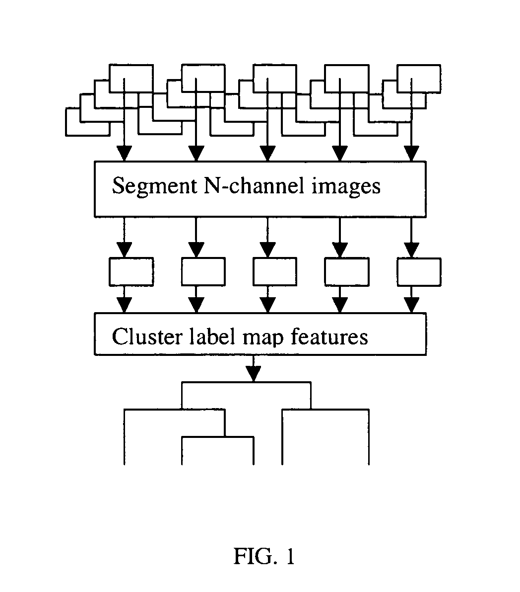
Image

Examples
example 1
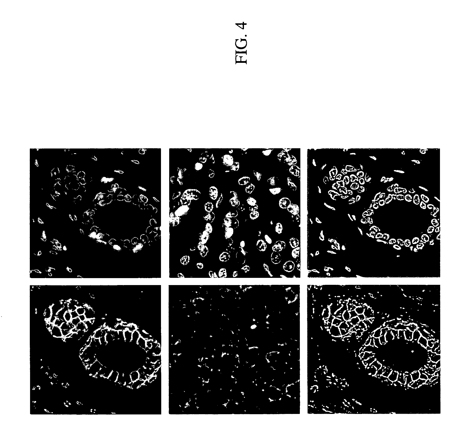
[0064]Four TMA's were obtained from Imgenex containing paraffin-embedded human breast tissue with both normal, infiltrating ductal carcinoma and metastasis in the lymph nodes. The four TMA's were produced from a single block of 60 patient cores. TMA slides of the samples were stained with conjugated antibodies using Cy3 and Cy5 fluorescence dyes and then photographed at 20× using a biological microscope. The stains were removed and the process repeated using the dyes listed in Table I below. For each staining step, the TMAs were counterstained with DAPI for registration of the nuclei. A final H&E stain was applied to add three more channels for analysis.
TABLE IStains for four TMAs from adjacent tissuesections of the same patients and the p-values for the correlation score separatingnormal and IDC breast cancerPStainsvalue1Cy3 β-catenin DAPI0.242Secondary cy3 keratin, DAPI, cy3 β-0.25catenin, smooth muscle actin, H&E3Cy3 β-catenin DAPI0.154Secondary Cy3 keratin, Secondary Cy5 β-N / Aca...
example 2
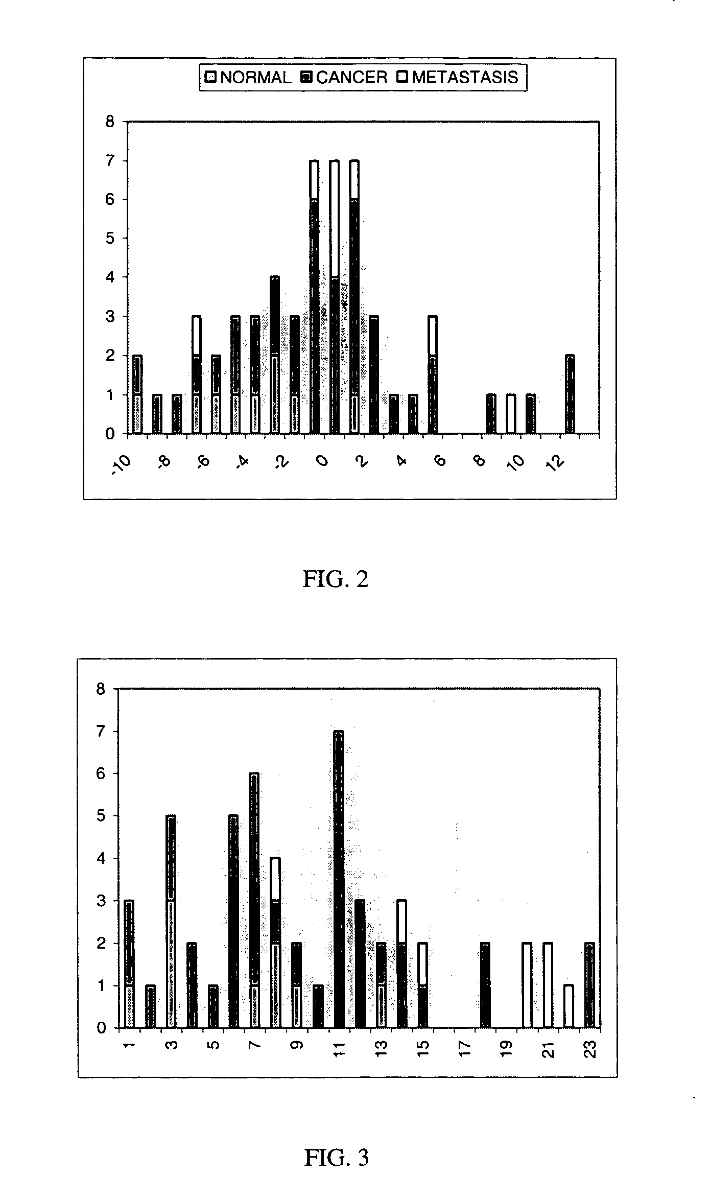
[0085]In this example, β-catenin translocation was again quantified in breast cancer with multiple registered biomarker images in formalin fixed paraffin-embedded breast tissue micro arrays. As further described below, the breast tissue samples (n=445) were filtered and correlated giving scores of −1.7 for normal (n=103), +0.2 for cancer (n=284) and +1.4 for metastasis (n=58). Normal tissue was discriminated against cancer (P=0.0001) and cancer against metastasis (P=0.06).
[0086]Seven TMA's were obtained from both Imgenex and Tristar containing paraffin-embedded human breast tissue with both normal and infiltrating ductal carcinoma. The tumor stage, patient age, diagnosis, ER, PR and P53 status were provided for the Imgenex 364 TMA's and the tumor stage and diagnosis were provided for the Imgenex 371 and Tristar TMA's.
[0087]The TMA slides were sequentially stained and bleached to increase the number of biomarkers on the same tissue sample in contrast to the conventional practice of u...
PUM
 Abstract
Description
Claims
Application Information