Kinetic biomarker for quantification of lymphoproliferation, clonal expansion, recruitment and trafficking in lymphoid tissues and of the in vivo actions of antigens and modulating thereon

a kinetic biomarker and lymphoid tissue technology, applied in the field of measuring proliferation, clonal expansion, recruitment and/or trafficking of immune cells, can solve the problems of unsatisfactory results, unsystematic and general unsatisfactory treatment efforts to reduce immune activation in hiv/aids, and achieve the effect of slowing the progression of immune deficiency and reducing immune activation

Inactive Publication Date: 2009-06-18
KINEMED
View PDF0 Cites 7 Cited by
  • Summary
  • Abstract
  • Description
  • Claims
  • Application Information

AI Technical Summary

Benefits of technology

The present invention describes a way to treat diseases that affect the body's ability to fight off viruses like HIV. These diseases are caused because cells from the lymph nodes get infected and can no longer function properly. This invention proposes using certain substances to stop these cells from getting sick and dying. By doing this treatment, it may be possible to improve the effectiveness of current therapies and even prevent further damage to the immune system.

Problems solved by technology

The technical problem addressed in this patent is how to treat chronic immune activation without causing further harm to already compromised immune systems, particularly in cases like HIV/AIDS where treatment options are limited. There is a risk of exacerbating the existing imbalance between immune activation and suppression through conventional methods, resulting in potential side effects and reduced efficacy. Therefore, new therapies aimed at reducing immune activation while maintaining effective immune functions are needed.

Method used


Image

  • Kinetic biomarker for quantification of lymphoproliferation, clonal expansion, recruitment and trafficking in lymphoid tissues and of the in vivo actions of antigens and modulating thereon
  • Kinetic biomarker for quantification of lymphoproliferation, clonal expansion, recruitment and trafficking in lymphoid tissues and of the in vivo actions of antigens and modulating thereon
  • Kinetic biomarker for quantification of lymphoproliferation, clonal expansion, recruitment and trafficking in lymphoid tissues and of the in vivo actions of antigens and modulating thereon

Examples


example 1

Protocols

[0270]The protocols in this Example 1 were followed for all experiments described below.

[0271]Reagents. Keyhole limpet hemocyanin (KLH) and all xenobiotics such as cyclosporine A and the others described below were from Sigma (St. Louis, Mo.) or from other well known commercial vendors of chemicals. Heavy water was from Cambridge Isotope Labs (Cambridge, Mass.). Tissue culture grade, endotoxin-free sterile PBS from Cellgro (City / State) was used for sham immunizations and as a diluent for KLH, which was stored in aliquots at −20° C. All other chemicals were from Sigma unless mentioned otherwise.

[0272]Animals. All animal procedures received prior written approval by KineMed's Animal Care and Use Committee. Animals (female C57BI / 6 and Balb / c mice, unless mentioned otherwise) were housed at KineMed's animal facility under specific pathogen-free conditions, subject to a 12 h light / 12 h dark cycle, and given water and standard chow ad libitum. They were purchased at 6 weeks of ag...

example 2

The Increase in PLN Cell f after Immunization is Antigen-Dependent

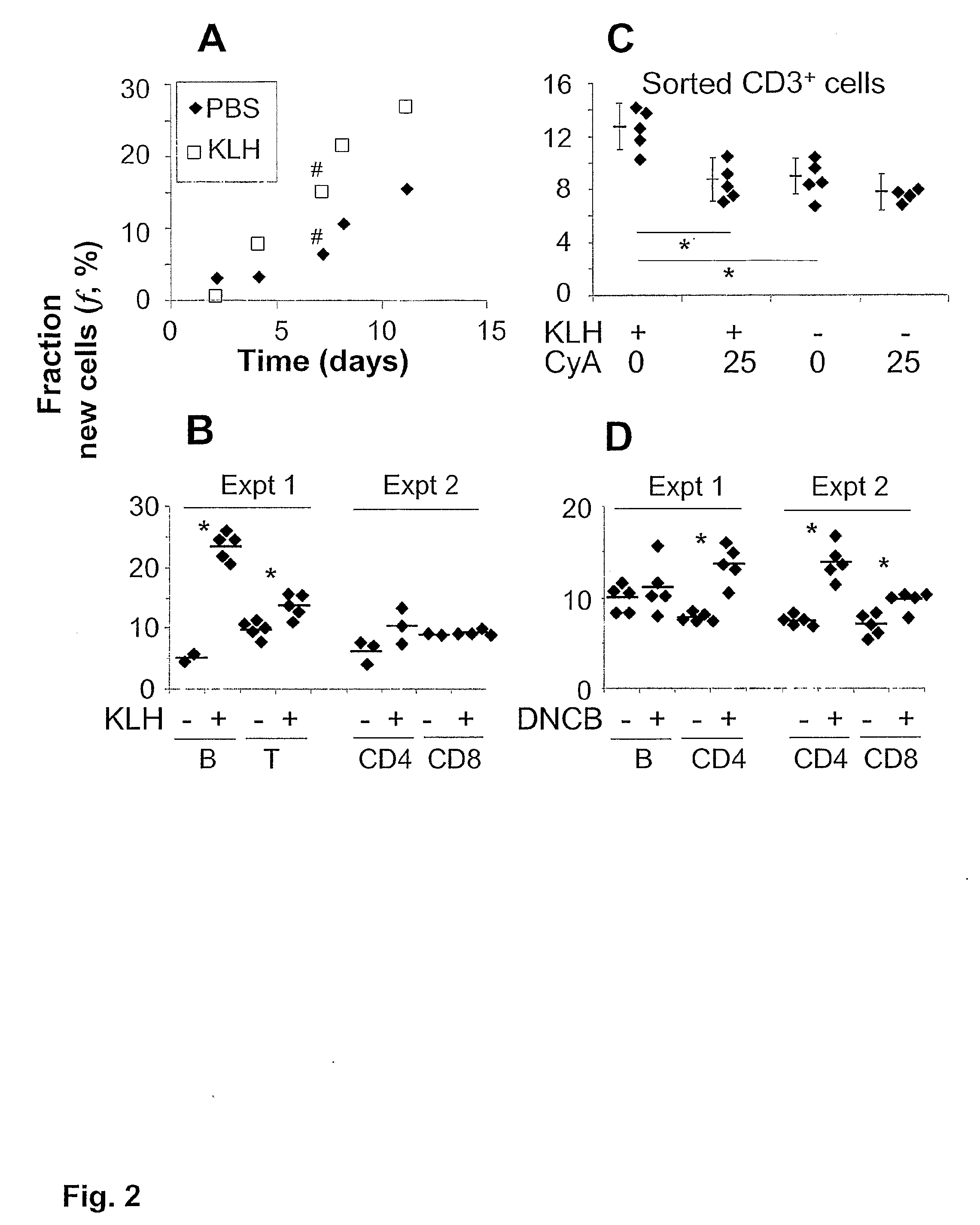
[0282]We next examined whether the increased f in PLN reflects antigen-dependent lymphoproliferation or preferential recruitment of proliferating over resting cells. First, we measured f in lymphocyte subsets (FIG. 2B). A pronounced increase in f was found in PLN B cells from KLH-immune mice. A smaller increase in f was detected in total and CD4+ T cells, but not in CD8+ T cells (FIG. 2B), consistent with the known ability of the former, but not the latter, to be primed by exogenous proteins. Moreover, this result suggests that proliferation of B and CD4+ T cells is not due to LPS contamination, because even low doses of LPS increase the percentage of proliferating CD8+ T cells.

[0283]Second, the KLH-stimulated increase in f of T cells was inhibited by cyclosporin A (FIG. 2C), a well known calcineurin antagonist that prevents nuclear translocation of NFATc after antigenic stimulation. By contrast, baseline f of T cells...

example 3

Differential Contributions from f (Clonal Expansion) and Cell Number to Lymphoproliferation

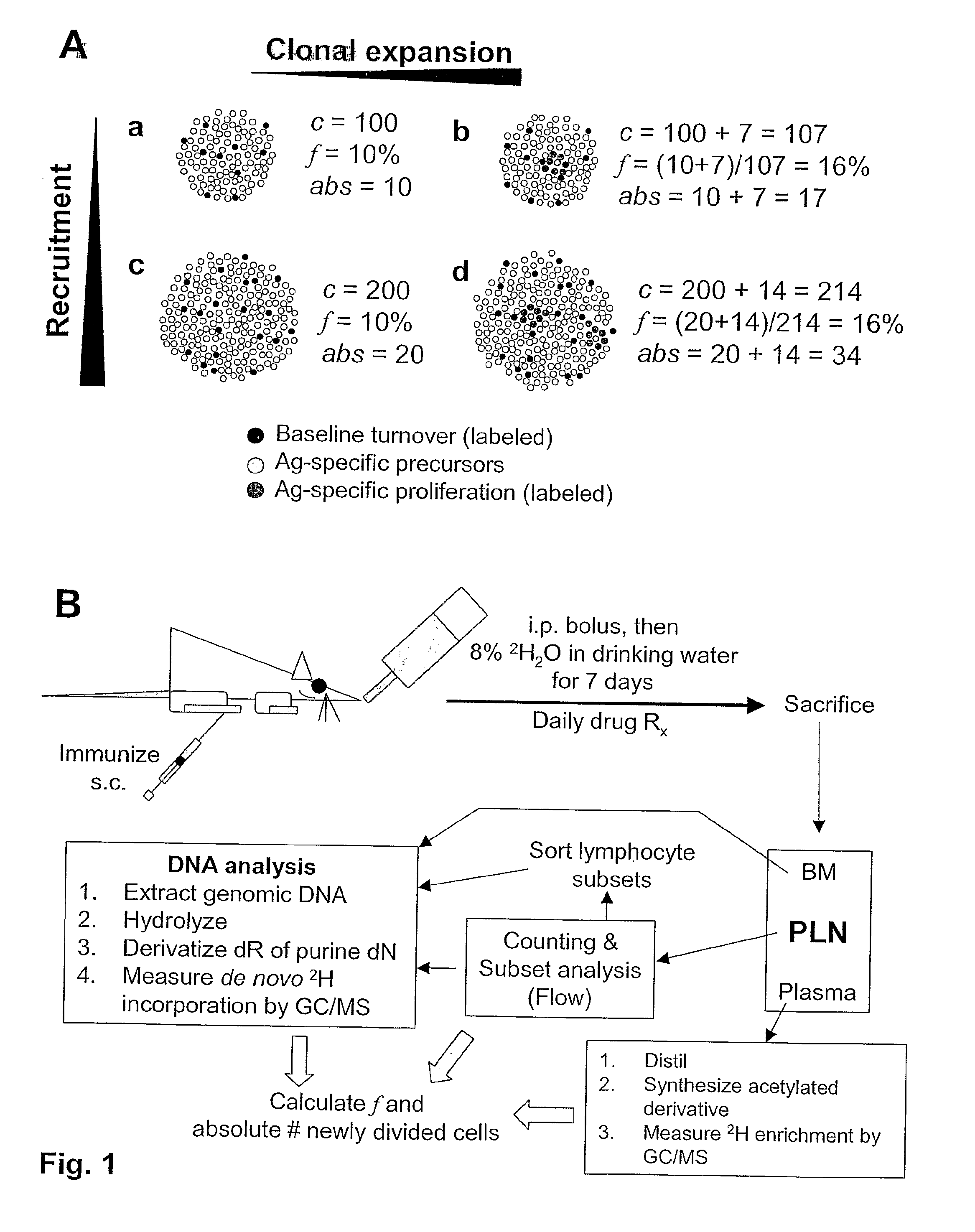
[0287]Absolute lymphoproliferation, i.e., the total number of LN cells that have divided during a period of 2H2O exposure, can be calculated as (LN cellularity×f). By this definition, absolute lymphoproliferation equals the net accrual of divided cells in the LN, due to constitutive cell proliferation and death, cell recruitment to the LN, local antigen-driven proliferation, and activation-induced death.

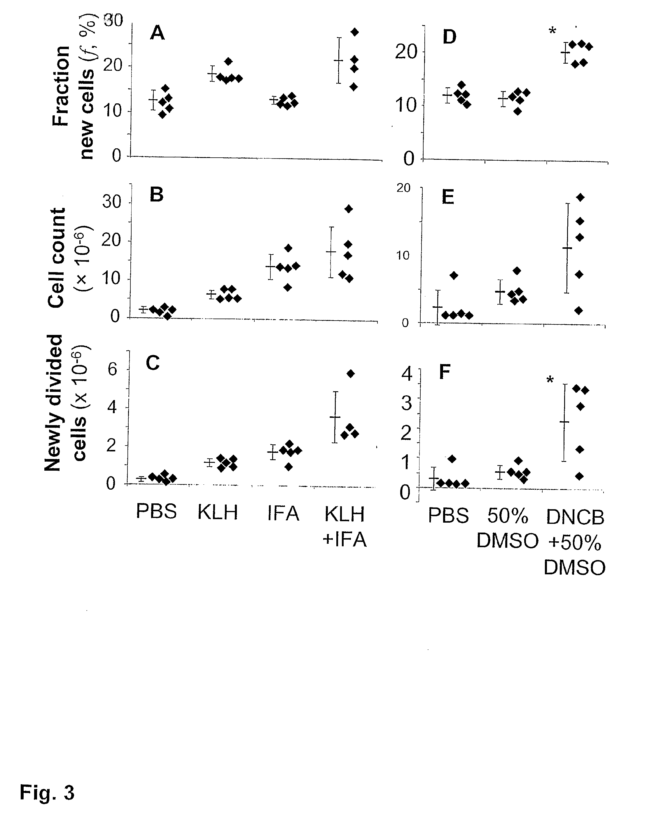
[0288]We determined the contributions from cellularity and f to absolute lymphoproliferation in draining LN 7 days after immunization of Balb / c mice with different doses of KLH (FIG. 5A-C). Cellularity was maximal around 25 μg KLH (FIG. 5A); a tenfold lower or higher antigen dose did not increase cellularity, compared to negative controls. In contrast, f was elevated to a dose-independent plateau over a 100-fold range of KLH doses (FIG. 5B). At KLH doses up to 2.5 μg, absolute lymphoproliferati...


PUM


Abstract

The methods of the present invention allow for the measurement of proliferation, clonal expansion trafficking and/or recruitment of lymphocytes into lymphoid tissue in an in vivo setting. Proliferation, clonal expansion, recruitment and/or trafficking of lymphocytes are measured by using stable isotopes to label newly synthesized DNA, isolating the newly-labeled DNA, and quantifying enrichment of the isolated DNA with mass spectrometry or other appropriate techniques. Such methods are useful in screening candidate drugs for stimulatory or inhibitory effects on proliferation, clonal expansion, recruitment and/or trafficking of lymphocytes. The methods also allow for the discovery of therapeutic agents in disorders of immune regulation such as HIV infection and for optimizing vaccine efficacy.

Description


Claims


Application Information

Patent Timeline
Owner KINEMED
Why Eureka
Try Eureka