System and method for the graphical presentation of the content of radiologic image study reports

a radiologic image and content technology, applied in the field of imaging technologies, can solve the problems of inaccurate and hastily compiled reports or records, affecting the patient's recovery time, and affecting the patient's recovery time, and achieve the effect of convenient perception

Inactive Publication Date: 2011-02-10
FUJIFILM MEDICAL SYST USA INC
View PDF9 Cites 11 Cited by
  • Summary
  • Abstract
  • Description
  • Claims
  • Application Information

AI Technical Summary

Problems solved by technology

Medical personnel spend a significant amount of their time addressing administrative tasks.
Time spent on performing such tasks diminish the time available for patients and in some instances lead to inaccurate and hastily compiled reports or records when personnel are faced with the need to see multiple patients.
There is currently no cohesive overview that can provide detailed information at a level that would be easy to comprehend and provide sufficient detail to a medical reader.
The most significant of these limitations being in the field of radiology.
These systems continue to be limited in their ability and features due to the inability to support radiological findings, the relationships between findings and the characteristics of the findings.
However, these systems suffer from a myriad of drawbacks.
Essentially these solutions have an inconsistent view of report content, and necessitate reader viewing and correlation of multiple text reports which can be quite tedious and cumbersome.
Presently, no systems are available to capably present in a graphical format, patient information that includes time and date, study types, evolution of findings and the significance of findings.
The limitations and lack of functionality present in currently available report systems and tools necessitate a comprehensive system that is capable of providing structured reports.

Method used


Image

  • System and method for the graphical presentation of the content of radiologic image study reports
  • System and method for the graphical presentation of the content of radiologic image study reports
  • System and method for the graphical presentation of the content of radiologic image study reports

Examples


Embodiment Construction

[0034]Generally, the system and methods described herein may be implemented in hardware, software or a combination thereof. The disclosed embodiments are intended to be illustrative since numerous modifications and variations thereof will be apparent to those of ordinary skill in the art.

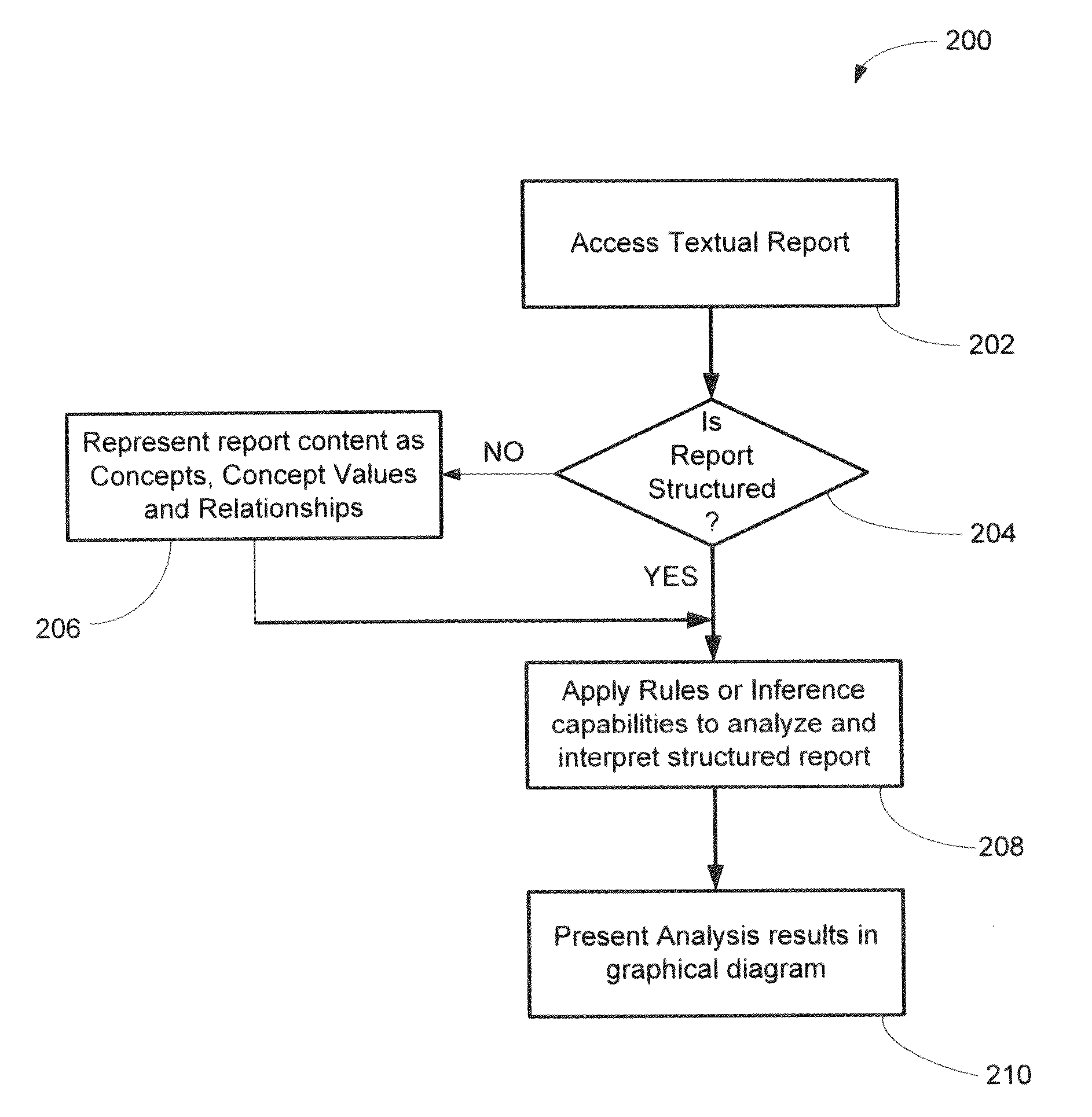
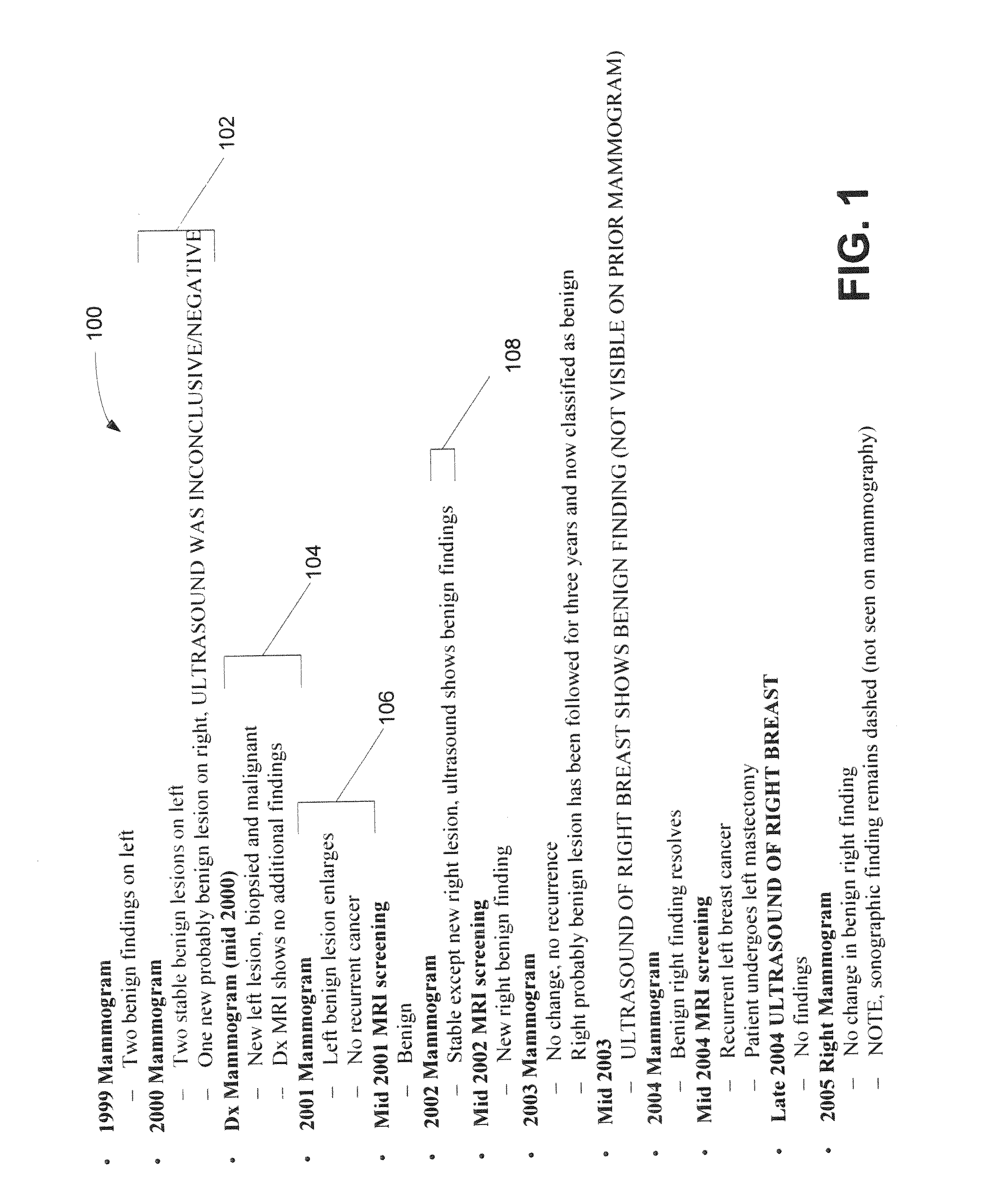
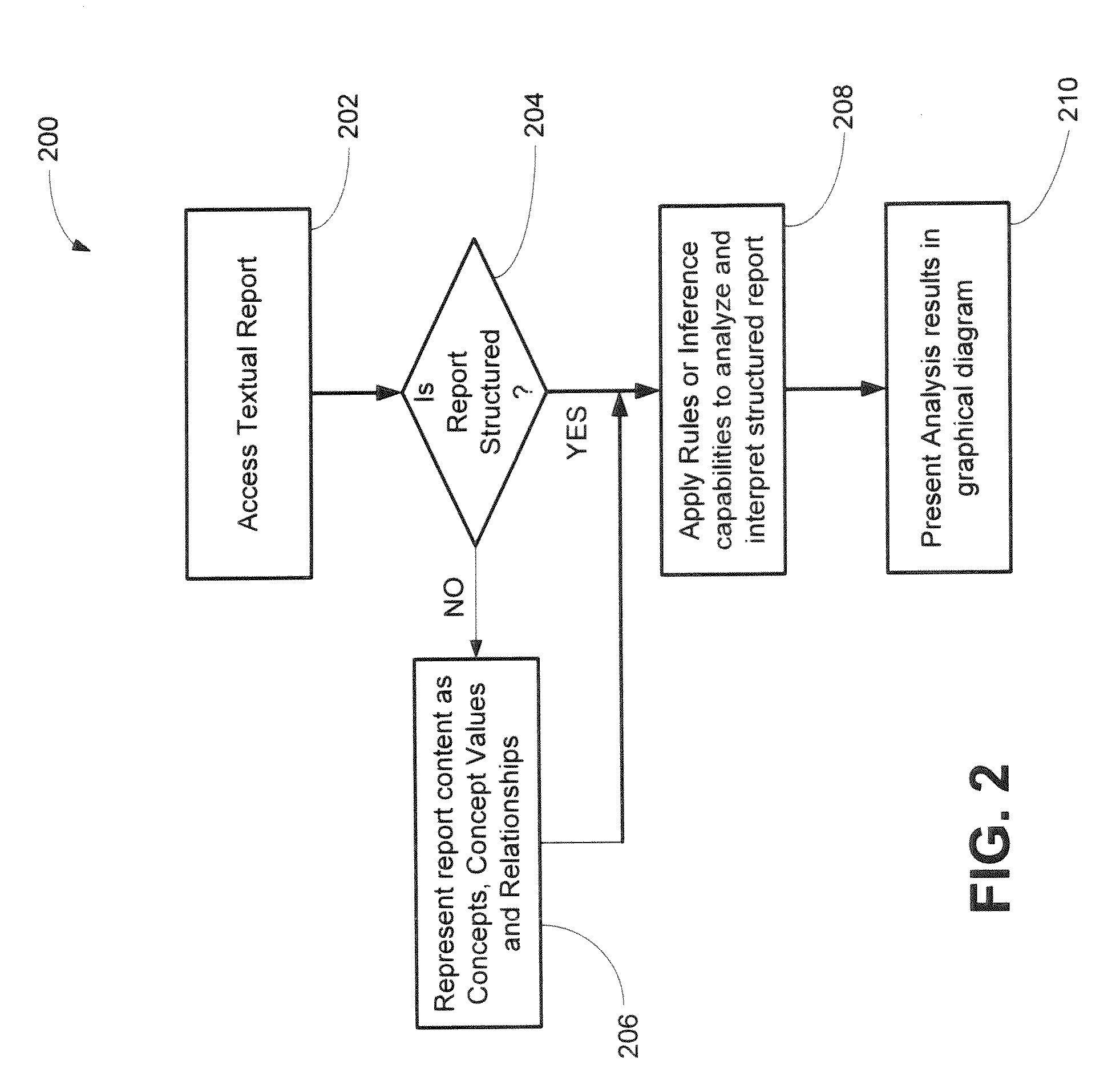
[0035]This document is organized as follows. In the first section, an overview of the techniques and implementation necessary to provide a graphical representation of radiological report contents in accordance with the present invention is provided and described. As would be appreciated by one skilled in the art, while the present invention is described with reference to radiological image study reports, the invention would be applicable to other textual reports and other fields. In the next section, an exemplary prose text to which the present invention may be applied is described, followed by a description of some exemplary steps that may be involved in analyzing and converting said text into a st...


PUM


Abstract

The present invention is directed in general to imaging technologies, and more specifically to a diagrammatic reporting methodology. A system and method are provided for graphically presenting the content of radiological image study reports. Even further, a system and method are provided for presenting the content of structured radiological reports including multiple imaging studies and their corresponding findings in a symbolic color-coded graphical diagram. Further still, the present invention may utilize an ontology of radiological knowledge to interpret report content to generate the information to be diagrammatically displayed in a symbolic color-coded graphical diagram.

Description

CROSS REFERENCE TO RELATED APPLICATIONS[0001]This application is a continuation-in-part of prior U.S. patent application Ser. No. 12 / 535,825, filed Aug. 5, 2009. The content of U.S. patent application Ser. No. 12 / 535,825 is hereby incorporated by reference in its entirety.TECHNICAL FIELD[0002]The present invention is directed in general to imaging technologies and more particularly to the reporting of radiological information. A system and method are provided for the graphical presentation of the contents of radiological image study reports. Even further, a system and method are provided for presenting the contents of structured radiological reports including multiple imaging studies and their corresponding findings in a single diagram. Further still, the present invention utilizes an ontology of radiological knowledge to interpret report content and generate information to be displayed in the graphical diagram.BACKGROUND OF THE INVENTION[0003]In medical imaging, Picture Archiving a...

Claims


Application Information

Patent Timeline
Patent Type & Authority Applications(United States)
IPC IPC(8): G06N5/02G06N5/04G06T11/20G06T7/00
CPCG06F19/3487G06F19/321G16H15/00G16H30/40
Inventor SALZ, DONALD E.MENDEL, JEFFREY B.HALE, CHARLES R.
Owner FUJIFILM MEDICAL SYST USA INC
Why Eureka
Try Eureka