Flexible, multi-channel microelectrode for recording laboratory animal eeg and method for recording laboratory animal eeg using the same

Active Publication Date: 2011-09-15
KOREA INST OF SCI & TECH
View PDF11 Cites 9 Cited by
  • Summary
  • Abstract
  • Description
  • Claims
  • Application Information

AI Technical Summary

Benefits of technology

[0007]Electroencephalography (EEG) of free moving laboratory animals, particularly mice, enables monitoring of the brain in vivo following a genetic or pharmaceutical manipulation. The microelectrode according to an embodiment disclosed herein needs no surgical operation for implantation and, when EEG is not recorded, wires may be easily detached from the laboratory animal. The microelectrode

Problems solved by technology

Further, since the mouse skull is relatively soft and about 200-730

Method used


Image

  • Flexible, multi-channel microelectrode for recording laboratory animal eeg and method for recording laboratory animal eeg using the same
  • Flexible, multi-channel microelectrode for recording laboratory animal eeg and method for recording laboratory animal eeg using the same
  • Flexible, multi-channel microelectrode for recording laboratory animal eeg and method for recording laboratory animal eeg using the same

Examples


Example

EXAMPLE

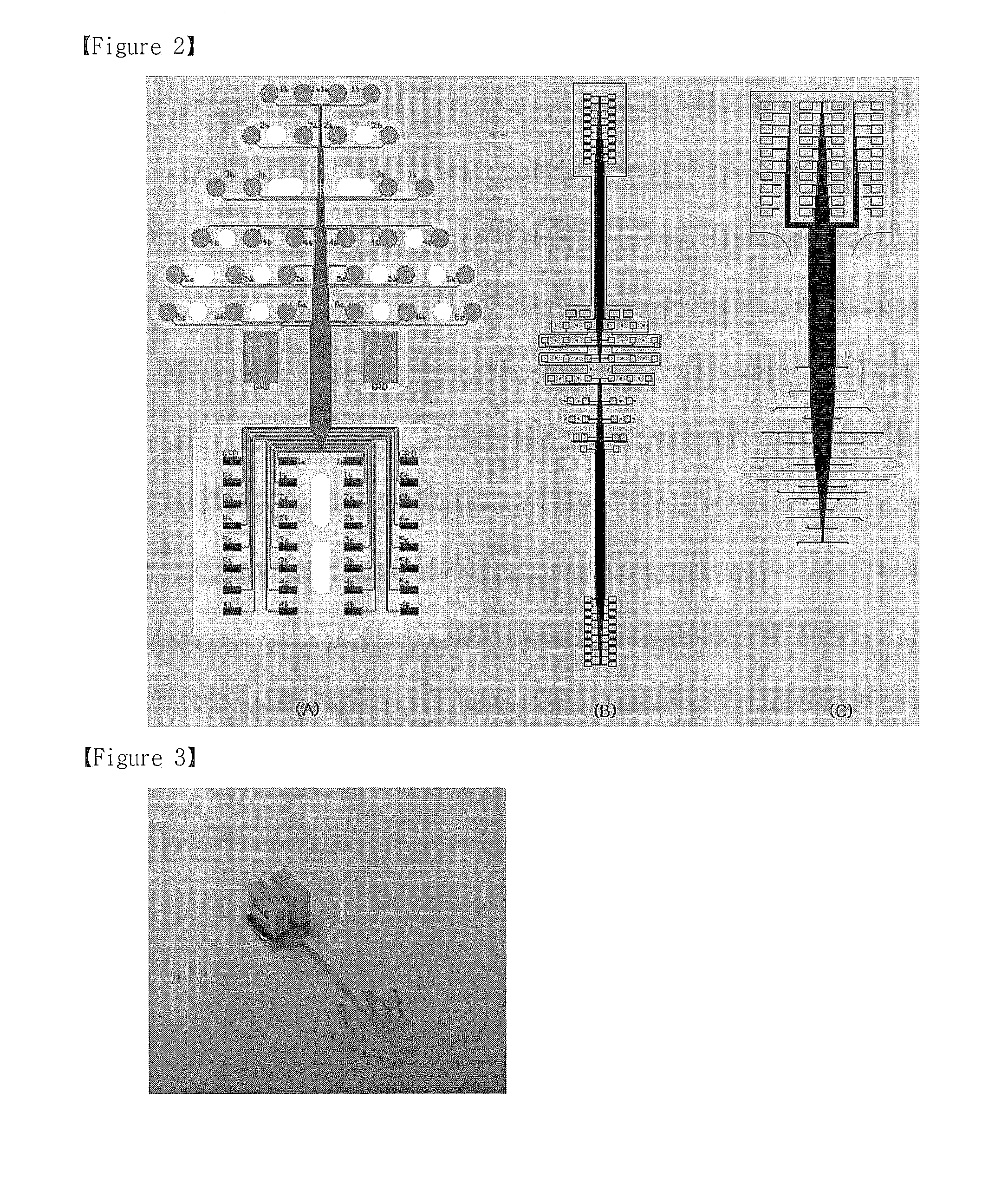
[0029]1. Design of Microelectrode

[0030]Electrodes for human electroencephalography (EEG) are usually located according to the 10-20 or 10-10 system recommended by the American EEG Society. The electrodes are uniformly positioned on two anatomical landmarks (the nasion and the inion) of the human scalp. Before positioning the electrodes, it is important to remove hair and apply a conductive gel between the electrode and the scalp to make the electrode characteristics uniform.

[0031]For mouse EEG, the electrode may be directly placed on the mouse skull after vertical incision of the scalp. However, the area of the exposed skull is limited. The infratemporal region of the skull is held by a network of muscle tissue transmitting a muscle signal. The surface of a mouse skull has landmarks such as the bregma (the point of meeting of the sagittal and coronal sutures) and the lambda. Traditionally, the bregma is used as a reference point. The distance between the bregma and the lambda...

Example

Test Example 1

Analysis of Electrode Characteristics

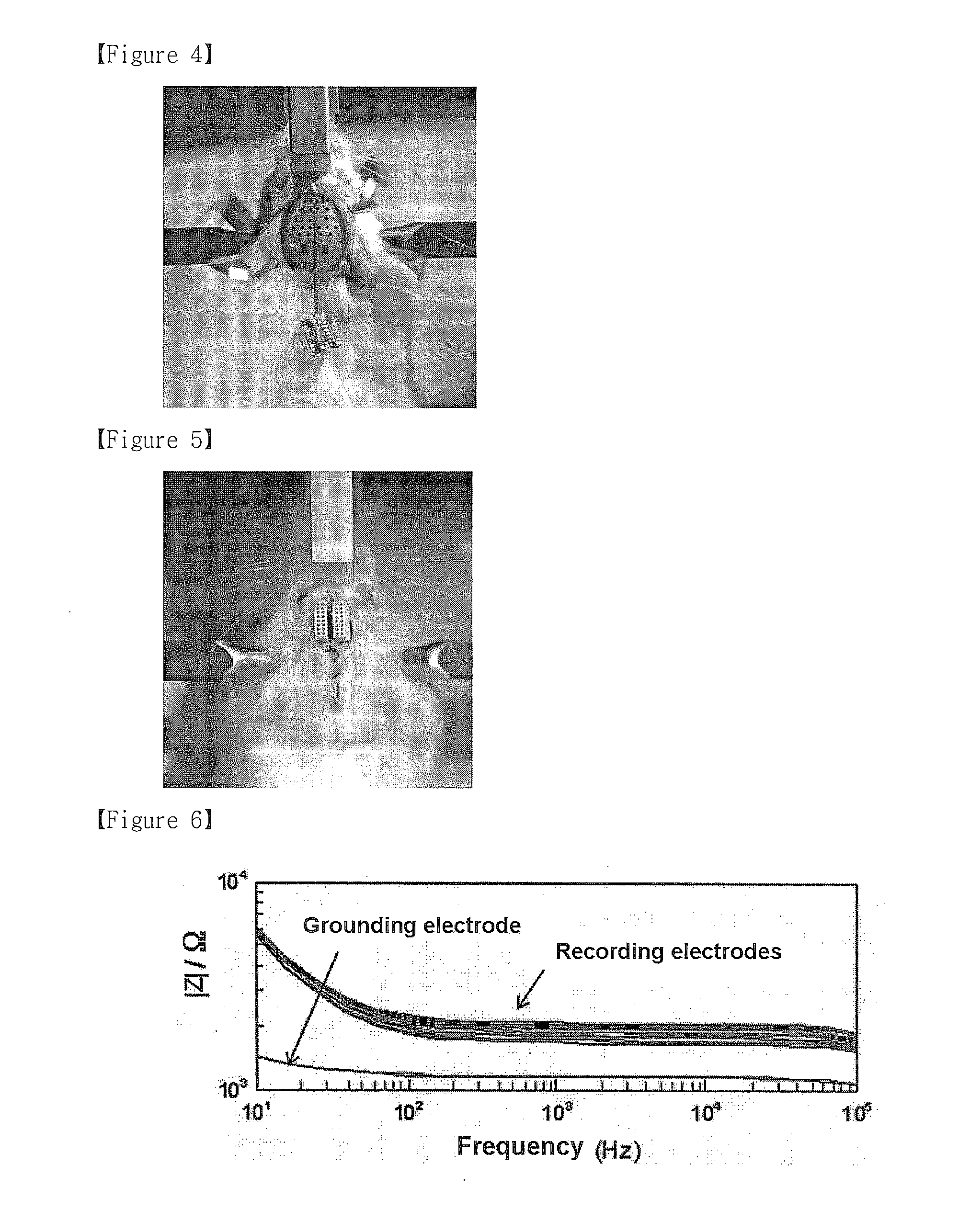
[0040]Electrochemical characteristics of the electrode were analyzed using an impedance spectrometer before and after Pt deposition (measurement amplitude: 50 mV; frequency range: 10-105Hz). The measurement was made using a three electrode setup including a Pt counter electrode (PT 1800, Schott Instruments, Mainz, Germany) and an Ag / AgCl reference electrode (B 2920, Schott Instruments, Mainz, Germany). The setup was coupled with an electrochemical interface (1287, Solartron Analytical, Farnborough, UK) and a frequency response analyzer (1255, Solartron Analytical, Farnborough, UK). The measurement was made in a physiological saline solution (0.9%) at room temperature. In order to remove organic residues on the electrode surface and to stabilize the impedance measurement, the electrode was cycled between −0.6 V and +0.9 V (scan rate: 0.1 V / s) prior to the analysis.

[0041]

[0042]FIG. 6 shows absolute value of impedance |Z| (Ω) and FIG. ...

Example

Test Example 2

In Vivo Test

[0043]Mouse EEG was recorded in vivo using an electrode according to an embodiment of this disclosure.

[0044]2-1. Comparison of EEG Signals from Existing Screw Electrode and Microelectrode According to the Disclosure

[0045]EEG signals from an existing screw electrode and a microelectrode according to an embodiment of this disclosure were compared at the same time. A mouse (8 weeks old, body weight 25 g) was anesthetized with Avertin (2% 20 μl / g, 20 mL / body weight kg) and placed on a stereotaxic apparatus (an apparatus used to examine the brain 3-dimensionally for brain surgery or study) (David Kopf Instruments, Model 902, Calif., USA). After incising an area of 2.0×2.5 cm with respect to the midline, the scalp was opened and fixed with a micro clamp. The skull was perforated and a screw electrode was implanted on the scalp (anteroposterior (AP): +2.5 mm, mediolateral (L): +2.5 mm). A pair of contacts of a microelectrode were positioned to be symmetric to each...


PUM


Abstract

Disclosed are a novel, elastic, biocompatible, micro-sized, polyimide-based multi-channel microelectrode for recording of electroencephalography (EEG) from a laboratory animal including mouse, and a method for recording of laboratory animal EEG using the microelectrode. The microelectrode may include 2 grounding electrodes and 32 recording electrodes. A connector for signal transmission easily connects the microelectrode to a signal acquiring apparatus. The total weight of the microelectrode, including the connector, does not exceed 150 mg. Laboratory animal EEG, including that of mouse, provides the advantage of monitoring the brain state of a freely moving animal following a genetic or pharmaceutical manipulation. The microelectrode can be implanted without surgery and may be detached from wires while EEG is not recorded from the laboratory animal. The microelectrode can successfully acquire broadband EEG signals from the skull of the laboratory animal and is effective in monitoring spatial and temporal pattern of brain activities of the laboratory animal.

Description

TECHNICAL FIELD[0001]Disclosed are a flexible, multi-channel microelectrode for recording of laboratory animal electroencephalography (EEG) and a method for recording of laboratory animal EEG using the microelectrode.BACKGROUND ART[0002]Genetically modified laboratory animals, particularly mice, are a powerful tool in the study of human diseases and treatments thereof. The benefit is extended to the field of neuroscience. Studies are ongoing on the function and behavior of the brain in molecular level, using genetically modified mice. Particularly, the recording of electroencephalography (EEG) of genetically modified mice is an important tool in understanding the mechanism of spontaneous oscillations of the brain or epilepsy in molecular or cellular level.[0003]In the recording of EEG of most laboratory animals, including mice, integrated activities over the whole brain is monitored using one or two channels. The existing EEG electrode is in the form of micro sized screw connected t...

Claims


Application Information

Patent Timeline
IPC IPC(8): A61B5/0478
CPCA61B5/0478A61B2503/40A61B2562/046A61B2562/0209A61B5/4094A61B2562/0215A61B5/291
Inventor CHOI, JI HYUNSHIN, HEE SUPKOCH, KLAUS PETER
Owner KOREA INST OF SCI & TECH
Why Eureka
Try Eureka