Implant for correcting skeletal mechanics

a technology applied in the field of medical equipment for enhancing and correcting skeletal mechanics, can solve the problems of no longer being able to support the first metatarsal, excessive strain on the soft tissues supporting this bone, and stretching of the soft tissues

Active Publication Date: 2012-04-19
GRAHAM MICHAEL E
View PDF1 Cites 4 Cited by
  • Summary
  • Abstract
  • Description
  • Claims
  • Application Information

AI Technical Summary

Benefits of technology

[0012]One of the primary purposes of the subject invention is to correct first ray deformities that arise to a significant extent from deformities at the metatarsocuneiform joint. This device will treat these deformities at their source. The inventor's device, a surgical implant, consists of a plate and a wedge. The plate is secured to both the metatarsal and cuneiform bones, preferably by bone screws. The wedge inserts, at least part-way, into the metatarsocuneiform joint, so as to contact at least a portion of the ends of the bones flanking this joint. A critical aspect of the invention is the removal of the tissue, including bone tissue, present in the metatarsocuneiform joint in a manner that allows restoration of the proper alignment of the first metatarsal bone relative to the first cuneiform bone. This typically involves removal of a pie shaped wedge of tissue within this joint and may also involve reshaping the ends of the bones. As described above, it is believed that a principal factor causing bone misalignment is deformities of the ends of bones.

Problems solved by technology

Excessive pronation (hyperpronation) of the foot leads to abnormal motion to the first metatarsal resulting in excessive strain on the soft tissues supporting this bone.
After a prolonged period of these excessive forces the soft tissues will stretch out and no longer be able to support the first metatarsal.
This instability leads to an abnormal deviation of the first metatarsal bone resulting in foot pathology.
The end result is that there will be instability at both the proximal and distal joints of the first metatarsal bone.
This deformation causes instability of the first metatarsal bone and contributes to the medial shift of the metatarsal bone.
The deviation of the first metatarsal bone leads to the formation of a bunion (hallux valgus) and can also lead to other deformities of the first ray.
The problem with non-surgical treatment options is that it is ineffective in eliminating the causative factor.
Also, every step leads to further deformity of the metatarsal bone.
Because the problem with this deformity is intrinsic, usually external remedies are ineffective in controlling the deforming forces.
These types of procedures straighten the metatarsal bone with respect to the hallux but leave instability at the first metatarsocuneiform joint.
Since the instability at the first metatarsocuneiform joint still exists, the first metatarsal bone will eventually deviate again and lead to the occurrence of an overall foot deformity.
These procedures lead to long recovery periods, at least six months, and can fail.
Unfortunately, this transfers the body's weight to the head of the second metatarsal bone instead of the first.
Often, further pathology ensues such as callus formation under the ball of the foot leading to further pain and possible ulceration.
This improper weight redistribution can lead to fractures in other parts of the foot.
The problem with these procedures is that the plates mimic a screw or staple and stabilize the fusion site between the two bones.
Consequently, there is still a shortening of the first metatarsal bone leading to the possibility of other ill effects.
Moreover, these plates are rather bulky and usually have to be removed after bone arthrodesis is achieved.

Method used


Image

  • Implant for correcting skeletal mechanics
  • Implant for correcting skeletal mechanics
  • Implant for correcting skeletal mechanics

Examples


Embodiment Construction

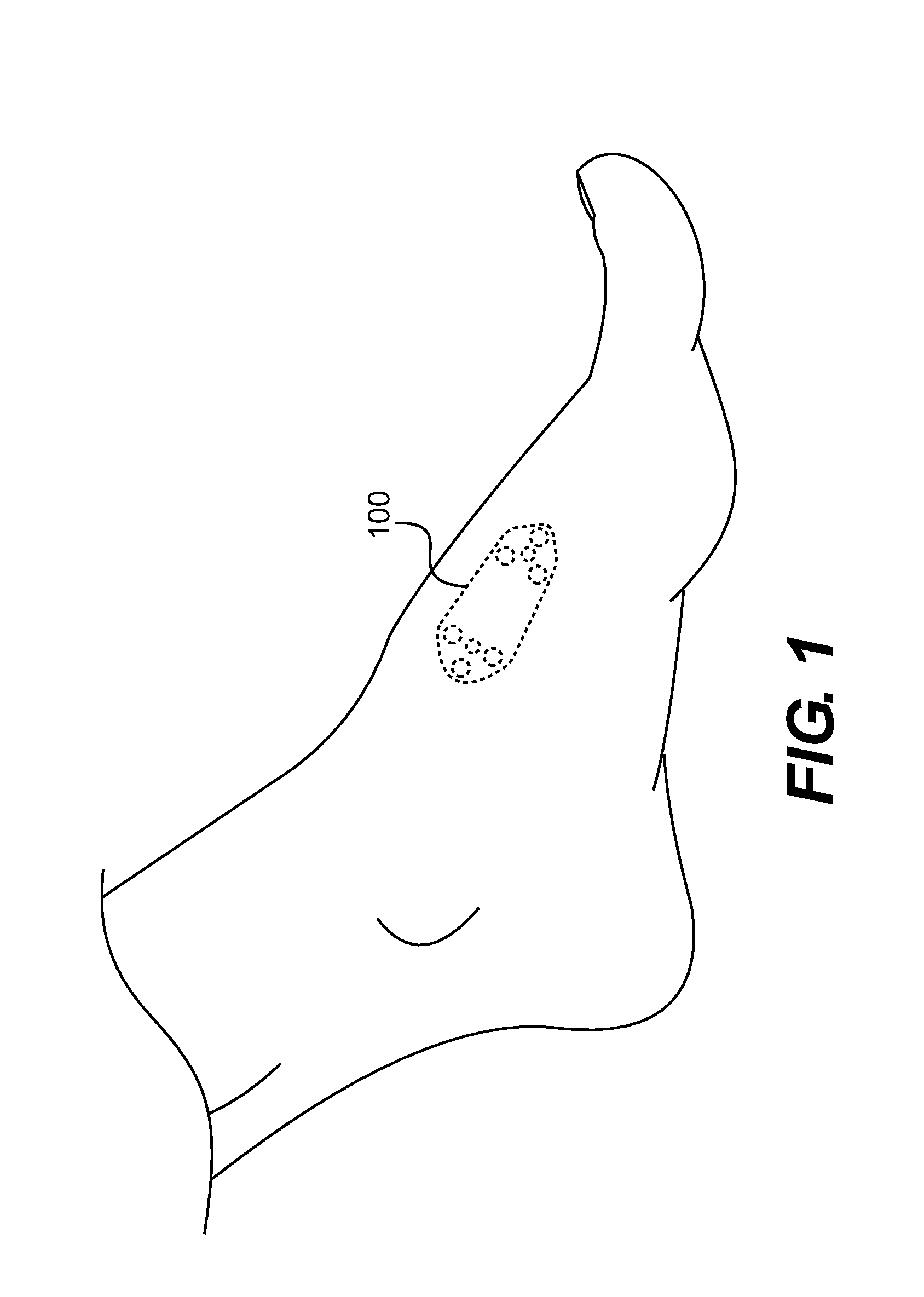
[0017]FIG. 1 illustrates a preferred location in which the subject implant for correcting skeletal mechanics 100 will be inserted. The implant 100 is placed at the inner, or medial, surface of a patient's foot over the metatarsocuneiform joint.

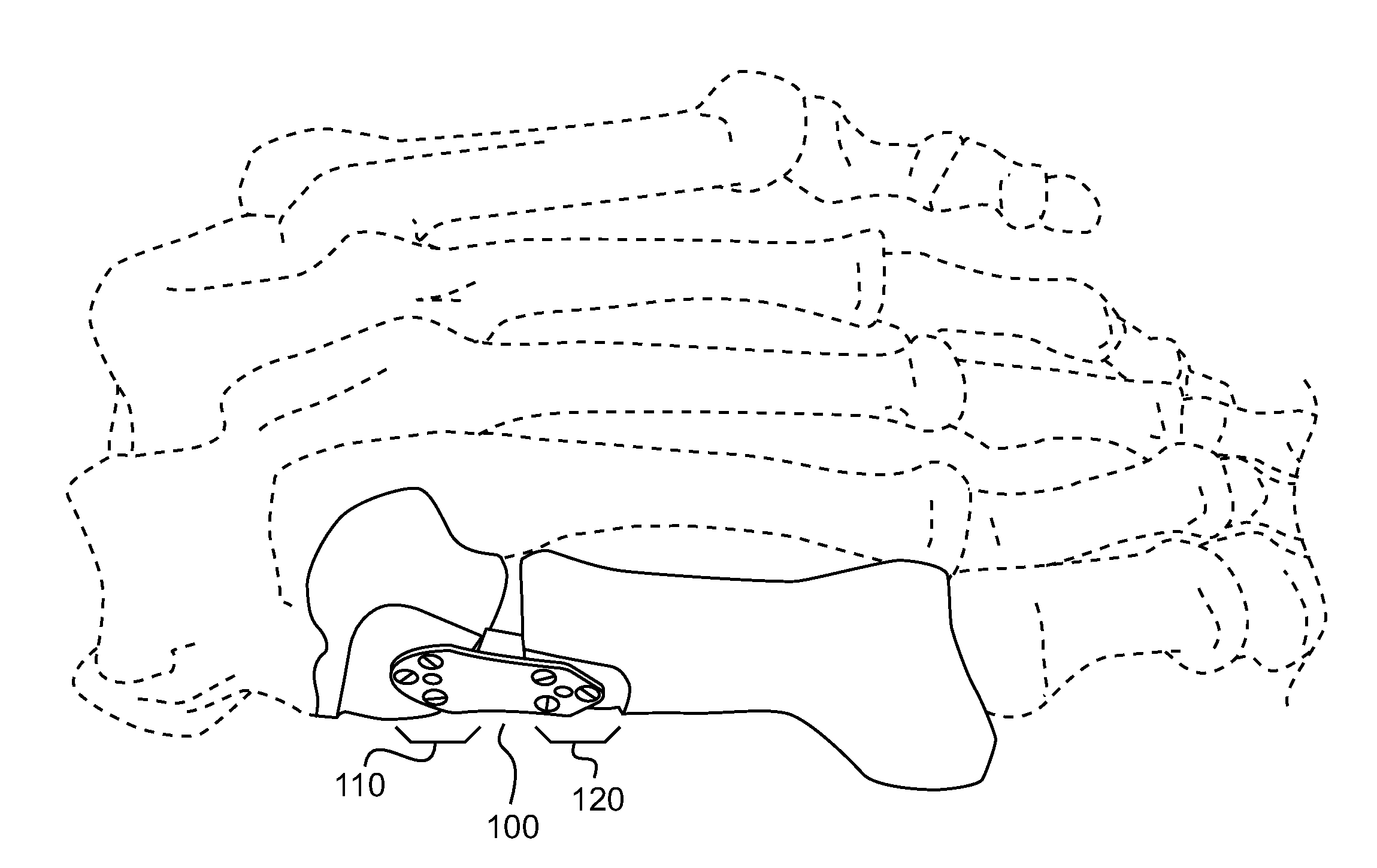
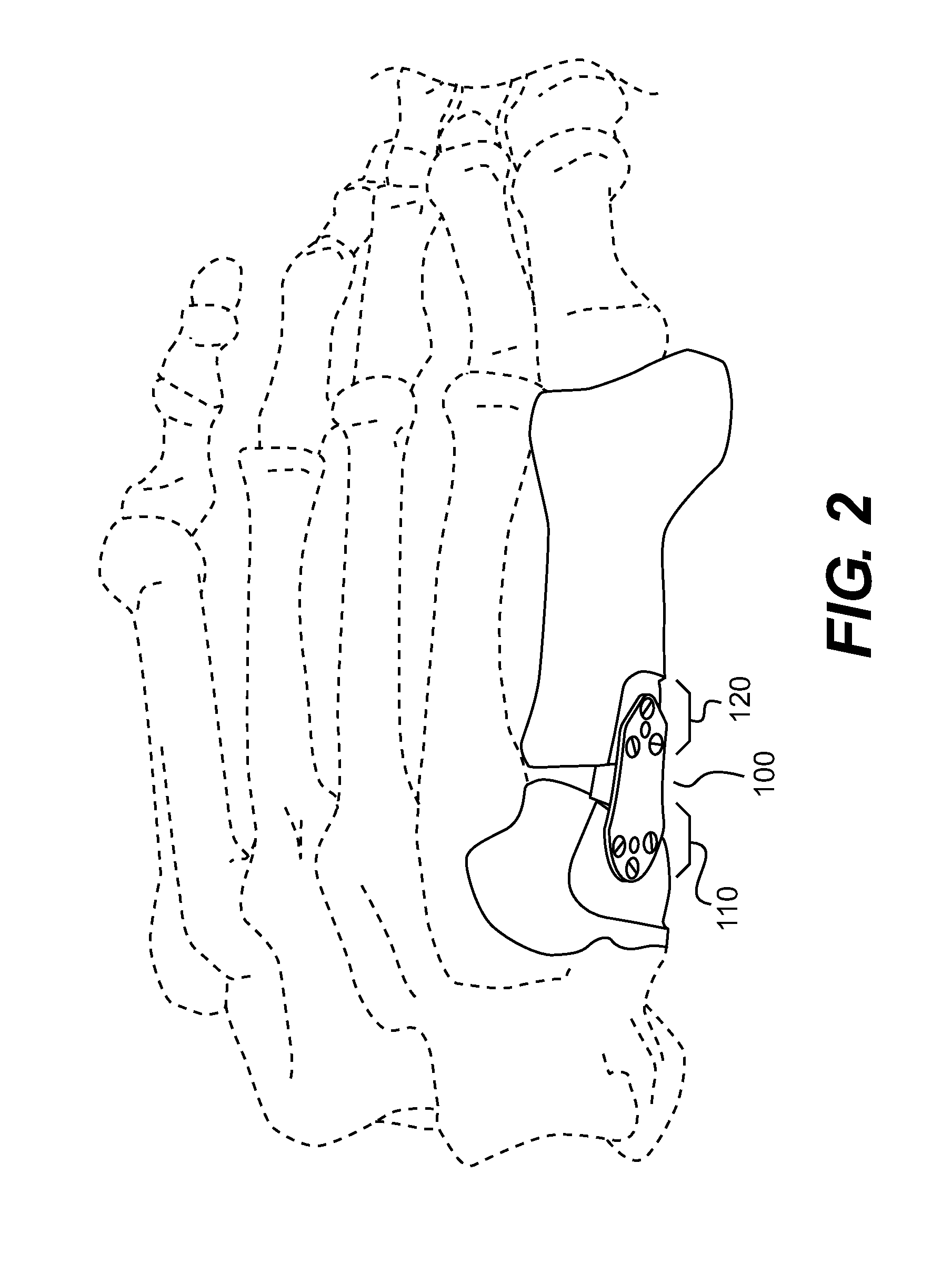
[0018]The device contains both a plate component 105 and a wedge component 130. See FIGS. 2-7 below. Turning to FIG. 2, the device is shown in place with the surrounding bone structure illustrated. The device is located such that the anterior portion of the plate is attached to a flush or flattened region on the side of the first metatarsal bone 120. The posterior portion is attached to a flush or flattened region on the side of the cuneiform bone 110. The wedge portion is inserted at least part way into the joint between these bones, the metatarsocuneiform joint.

[0019]FIGS. 3-7 show different detail views of a preferred implant. As stated above, the implant 100 is constructed with a plate component 105 and a wedge component 130. The wedge com...


PUM


Abstract

Deformities present on the end of a bone, for example the end of the metatarsal bone making up part of the metatarsocuneiform joint, can lead to deformities such as bunions. These deformities are treatable with an implant that comprises a plate with a wedge extending perpendicular from the plate. Following removal of cartilage from the joint, deformed portions at the end of the bone are removed and the wedge is inserted in the joint and held in place when the plate is attached to the bones flanking the joint. This effectively fuses the two bones together. The wedge can be shaped in various ways depending on the particular deformity present.

Description

BACKGROUND OF THE INVENTION[0001]This invention relates to a medical apparatus for enhancing and for correcting skeletal mechanics. More specifically, this invention relates to the correction of certain bone alignment deformities that impair optimal biped mechanics of the foot.[0002]Excessive pronation (hyperpronation) of the foot leads to abnormal motion to the first metatarsal resulting in excessive strain on the soft tissues supporting this bone. After a prolonged period of these excessive forces the soft tissues will stretch out and no longer be able to support the first metatarsal. This instability leads to an abnormal deviation of the first metatarsal bone resulting in foot pathology. The deforming forces acting on the first metatarsal leads to three possible deviations: (1) pure medial deviation of the metatarsal, (2) dorsal deviation (dorsiflexion) of the metatarsal, and (3) the combination of the previous two, dorsomedial deviation. When the first metatarsal deviates medial...

Claims


Application Information

Patent Timeline
Patent Type & Authority Applications(United States)
IPC IPC(8): A61B17/80A61B17/88
CPCA61B17/809A61B17/8061
Inventor GRAHAM, MICHAEL E.
Owner GRAHAM MICHAEL E
Why Eureka
Try Eureka