
Hip joint device and method

Active Publication Date: 2012-04-26
FORSELL PETER
View PDF7 Cites 5 Cited by
  • Summary
  • Abstract
  • Description
  • Claims
  • Application Information

AI Technical Summary

Benefits of technology

[0049]According to another embodiment, the anchoring portion can be placed in a first and second state, and said anchoring portion could be adapted to, in the second state, further engage at least one of the cancellous bone of the collum femur, the cortical bone of the collum femur and an artificial material placed inside of the collum femur, for further stabilizing the medical device.

Problems solved by technology

There are both natural and pathological processes leading to deteriorated joint function.
With age and wear, the articular cartilage becomes less effective as a shock absorber and a lubricated surface.
This abnormal wearing of the cartilage also results in a decrease of the joints lubricating fluid called Synovial fluid.
To get access to the joint, the supporting Capsule attached to Femur and Ilium needs to be penetrated, making it difficult to get a fully functional joint after the surgery.

Method used


Image

  • Hip joint device and method
  • Hip joint device and method
  • Hip joint device and method

Examples


Embodiment Construction

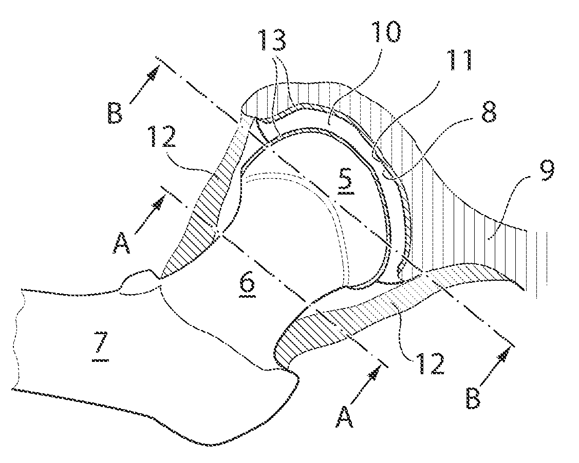
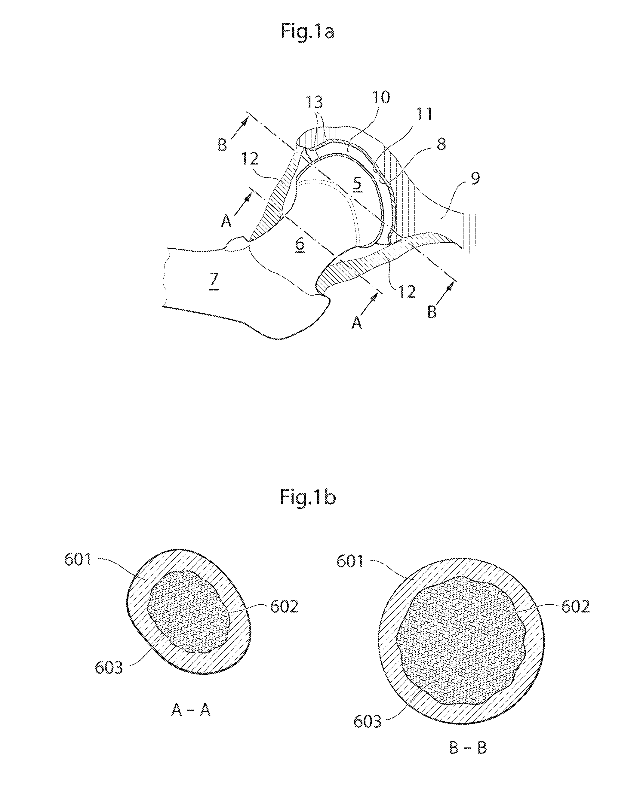
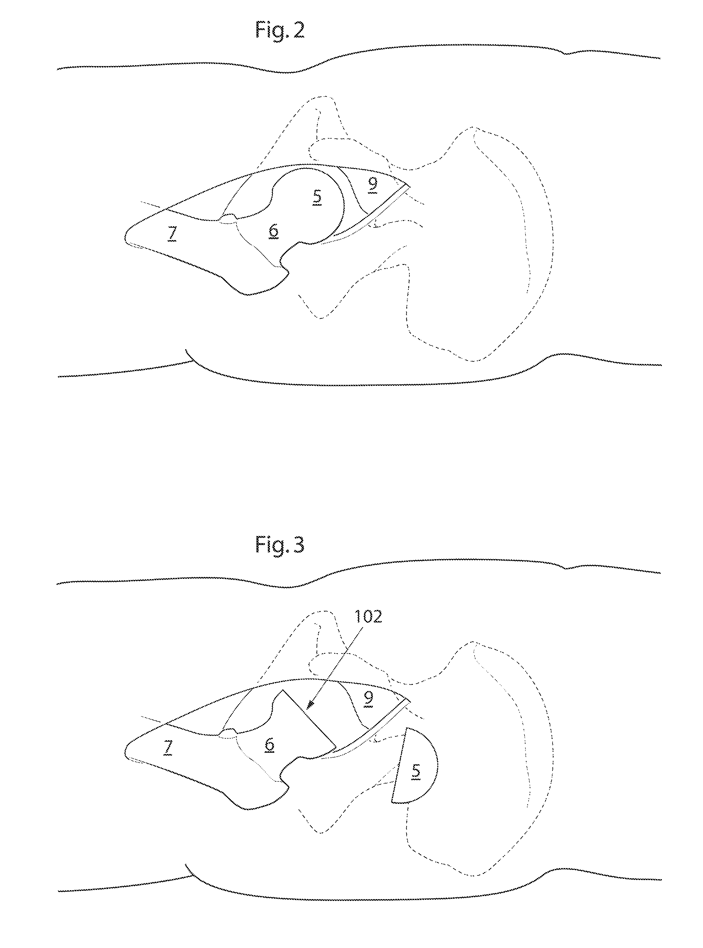
[0091]In the following a detailed description of preferred embodiments of the present invention will be given. In the drawing figures, like reference numerals designate identical or corresponding elements throughout the several figures. It will be appreciated that these figures are for illustration only and are not in any way restricting the scope of the invention. Thus, any references to direction, such as “up” or “down”, are only referring to the directions shown in the figures. Also, any dimensions etc. shown in the figures are for illustration purposes.

[0092]Functional hip movements are to be understood as movements of the hip that at least partly correspond to the natural movements of the hip. On some occasions the natural movements of the hip joint might be somewhat limited or altered after hip joint surgery, which makes the functional hip movements of a hip joint with artificial surfaces somewhat different than the functional hip movements of a natural hip joint.

[0093]The fun...


PUM


Abstract

A medical device for implantation in a hip joint of a human patient is provided. The natural hip joint having a ball shaped caput femur as the proximal part of the femoral bone with a convex hip joint surface towards the centre of the hip joint and a bowl shaped acetabulum as part of the pelvic bone with a concave hip joint surface towards the centre of the hip joint. The caput femur has a centrally placed longitudinal extension, extending through the center of the caput and collum femur, aligned with the collum femur, defined as the caput and collum femur center axis. The medical device comprising; an artificial acetabulum, comprising a concave surface towards the centre of the hip joint. The artificial concave acetabulum is adapted to, when implanted, be fixated to the femoral bone of the human patient, and be in movable connection with an artificial caput femur fixated to the pelvic bone of the patient.

Description

[0001]The present invention relates generally to a medical device for implantation in a hip joint, and a method of providing said medical device.BACKGROUND ART[0002]The hip joint is a synovial joint, joining the pelvis to the proximal portion of the femoral bone. Synovial joints are the most common types of joints in mammals, and are typical of nearly all limb joints. The contacting surfaces of said the pelvic, the acetabulum, and the contacting surface of the femoral bone, the caput femur, are smooth and rounded, and covered by articular cartilage. A synovial membrane, encapsulates the joint, forming a hip joint cavity, which contains synovial fluid. Outside the synovial membrane is a fibrous capsule and ligaments, forming an articular capsule.[0003]There are both natural and pathological processes leading to deteriorated joint function. With age and wear, the articular cartilage becomes less effective as a shock absorber and a lubricated surface. Different degenerative joint disea...

Claims


Application Information

Patent Timeline
IPC IPC(8): A61F2/32
CPCA61F2/32A61F2/36A61F2002/30471A61F2/34A61F2/3601A61B17/1637A61B17/1664A61B17/74A61B17/86A61F2002/30507A61F2002/30558A61F2002/30563A61F2002/30565A61F2002/30579A61F2002/30878A61F2002/30886A61F2002/3241A61F2002/3483A61F2002/3615A61F2002/3631A61F2002/4631A61F2002/4635A61F2002/4677A61F2/3603A61F2240/002A61F2/3607A61F2/3609A61F2002/365
Inventor FORSELL, PETER
Owner FORSELL PETER
Learn More