
Surgical Ligation Clip and Applicator Device

a technology of which is applied in the field of surgical ligation clip and applicator device, can solve the problems of disadvantageous risk of unintended laceration after being secured onto target tissues, current available clips failing, and unsuitable for use on prostatic vascular pedicles, so as to prevent the closure of the ligation clip, avoid undesired hemorrhaging, and avoid undesired hemorrhaging

Inactive Publication Date: 2012-09-06
JENKINS CLINIC
View PDF0 Cites 117 Cited by
  • Summary
  • Abstract
  • Description
  • Claims
  • Application Information

AI Technical Summary

Benefits of technology

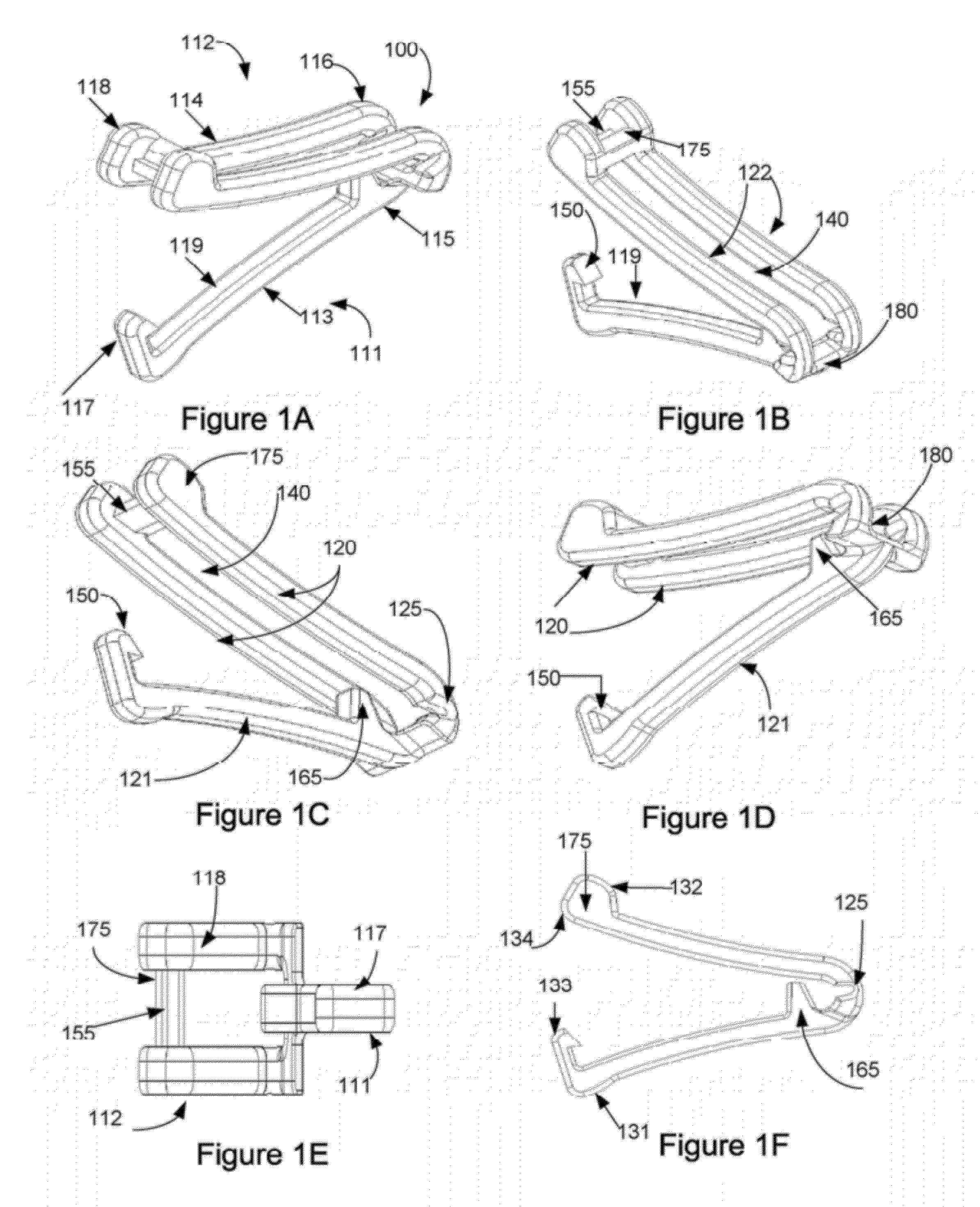
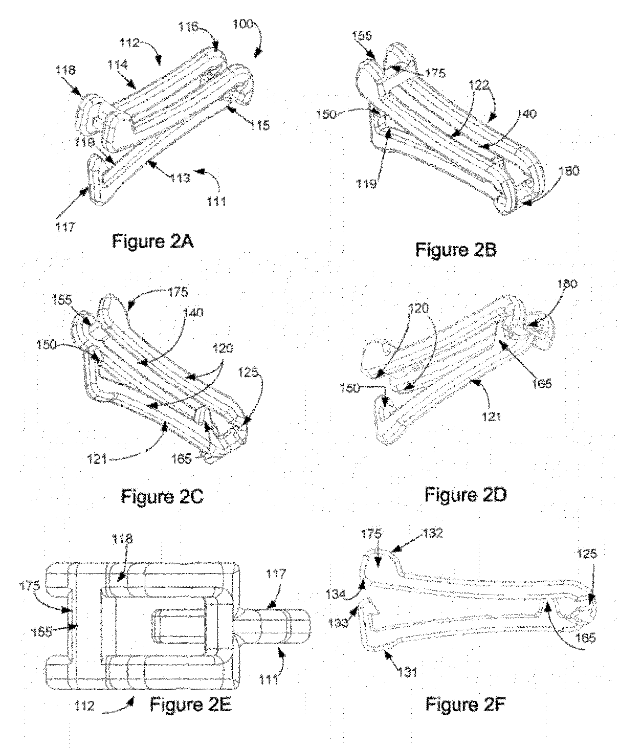
[0009]The surgical tissue ligation clip of the present invention further has a proximal tissue shield extending inwardly from the proximal portion of the first or second leg members past the tissue clamping inner surface of the opposite leg member when the leg members are in a partially closed position. The proximal tissue shield prevents tissue that is being clamped between the inner surfaces of the leg members from being pinched and cut at the hinge to avoid undesired hemorrhaging, disassociating the hinge, or preventing closure of the ligation clip for implantation.
[0010]The surgical tissue ligation clip can further have a distal tissue shield extending outwardly from side to side of the distal portion of the outer surface of the second leg member proximal to locking element. The distal tissue shield prevents tissue being clamped between the inner surfaces of the leg members from being pinched between the hook member and locking element to avoid undesired hemorrhaging, interfering with securely closing the ligation clip for implantation, or disassociating the hinge. Tissue shielding and clearing away from the ligation clip mechanism also provides improved tactile feedback to the user when the clip is closed.
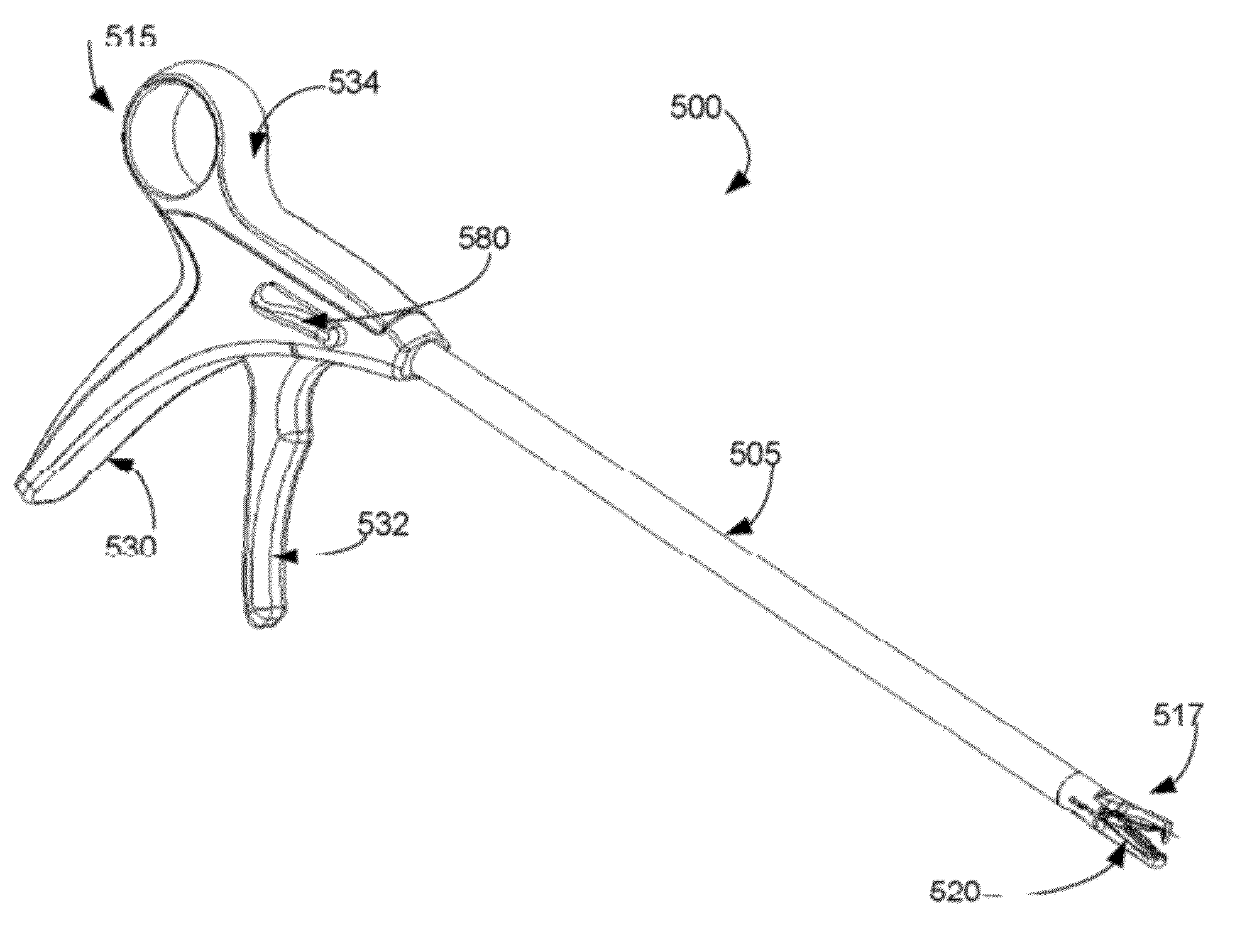
[0011]The present invention further provides a minimally invasive applicator for a surgical ligation clip. The applicator can be configured for single clip delivery, or be adapted for receiving a plurality of surgical ligation clips individually arranged in line for movement therethrough. The elongated surgical ligation clip chamber has a proximal end, a central storage region and an open distal end. The surgical ligation clip applicator further has a pair of jaws disposed on the open distal end of the chamber having a sharpened incising tooth member extending from at least one side towards the opposite jaw. The jaws selectively articulate between an open position for retaining an open ligation clip therebetween, and a closed position for penetrating interfering tissue with the sharpened incising tooth member between an opposing pair of jaws, and securely closing the ligation clip onto the target tissue.

Problems solved by technology

Currently available surgical ligation clips and delivery means are not ideally adapted for use on prostatic vascular pedicles.
Furthermore, the bundling of excess tissue associated with the thick pedicle vasculature around the ligation clip hinge or the ligation clip closure mechanism frequently causes currently available clips to fail through breakage or lack of secure engagement.
Existing clips that have sharpened features disposed thereon for tissue penetration also present a disadvantageous risk of unintended laceration after being secured onto target tissues.
Existing systems for ligation clip placement also inadequately protect ancillary tissue laceration not only after being secured to the target tissue, but upon delivery after exiting the trocar or other laproscopic delivery and applicator devices.
Existing clips also frequently fail to remain securely engaged within the applicator tool during delivery or placement and can misfire or inadvertently fall into the surgical field.

Method used


Image

  • Surgical Ligation Clip and Applicator Device
  • Surgical Ligation Clip and Applicator Device
  • Surgical Ligation Clip and Applicator Device

Examples


Embodiment Construction

[0048]Embodiments of the invention now will be described more fully hereinafter with reference to the accompanying drawings, in which embodiments of the invention are shown. This invention may, however, be embodied in many different forms and should not be construed as limited to the embodiments set forth herein; rather, these embodiments are provided so that this disclosure will be thorough and complete, and will fully convey the scope of the invention to those skilled in the art. Like numbers refer to like elements throughout. The singular forms “a,”“an,” and “the” can refer to plural instances unless context clearly dictates otherwise or unless explicitly stated.

[0049]The various embodiments described herein provide exemplary devices, systems, methods of use and manufacture of surgical ligation clips and applicators for patient tissue in need thereof. The present invention provides a surgical tissue ligation clip, comprising first and second leg members each having a central port...


PUM


Abstract

A surgical tissue ligation clip and an applicator device for delivery of multiple ligation clips is provided. The surgical ligation clips have first and second leg members joined by a resilient hinge at one end and a deflectable hook member and a locking element on the opposite end. The surgical tissue ligation clip has a proximal tissue shield that prevents tissue from being pinched at the hinge, and / or a distal tissue shield that prevents tissue from being pinched between the hook member and locking element and interfering with securely closing the ligation clip for implantation. A minimally invasive applicator for the surgical ligation clip is provided for delivering individually arranged ligation clips in line, incising target tissue and delivering a plurality of surgical ligation clips at the tissue site.

Description

CROSS REFERENCE TO RELATED APPLICATION[0001]This application claims the priority benefit of U.S. Provisional Patent Application No. 61 / 449,247 filed on Mar. 4, 2011. The contents of this application is hereby incorporated by reference in its entirety.FIELD OF THE INVENTION[0002]The present invention relates generally to vessel sealing devices, and particularly to a system, apparatus, and methods for providing a surgical clip device for ligating tissues during surgical procedures.BACKGROUND OF THE INVENTION[0003]A radical prostatectomy is frequently the recommended procedure for treating cancerous prostate tissue. The primary blood supply for the prostate are the prostatic pedicles. In the process of removing the prostate, the pedicles must be ligated and divided. Careful hemostasis of the prostatic vascular pedicles is required to avoid collateral injury to the prostate and adjacent neural tissue, which is necessary to preserve continence and potency after recovery, and to create ne...

Claims


Application Information

Patent Timeline
Patent Type & Authority Applications(United States)
IPC IPC(8): A61B17/10A61B17/03
CPCA61B17/1285A61B17/1227
Inventor MALIZIA, ANTHONY A.MALIZIA, ELIZABETH L.VANHIEL, BRIANCHARLES, KIRKCARROLL, MAUREENBOST, FRANKLIN
Owner JENKINS CLINIC
Learn More