Methods and Systems to Measure Corneal Epithelial Thickness and Power, Stromal Thickness, Subepithelial Corneal Power and Topography for Disease Diagnosis

a technology of epithelial thickness and power, which is applied in the field of ophthalmology, can solve problems such as unsuitable methods, and achieve the effects of enhancing image quality, sufficient data density, and reliably determining corneal epithelial and stromal properties

Active Publication Date: 2013-05-23
UNIV OF SOUTHERN CALIFORNIA
View PDF7 Cites 23 Cited by
  • Summary
  • Abstract
  • Description
  • Claims
  • Application Information

AI Technical Summary

Benefits of technology

The patent describes a method for generating corneal images using a scanning pattern that provides sufficient data density and distribution over the cornea. The scanning pattern can be a radial spoke pattern or a raster pattern. By repeating the scanning multiple times using the same pattern, duplicate scans can be generated to enhance image quality or detect eye movement during scans. The technical effect of this method is to improve the accuracy and reliability of corneal imaging for diagnosis and treatment purposes.

Problems solved by technology

The technical problem addressed in this patent text is the need for a fast, reliable, and convenient method to measure the thickness and refractive power of the corneal epithelium and stroma, which can aid in the detection and diagnosis of keratoconus and other corneal ectatic disorders. This is important for planning and performing laser corneal surgeries and minimizing regression after surgery. Current methods such as optical coherence tomography (OCT) and confocal microscopy have limitations in terms of speed and the number of points measured, and there is no known method of using FD-OCT to generate corneal epithelial and stromal maps.

Method used


Image

  • Methods and Systems to Measure Corneal Epithelial Thickness and Power, Stromal Thickness, Subepithelial Corneal Power and Topography for Disease Diagnosis
  • Methods and Systems to Measure Corneal Epithelial Thickness and Power, Stromal Thickness, Subepithelial Corneal Power and Topography for Disease Diagnosis
  • Methods and Systems to Measure Corneal Epithelial Thickness and Power, Stromal Thickness, Subepithelial Corneal Power and Topography for Disease Diagnosis

Examples


example

[0089]Corneal Epithelial Thickness Mapping in Normal and Keratoconic Eyes with Fourier-Domain Optical Coherence Tomography

[0090]One purpose of this example is to map corneal epithelial thickness in normal and keratoconic eyes with optical coherence tomography (OCT).

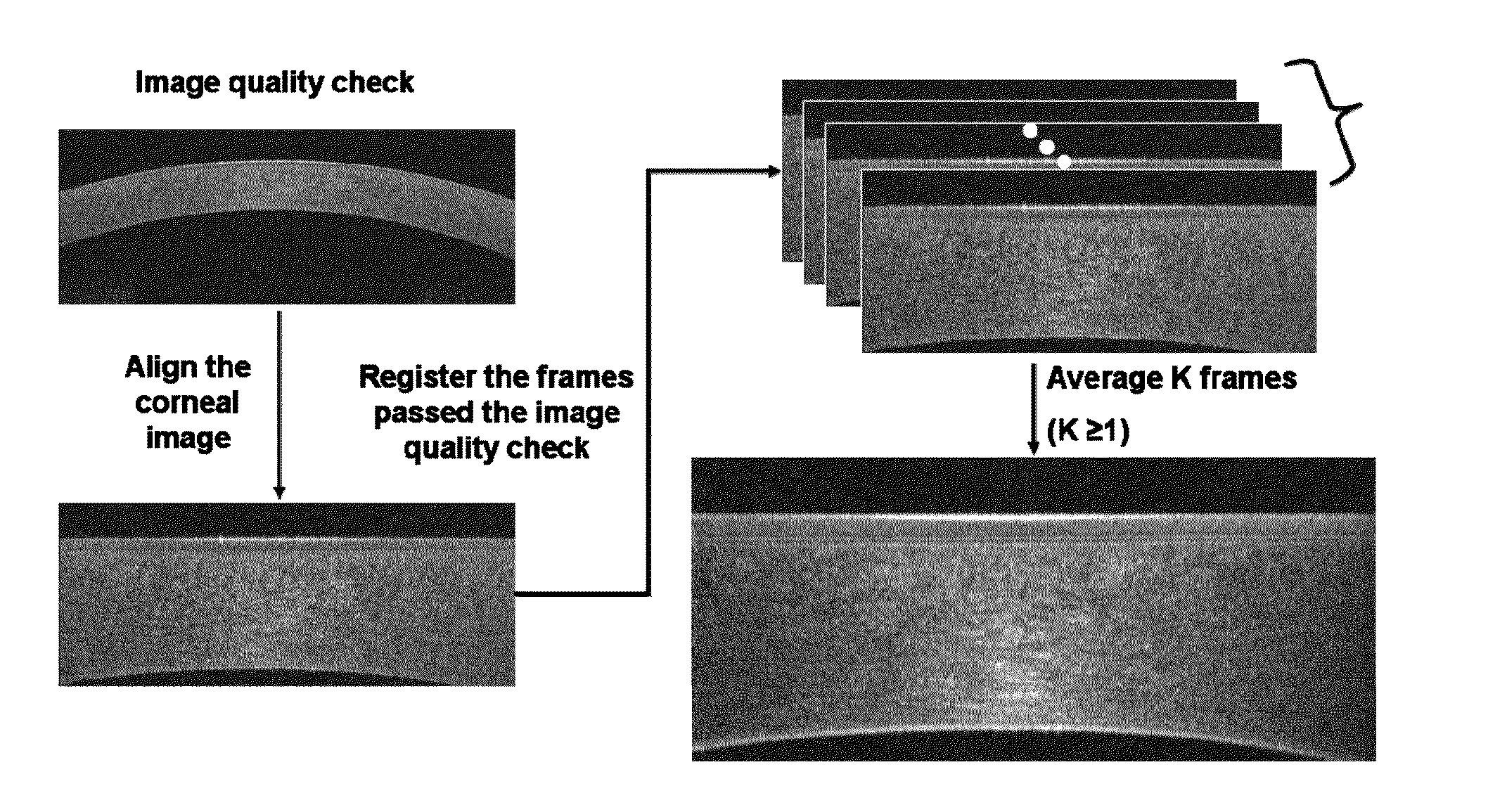
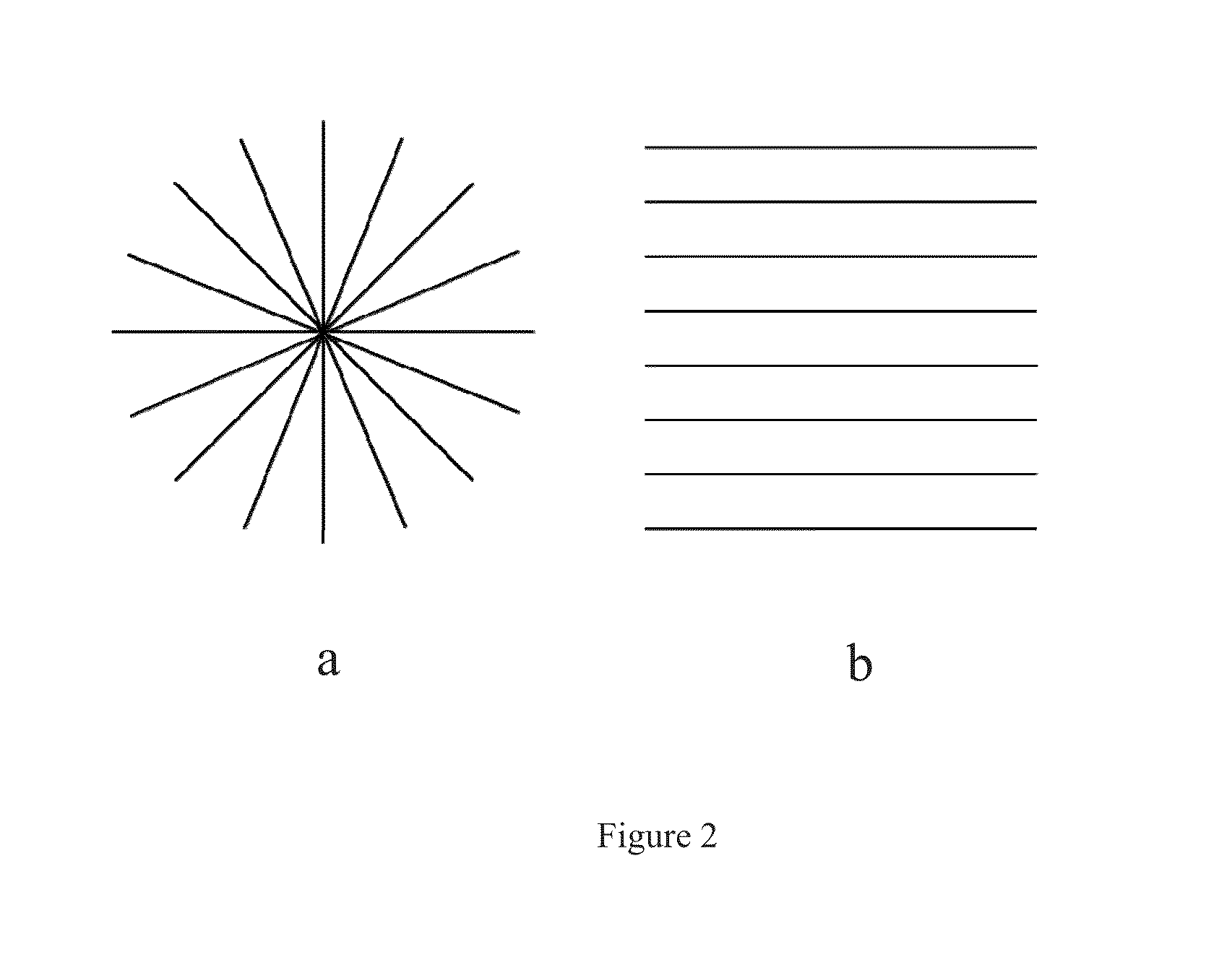
[0091]Methods. A Fourier-domain OCT system with 26,000 axial-scans / second scan speed and 5 μm axial resolution was used. A pachymetry scan pattern (8 radials, 1024 axial-scans each, 6 mm diameter) centered at the pupil center was used to image the cornea, A computer algorithm was developed to generate the epithelial thickness (tear film included) map automatically.

[0092]The map was divided into 3 zones by diameter: central 2 mm, superior 2-5 mm, and inferior 2-5 mm. The average epithelial thickness from each zone was calculated. Normal and keratoconic eyes (24 eyes each) were scanned 3 times. The repeatability of the measurement was evaluated by pooled standard deviation (SD).

[0093]Results. The central, superior, and infe...


PUM


Abstract

This invention discloses methods and systems for measuring corneal epithelial thickness and power, stromal thickness, subepitheila corneal power and topography. The systems and methods disclosed herein are non-invasive, non-contact and automated imaging methods which preferably makes use of Fourier-domain optical tomography. Also disclosed herein are scanning patterns and image analysis methods for utilizing and analyzing Fourier-domain optical coherence tomography images to obtain information about conical epithelial and stromal properties as well as parameters useful for evaluating the properties. The methods and systems described herein are useful as eye disease diagnostic tools and eye surgery planning tools.

Description


Claims


Application Information

Patent Timeline
Owner UNIV OF SOUTHERN CALIFORNIA
Why Eureka
Try Eureka