
Method for the reduction of artifacts in image data sets and computing facility
a technology of image data and computing facilities, applied in the field of artifact reduction of image data sets and computing facilities, can solve the problems of mainly affecting not always able to completely eliminate artifacts, and often remain in image data, so as to increase the homogeneity of image data sets and reduce the effect of homogeneity artifacts
- Summary
- Abstract
- Description
- Claims
- Application Information
AI Technical Summary
Benefits of technology
Problems solved by technology
Method used
Image

Examples
Embodiment Construction
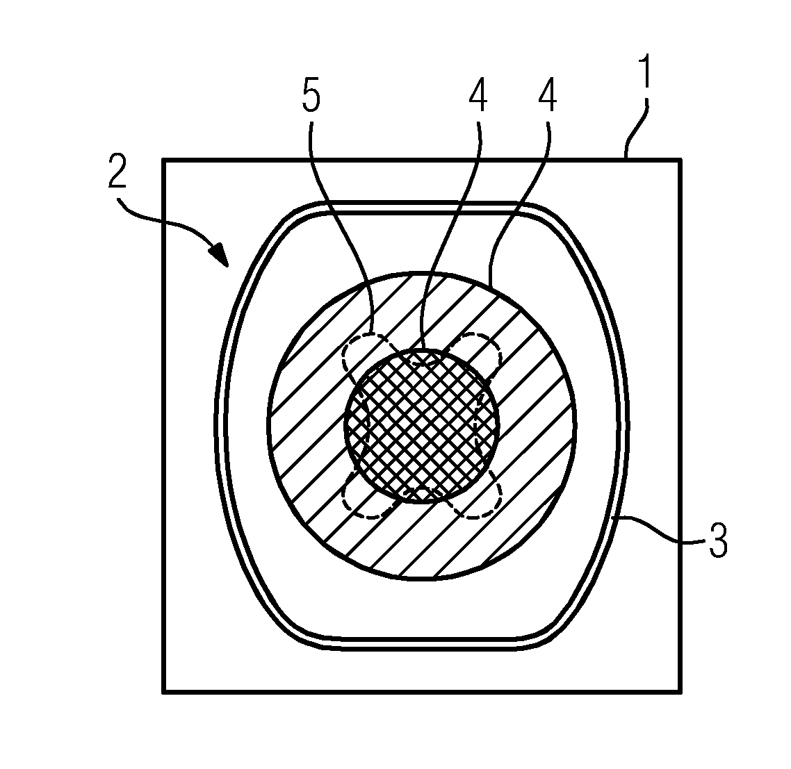
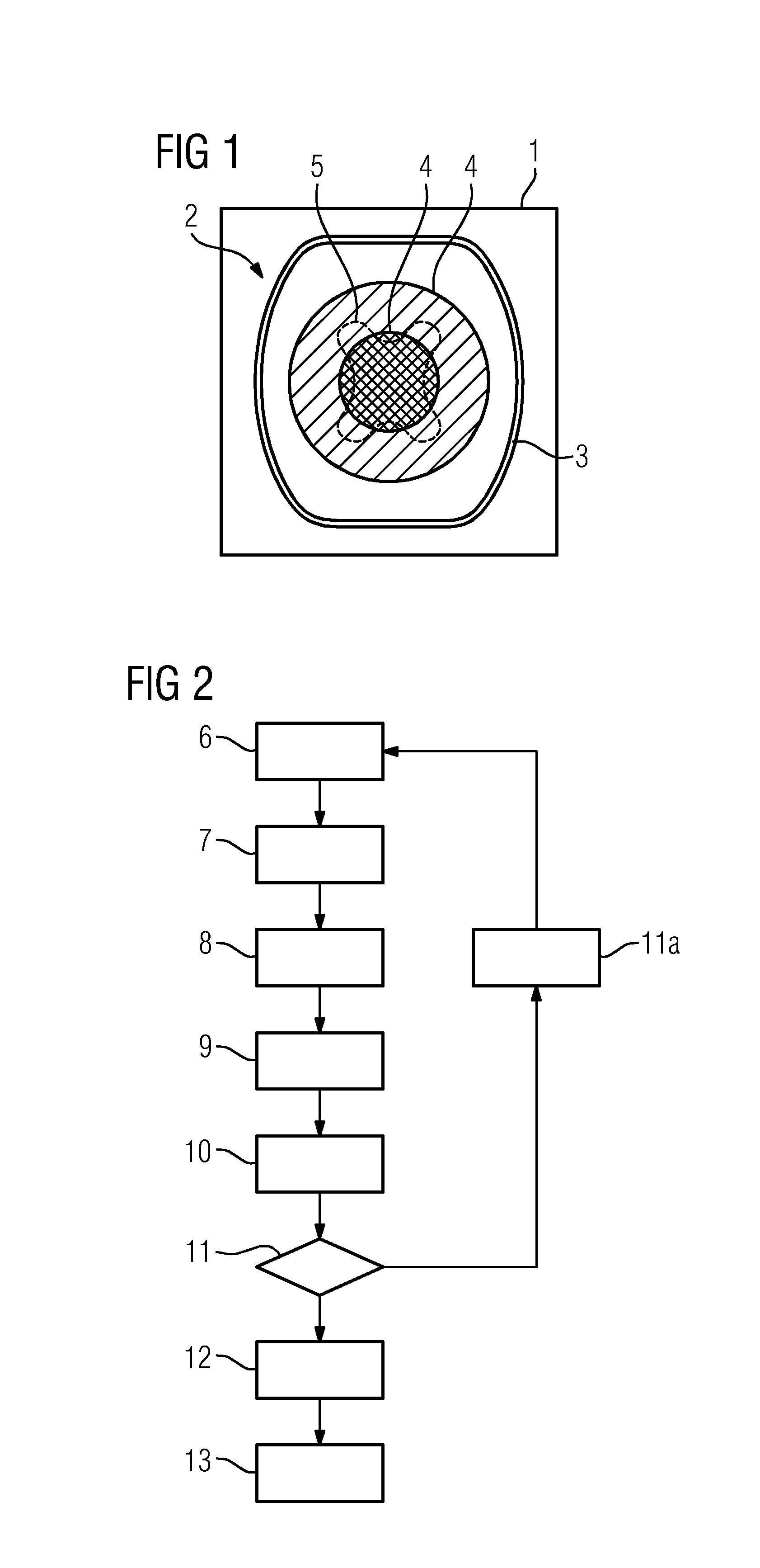
[0043]FIG. 1 illustrates the problem underlying the invention by means of an image slice 1, to be shown, of a three-dimensional image data set, which was reconstructed from two-dimensional projection images having a C-arm x-ray device. The image slice can, for example, be formed as part of a multiplanar reconstruction (multi planar reformation), wherein the dividing direction of the three-dimensional image data set is the axial direction. The three-dimensional image data set shows the head 2 of a patient, in the case of which the soft tissue which lies inside the head is outwardly adjacent to the cranium 3 (and a layer of skin, which is not shown in more detail here). As is indicated by the regions 4, which in reality are, of course, not closely adjacent, the soft tissue is represented ever more clearly towards surface, wherein it is normally a fluid method. This means that the attenuation value for one and the same material class, here also soft tissue, appears to increase ever mor...
PUM
 Abstract
Description
Claims
Application Information