
Disposable eyepiece system for an ultrasonic eye scanning

Active Publication Date: 2015-08-27
ARCSCAN INC
View PDF8 Cites 10 Cited by
  • Summary
  • Abstract
  • Description
  • Claims
  • Application Information

AI Technical Summary

Benefits of technology

[0044]The ciliary body is the circumferential tissue inside the eye composed of the ciliary muscle and ciliary processes. There are three sets of ciliary muscles in the eye, the longitudinal, radial, and circular muscles. They are near the front of the eye, above and below the lens. They are attached to the lens by c

Problems solved by technology

Except for on-axis measurements, dimensions and locations of eye components behind the iris cannot be fully determined by optical means.
Ultrasonic imaging can be used to provide the required accurate images of the natural lens especially where the zonules attach the natural lens to the ciliary body which is well off-axis and behind the iris and therefore not readily accessible to optical imaging.
U.

Method used


Image

  • Disposable eyepiece system for an ultrasonic eye scanning
  • Disposable eyepiece system for an ultrasonic eye scanning
  • Disposable eyepiece system for an ultrasonic eye scanning

Examples


Example

DETAILED DESCRIPTION OF THE DRAWINGS

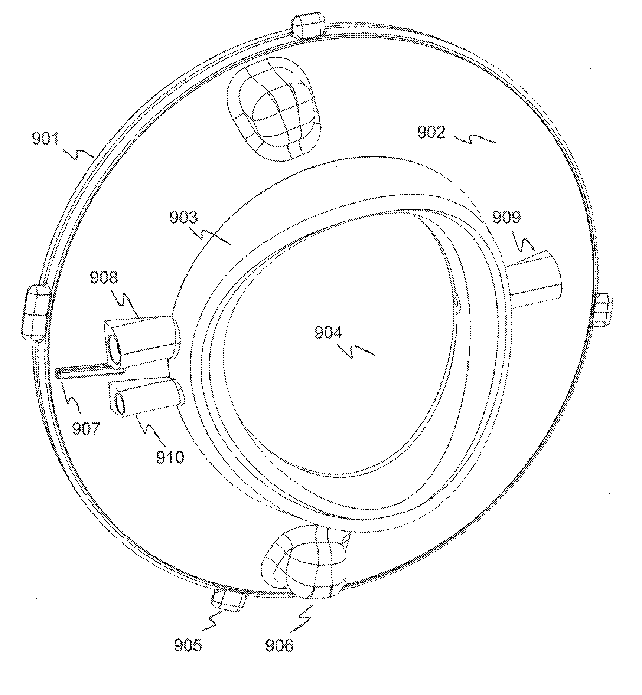
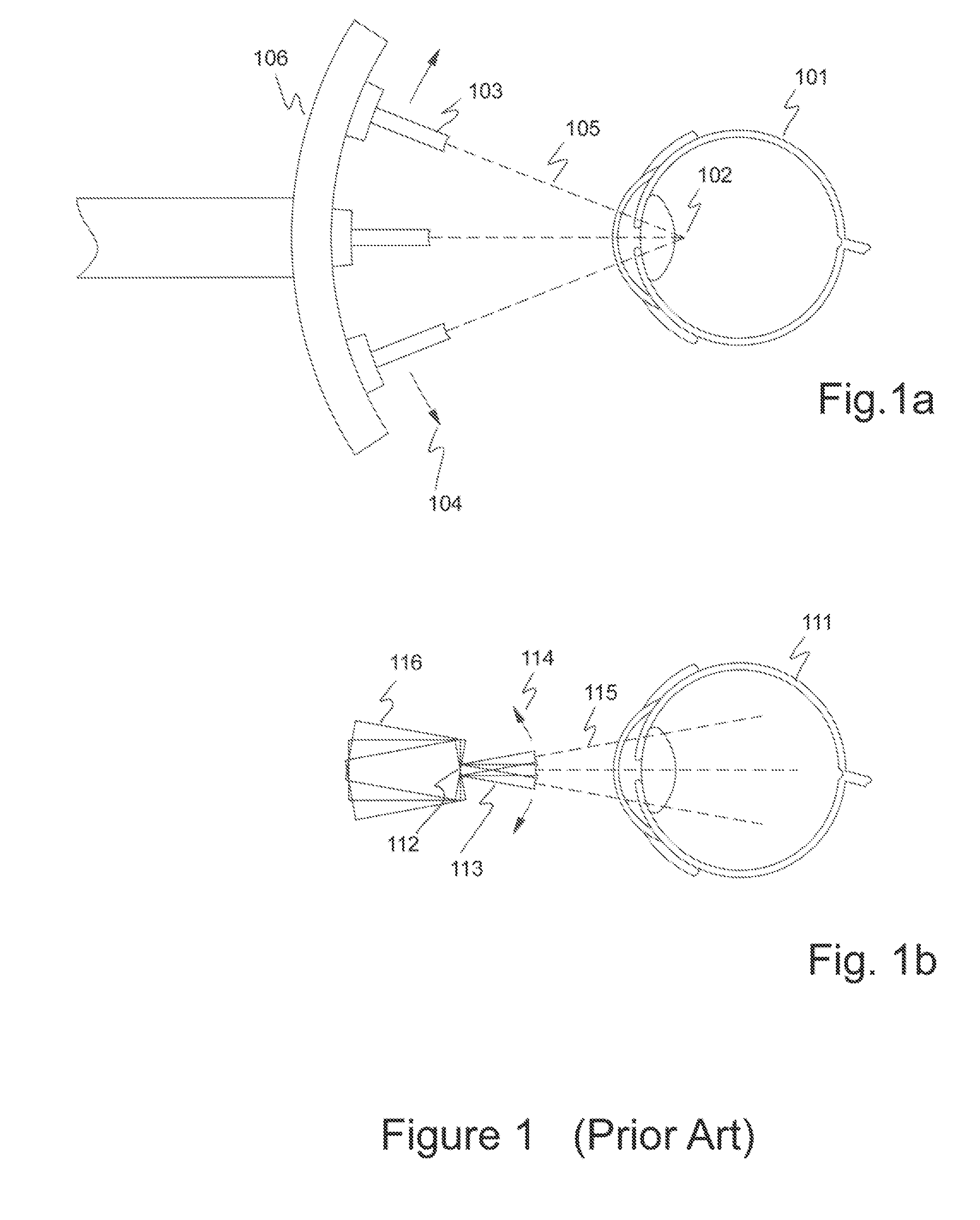
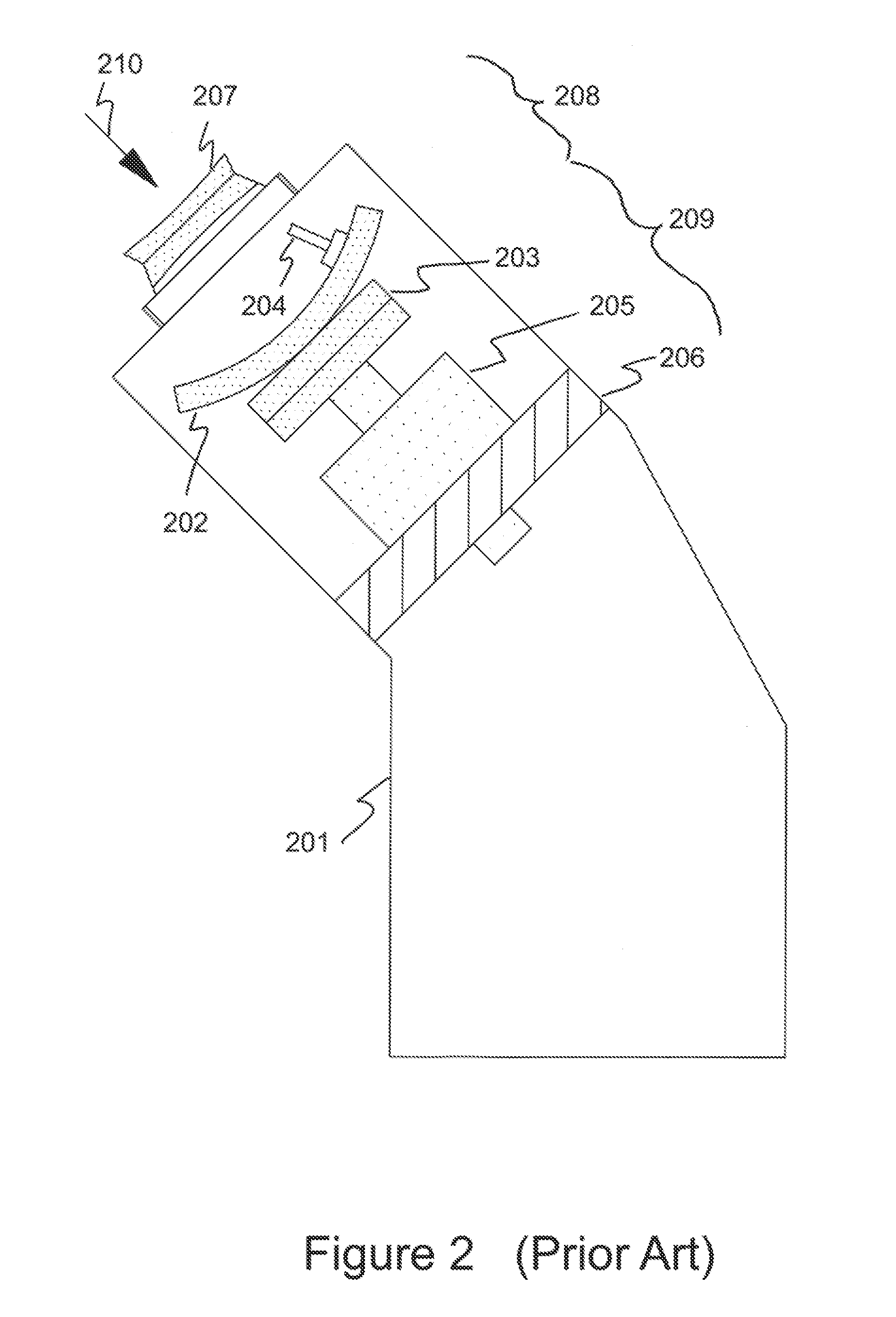
[0106]The main elements of a human eye are shown, for example, in “Optics of the Human Eye”, D. A. Atchison, G. Smith, Robert Stevenson House, Edinburgh, ISBN 0 7506 3775 7, first printed in 2000. The cornea, which is optically transparent, is located at the front of the eye and is located in the anterior chamber. The anterior and posterior surfaces of a normal cornea and the internal layers, such as Bowman's layer, within a normal cornea are specular surfaces. The iris separates the anterior chamber from the posterior chamber. The back of the lens forms the rear of the posterior chamber. The natural lens sits directly behind the iris. Only the central part of the lens, which is behind the pupil, can be seen optically. The anterior and posterior surfaces of a normal lens are specular surfaces. The cornea, iris and lens comprise the main optical refractive components of the eye. The anterior and posterior chambers comprise the anterior segment of t...


PUM


Abstract

A disposable eye piece system, including the eye piece and other disposable components, for a precision ultrasonic scanning apparatus is disclosed. The eye piece includes a fill port, a vent port and a drain port molded into the base of the eye piece. An RF Identification chip may be molded into the plastic base of the eye piece or, alternately into the conformable face seal of the eye piece. The components of a disposable eye piece system form an integrated disposable package that can be assembled, packaged, transported and used while maintaining the eye piece and saline solution in a sterile condition. The protective shipping container 1) protects the eye piece from damage, human contact and exposure to ambient air during shipping and storage and 2) allows the scanning physician or technician to attach the eye piece to the scanning machine without compromising the sterile condition of the eye piece.

Description

CROSS-REFERENCE TO RELATED APPLICATIONS[0001]This application claims the benefits, under 35 U.S.C.§119(e), of U.S. Provisional Application Ser. No. 61 / 943,667 entitled “Disposable Eyepiece System for an Ultrasonic Eye Scanning Apparatus” filed Feb. 24, 2014 which is incorporated herein by reference.FIELD OF INVENTION[0002]The present invention relates to ultrasonic imaging of biological materials such as the cornea and natural lens of the eye and in particular relates to a disposable eye piece system.BACKGROUND OF THE INVENTION[0003]Ultrasonic imaging has found use in accurate measurement of structures of the eye such as, for example, the cornea and lens capsule. Such measurements provide an ophthalmic surgeon valuable information that can be used to guide various surgical procedures for correcting refractive errors such as LASIK and lens replacement. They also provide diagnostic information after surgery has been performed to assess the geometrical location of corneal features such...

Claims


Application Information

Patent Timeline
IPC IPC(8): A61B8/10
CPCA61B8/10A61B8/4209A61B8/4422A61B8/14
Inventor HEATH, GARY B.LEVIEN, ANDREW K.WATSON, JOHN D.
Owner ARCSCAN INC
Learn More