Pdgfrbeta-fc fusion proteins and uses thereof

a technology of fusion proteins and pdgfrbeta, which is applied in the field of platelet-derived growth factors, can solve the problems of plasma proteins leakage into the vitreous of patients, inability to limit and inability to achieve the goal of reducing the formation of new vessels in the retina in patients

Inactive Publication Date: 2016-01-14
BAYER PHARMA AG
View PDF0 Cites 2 Cited by
  • Summary
  • Abstract
  • Description
  • Claims
  • Application Information

AI Technical Summary

Benefits of technology

[0035]In some embodiments, the relative increase of in vitro potency measured as IC50 for phosphorylation of Protein Kinase B (AKT) in a PDGF-BB-mediated phosphorylation assay for any of the PDGFRbeta-Fc fusion proteins having a linker described herein as compared to a linker-less PDGFRbeta-Fc fusion protein is at least 2, 3, 4, 5, 6, 7, 8, 9, 10, 12, 14, 16, 18 or 20 times greater than the increase in binding affinity of the PDGFRbeta-Fc fusion protein for the PDGF ligand -BB compared to the linker-less PDGFRbeta-Fc fusion protein.

Problems solved by technology

As a consequence, fluid and soluble plasma components drain into surrounding tissue, leaving more distant areas in shortage of oxygen and nutrients.
Neovascular disease of the retina causes loss of vision and blindness via the formation of new vessels in the retina and increased vascular permeability resulting in leakage of plasma proteins into the vitreous of patients.
Therapeutic approaches to limit the formation of new vessels in the retina are so far not successful in all patients.
In fact, in more than 50% of the reported cases visual acuity does not improve or declines again after initial improvement.
A downside of targeting PDGF signaling in angiogenesis has been that several studies in preclinical models demonstrated that reducing the pericyte coverage of blood vessels increases vascular leakage, i.e. the leakage of serum protein into the surrounding tissues.
Full IgG antibodies have an exceptionally long half-life, while small antibody derivatives or other biologics formats often suffer from rapid elimination from circulation.
However, little is known about the half-life extension of proteins in the retina.

Method used


Image

  • Pdgfrbeta-fc fusion proteins and uses thereof
  • Pdgfrbeta-fc fusion proteins and uses thereof
  • Pdgfrbeta-fc fusion proteins and uses thereof

Examples


example 1

Generation of Different Recombinant PDGFRbeta Fc Constructs

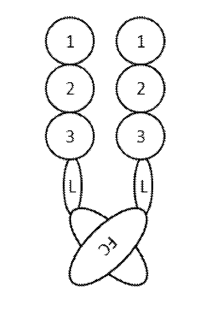
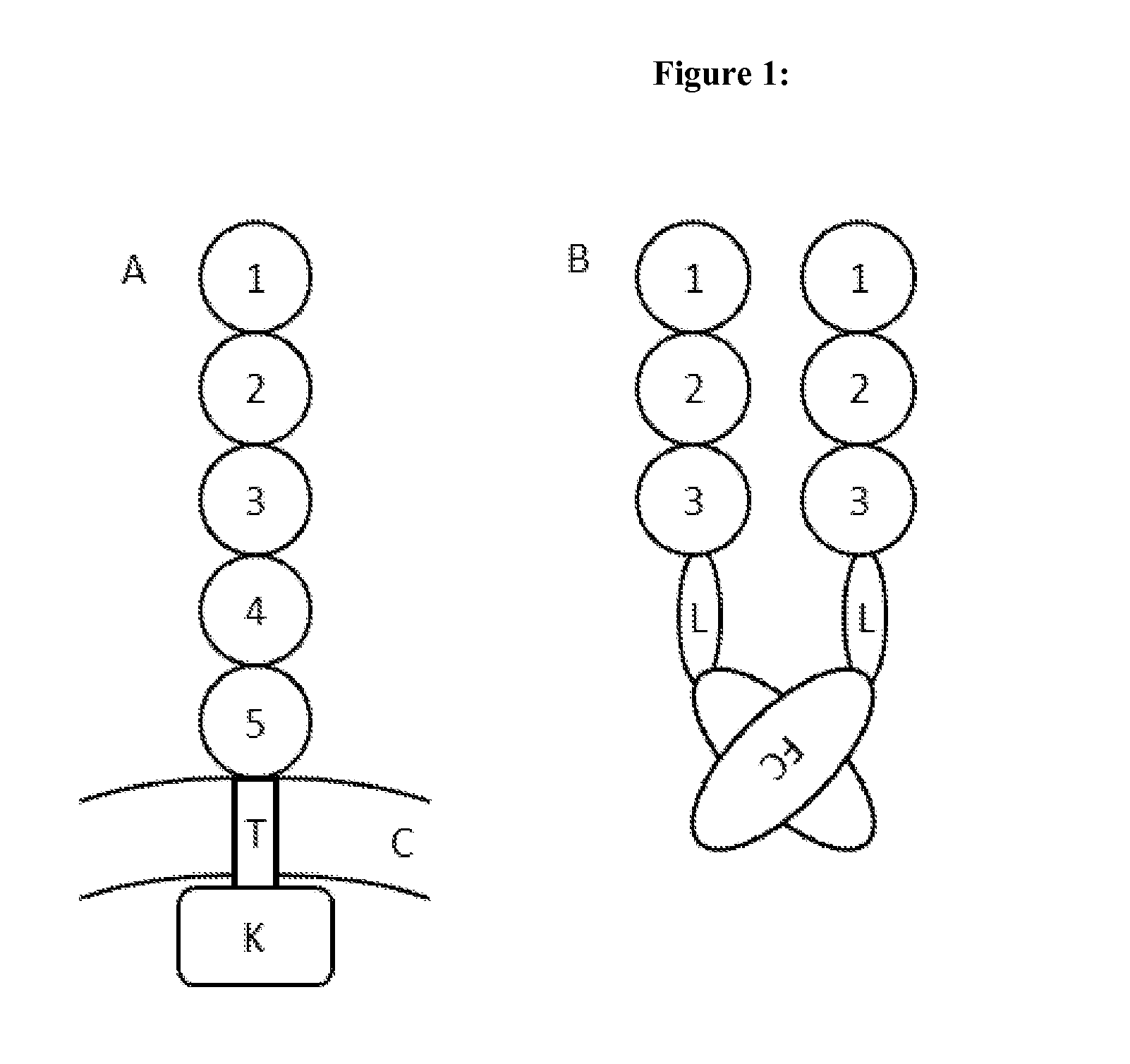
[0155]To generate different Fc fusion proteins (SEQ ID NO: 15-25) the extracellular domains 1-3 of PDGFRbeta, derived from UNIPROT ID P09619 (SEQ ID NO: 2 and FIG. 1A) were C-terminally fused to a human IgG1 Fc-part (SEQ ID NO: 14). This was done either directly (SEQ ID NO: 15) or via different amino acid linkers (SEQ ID NO: 16-25 see FIG. 1 B). The linker was a GGGGS linker selected from SEQ ID NO: 7-13, i.e.

SEQ ID NO 7:GGGGS (1x GGGGS),SEQ ID NO 8:GGGGSGGGGS (2x GGGGS),SEQ ID NO 9:GGGGSGGGGSGGGGS (3x GGGGS),SEQ ID NO 10: GGGGSGGGGSGGGGSGGGGS (4x GGGGS),SEQ ID NO 11:GGGGSGGGGSGGGGSGGGGSGGGGS (5x GGGGS),SEQ ID NO 12:GGGGSGGGGSGGGGSGGGGSGGGGSGGGGS (6x GGGGS),SEQ ID NO 13:GGGGSGGGGSGGGGSGGGGSGGGGSGGGGSGGGGS (7x GGGGS),

or a linker selected from SEQ ID NOs: 4-6, i.e SEQ ID NO 4: EPKSC, SEQ ID NO 5: EPKSS or SEQ ID NO 6: GGGGG. The gene of the respective Fc fusion protein was cloned into a suitable expression vector based on a CM...

example 2

BiaCore

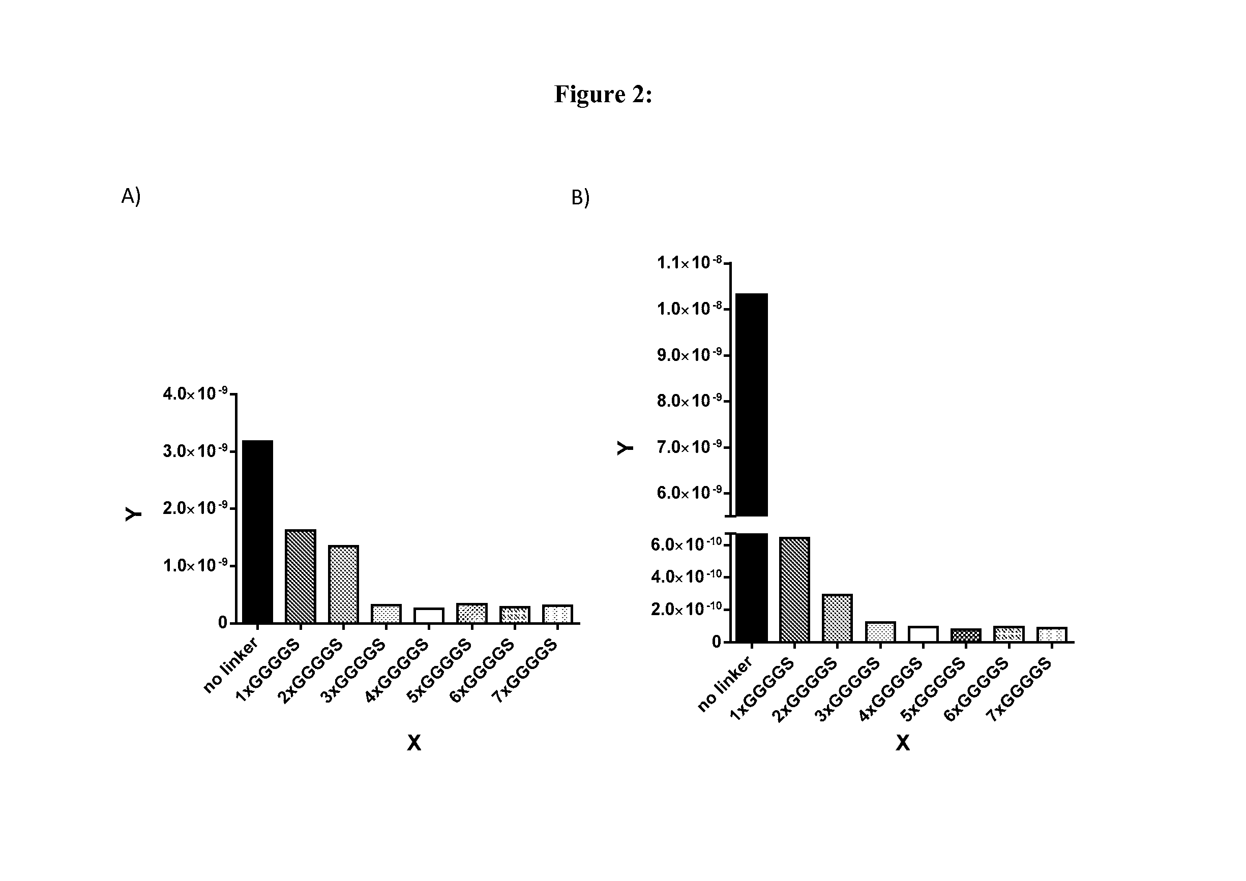
[0157]Binding affinities (KD values) of PDGFRbeta-Fc fusion proteins were measured by using surface plasmon resonance assays. Experiments were performed using a Biacore T200 instrument (GE Healthcare Biacore, Inc.) with Series S Sensor Chips CM5 (GE Healthcare Biacore, Inc.). Binding assays were carried out at 25° C. with assay buffer HBS-EP+ supplemented with BSA (Sigma) and NaN3 (10 mM HEPES pH 7.4, 500 nM NaCl, 1 mg / ml BSA, 0.05% SP20, 0.05% NaN3). Fc fusions were captured with an anti-hIgG capture antibody covalently immobilized to the chip surface via amine coupling chemistry. Reagents for amine coupling (1-ethyl-3-(3-dimethylaminopropyl)carbodiimide hydrochloride (EDC), N-hydroxysuccinimide (NHS), ethanolamine-HCl pH 8.5) were used from the Amine Coupling Kit (GE Healthcare, product code BR-1000-50). anti-hIgG capture antibody and immobilization buffer (10 mM sodium acetate pH 5.0) was used from the Human Antibody Capture Kit (GE Healthcare, BR-1008-39). The sensor chip...

example 3

Cell-Based Assays to Determine In Vitro Potency of PDGFRbeta-Fc Fusion Proteins

[0161]PDGFRbeta-Fc fusion proteins have been characterized in vitro using Normal Human Dermal Fibroblasts (NHDF) (NHDF juvenile foreskin, Promocell, Catalog Number: C-12300, cultivated according to provider's manual, (http: / / www.promocell.com / fileadmin / promocell / PDF / C-12300.pdf) cell-based assays in which two downstream signaling events after stimulation with human PDGF-BB (Recombinant Human PDGF-BB, CF, R&D Systems, Cat. No. 220-BB-010) were assessed:[0162]pPDGFRbeta Tyr751 phosphorylation (Meso Scale Phospho PDGFR-beta (Tyr751) Assay Whole Cell Lysate Kit K150DVD-1)[0163]pAKT Ser473 phosphorylation (Meso Scale Phospho (Ser473) Total AKT Assay Whole Cell Lysate Kit K15100D-1)

[0164]NHDFs were starved for 3 h with serum-free culture medium, stimulated for 30 min with 6.3 ng / ml (for pAKT assays) and 50 ng / ml (for pPDGFRbeta assays) PDGF-BB (R&D Systems, Recombinant Human PDGF-BB, CF; 220-BB-010), which was ...


PUM

PropertyMeasurementUnit
pHaaaaaaaaaa
Tmaaaaaaaaaa
concentrationaaaaaaaaaa

Abstract

The present disclosure provides PDGFRbeta-Fc fusion proteins, or biologically active fragments thereof, comprising an extracellular domain of PDGFRbeta, or a biologically active fragment thereof, a linker and a Fc domain, wherein said fusion protein binds one or more of platelet-derived growth factor ligands with high affinity. The PDGFRbeta-Fc fusion proteins, or biologically active fragments thereof, accordingly, can be used to treat pathological neovascularization and fibrosis, e.g. cancer, ocular neovascular disorders or nephropathies. The disclosure also provides methods for treating ocular neovascular disorders with these fusion proteins without increasing vascular leakage. Such PDGFRbeta-Fc fusion proteins, or biologically active fragments thereof, exhibit increased terminal half time in the eye. Also provided are nucleic acid sequences encoding the foregoing PDGFRbeta-Fc fusion proteins, or biologically active fragments thereof, vectors containing the same, pharmaceutical compositions and kits with instructions for use.

Description

SEQUENCE LISTING[0001]The instant application contains a Sequence Listing which has been submitted via EFS-Web and is hereby incorporated by reference in its entirety. Said ASCII copy, created on Jul. 10, 2014, is named BCPP—236—101_Sequence_Listing_PTO.txt, and is 95,346 bytes in size.BACKGROUND OF THE DISCLOSURE[0002]The Platelet-Derived Growth Factor (PDGF) family consists of four members (A-D) that share a cluster of 8 cysteine residues. PDGFs form biologically active homodimers and one heterodimer (PDGF-AB) via disulfide bonds. They bind to two closely related receptor tyrosine kinases, PDGFRalpha and -beta, which homo- or heterodimerize upon ligand binding. The specificity of the different PDGF ligands for both receptors varies, with PDGF-BB and -DD binding PDGFRbeta, and PDGF-BB, PDGF-CC and -AA binding PDGFRalpha. PDGF-BB, PDGF-AB and to a weaker extent PDGF-CC and -DD were also reported to bind and activate the heterodimeric PDGFRalpha / beta (Heldin and Westermark, Physiol R...

Claims


Application Information

Patent Timeline
Patent Type & Authority Applications(United States)
IPC IPC(8): C07K14/49
CPCC07K2319/30C07K14/49
Inventor LINDEN, LARSSCHLANGE, THOMASWILMEN, ANDREASTRAUTWEIN, MARKSCHOMBER, TIBORBOTTGER, MICHAELKLAR, JURGENGREVEN, SIMONE
Owner BAYER PHARMA AG
Why Eureka
Try Eureka