Diagnostic apparatus for lesion, image processing method in the same apparatus, and medium storing program associated with the same method

a technology of diagnostic apparatus and image processing method, which is applied in the direction of image analysis, image enhancement, instruments, etc., can solve the problems of difficult to obtain satisfactory diagnostic accuracy, difficult to distinguish between benign tumors and malignant tumors, and difficult to distinguish between moles and spots

Active Publication Date: 2016-06-30
CASIO COMPUTER CO LTD
View PDF0 Cites 3 Cited by
  • Summary
  • Abstract
  • Description
  • Claims
  • Application Information

AI Technical Summary

Problems solved by technology

However, not only discrimination between a mole and a spot but also discrimination between a benign tumor and a malignant tumor are substantially difficult with a naked eye inspection and even a magnifying glass inspection.
In this case, as an image conversion is uniformly performed regardless of the type of lesion or the stage or degree of progression, satisfactory diagnostic accuracy is hard to obtain, and the diagnosis actually depends on a skill of a physician or clinician.

Method used


Image

  • Diagnostic apparatus for lesion, image processing method in the same apparatus, and medium storing program associated with the same method
  • Diagnostic apparatus for lesion, image processing method in the same apparatus, and medium storing program associated with the same method
  • Diagnostic apparatus for lesion, image processing method in the same apparatus, and medium storing program associated with the same method

Examples


embodiment

EFFECT OF EMBODIMENT

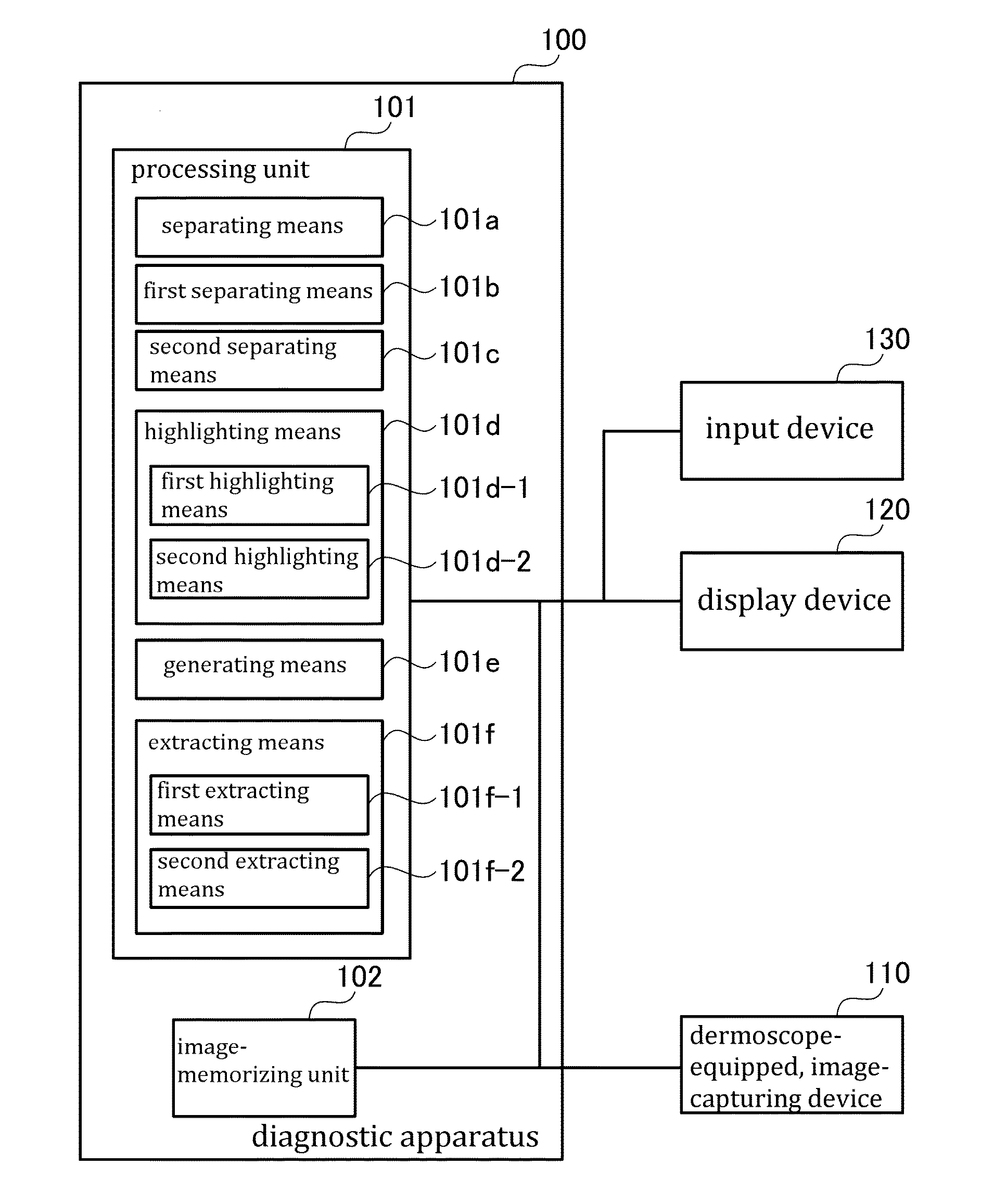
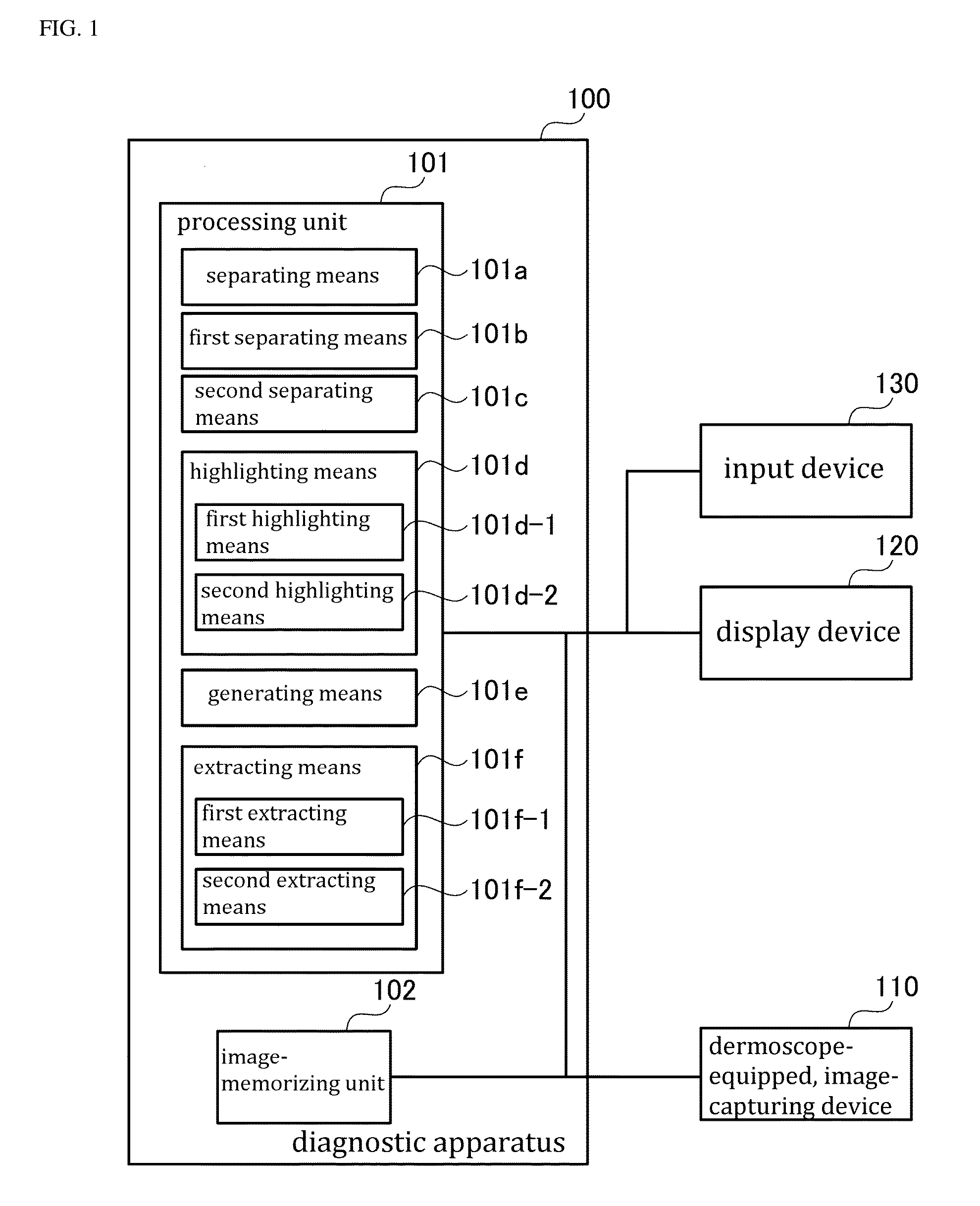
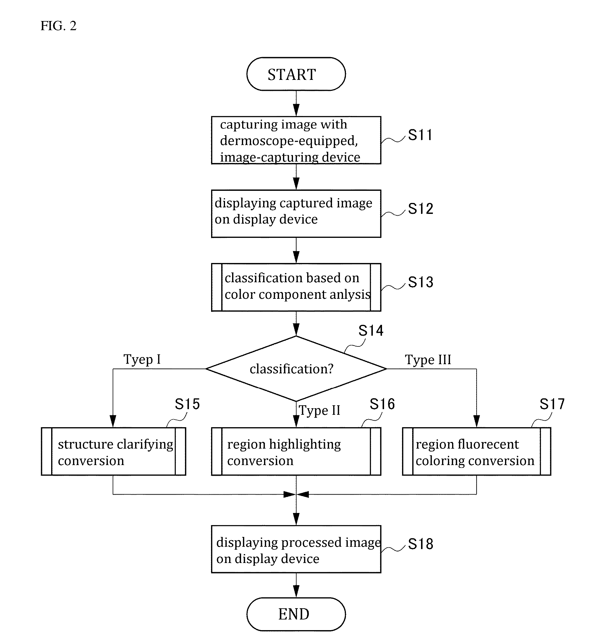
[0108]As described previously, in accordance with the diagnostic apparatus 100 of the embodiment of the invention, the classifying means 101a of the processing unit 101 classify the captured image of the affected area to be diagnosed based on the stage of the progression or pattern of the lesion, and the image conversion processing corresponding to the classification as thus obtained is performed. For example, if the stage of the progression is classified as the low stage, the structure clarifying conversion processing is performed; if the stage of the progression is classified as the middle stage, the region highlighting conversion processing is performed; and if the stage of the progression is classified as the high stage, the region fluorescent coloring conversion processing is performed. As such, an appropriate or optimal processed image can be obtained depending on the stage of the progression or type of the lesion, thereby the physician to easily make a dia...


PUM


Abstract

The invention provides a method of processing an image in a diagnostic apparatus 100 of diagnosing a lesion using a captured image of an affected area to be diagnosed, comprising the steps of: (i) classifying the captured image based on a stage of a progression of the lesion (Step S13 and S14); and (ii) performing an image conversion processing, which corresponds to a classification obtained as a result of step (i), on the captured image to generate a converted image (Step S15-17).

Description

CROSS-REFERENCE TO RELATED APPLICATIONS[0001]This application claims priority from Japanese Patent Application No. 2014-263717 filed on Dec. 26, 2014, which is incorporated herein by reference.TECHNICAL FIELD[0002]The present invention relates to a diagnostic apparatus for a lesion, and an image processing method in the same apparatus, and a medium storing program associated with the same method.BACKGROUND ART[0003]Generally, visual inspection is necessarily performed to diagnose a cutaneous legion, thereby obtaining an amount of information. However, not only discrimination between a mole and a spot but also discrimination between a benign tumor and a malignant tumor are substantially difficult with a naked eye inspection and even a magnifying glass inspection. For the reasons, dermoscopic inspection in which a dermoscope-equipped camera is used to capture an image of a disease has been conventionally performed.[0004]The dermascope is a noninvasive diagnostic device in which a dise...

Claims


Application Information

Patent Timeline
Patent Type & Authority Applications(United States)
IPC IPC(8): G06T7/00G06T7/40G06K9/00G06T5/00
CPCG06T7/0012G06T5/003G06T5/007G06T7/408G06T2207/30096G06K9/00536G06T2207/10024G06T2207/10056G06T7/0081G06T2207/20028G06T2207/30088G06T2207/30101G06T7/11G06T7/90G06F2218/12
Inventor HOUJOU, YOSHIHARUAOKI, NOBUHIROMINEO, SHIGEKI
Owner CASIO COMPUTER CO LTD
Why Eureka
Try Eureka