Apparatus, method, and non-transitory computer-readable medium for supporting viewing examination images

a computer-readable medium and examination image technology, applied in the field of apparatus, a method, and a non-transitory computer-readable medium for supporting examination images, can solve the problems of requiring extreme time and effort, and the keyword setting of each examination image requires much time and effort, so as to achieve the effect of being viewed easily and quickly

Active Publication Date: 2016-10-06
FUJIFILM CORP
View PDF5 Cites 27 Cited by
  • Summary
  • Abstract
  • Description
  • Claims
  • Application Information

AI Technical Summary

Benefits of technology

[0025]According to the aspects of the present invention, the at least one attached image, which is attached to the medical report, is extrac...

Problems solved by technology

However, setting the keyword for each examination image needs much time and effort.
In particular, for example, in the...

Method used


Image

  • Apparatus, method, and non-transitory computer-readable medium for supporting viewing examination images
  • Apparatus, method, and non-transitory computer-readable medium for supporting viewing examination images
  • Apparatus, method, and non-transitory computer-readable medium for supporting viewing examination images

Examples


Embodiment Construction

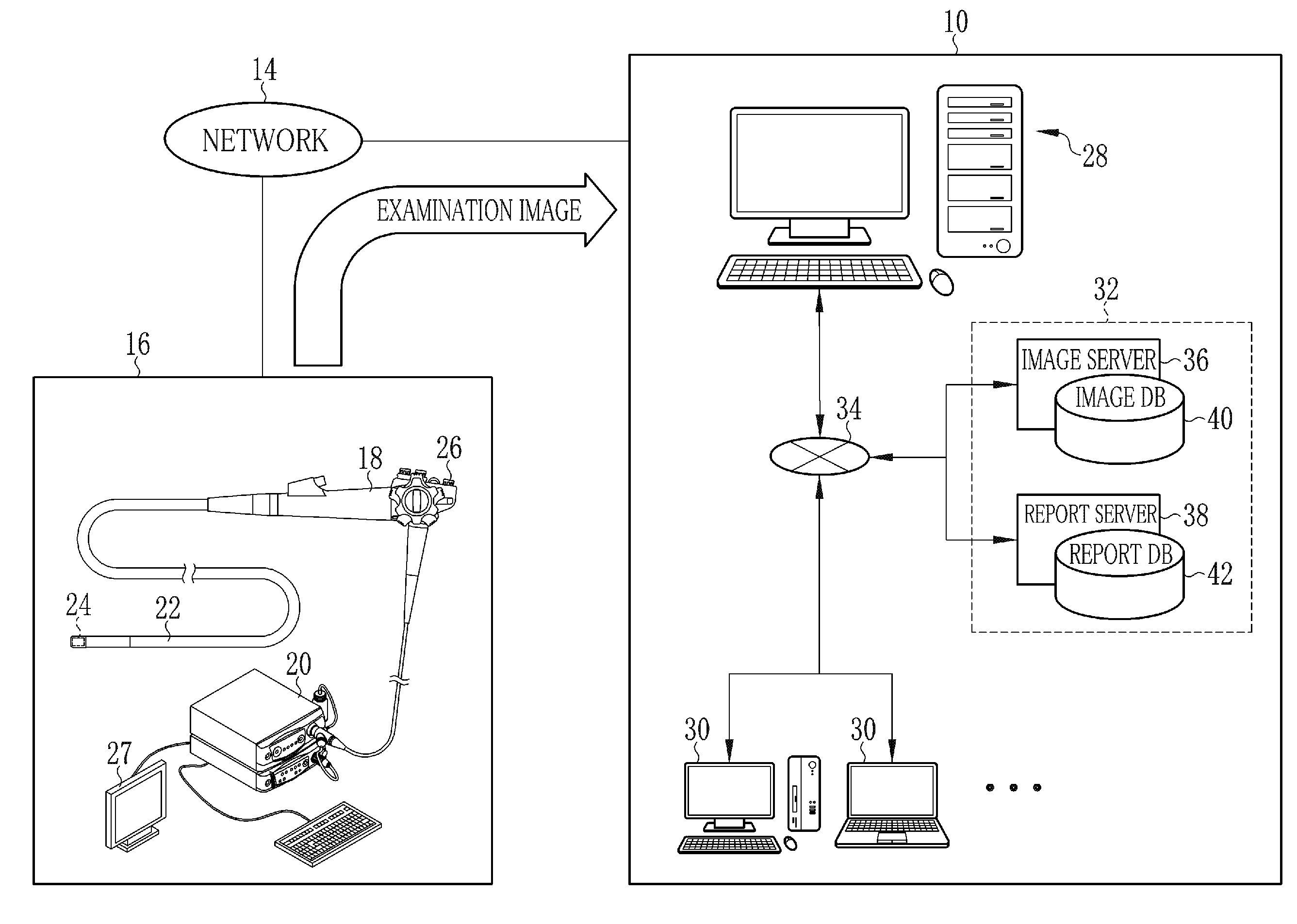
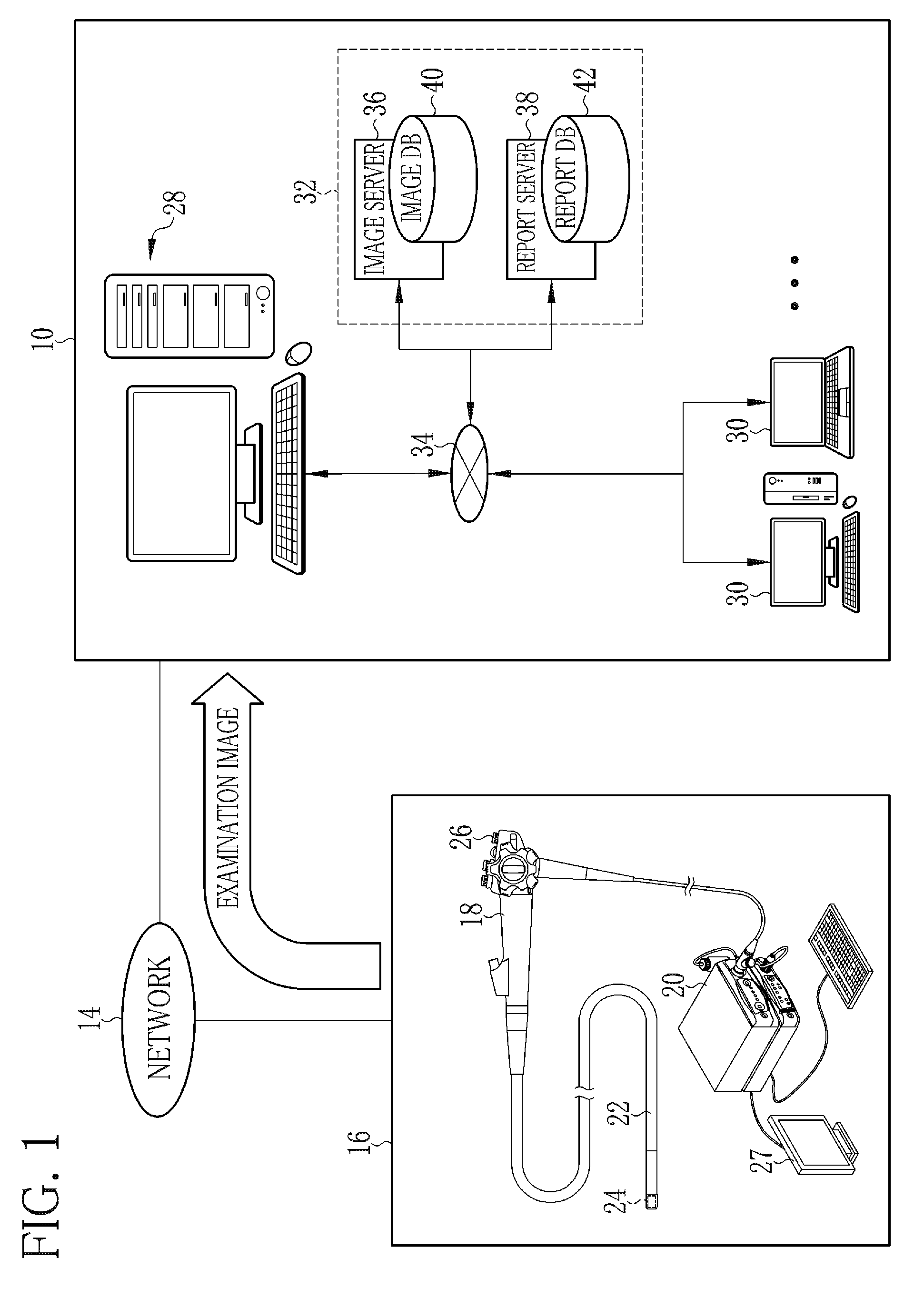
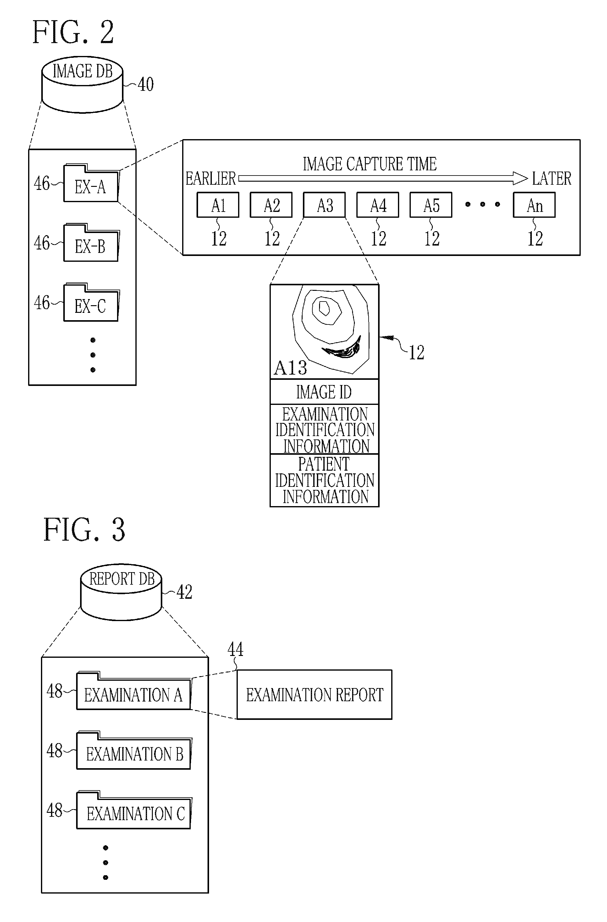
[0039]An image view support system 10 (see FIG. 1) is a computer system that supports viewing examination images 12 (see FIG. 2) obtained in at least one endoscopic examination (hereinafter simply referred to as the examination). The image view support system 10 is connected to an endoscope system 16 through a network 14. The network 14 is, for example, a LAN (local area network) in a hospital.

[0040]The endoscope system 16 is used for performing the examination. The examination is performed by, for example, an endoscopist upon a request of a patient's doctor (clinician). The two or more examination images 12 are obtained or captured in the examination, which will be described below. The endoscopist selects at least one of the examination images 12 and prepares an examination report 44 (see FIG. 4), to which the at least one selected examination image 12 is attached. The examination report 44 and the examination images 12, which are obtained in the examination, are viewed by the clin...


PUM


Abstract

Based on an examination report of an endoscopic examination designated, an attached image extraction unit reads image identification information, which identifies an examination image attached to the examination report. Based on the image identification information, the attached image extraction unit extracts the examination image (that is, an attached image) attached to the examination report, from examination images corresponding to the designated endoscopic examination. The examination images are read from an image folder of an image database (DB). A screen display controller performs control to display the extracted attached image on a display screen.

Description

CROSS-REFERENCE TO RELATED APPLICATIONS[0001]The present application claims priority under 35 U.S.C. §119 to Japanese Patent Application No. 2015-070157, filed Mar. 30, 2015. Each of the above applications is hereby expressly incorporated by reference, in its entirety, into the present application.BACKGROUND OF THE INVENTION[0002]1. Field of the invention[0003]The present invention relates to an apparatus, a method, and a non-transitory computer-readable medium for supporting viewing examination images obtained in at least one endoscopic examination.[0004]2. Description Related to the Prior Art[0005]Endoscopes for capturing images of a patient's body cavity are known. An insertion section of the endoscope is inserted into the patient's body cavity and the images are captured by an imaging unit, which is incorporated in a distal end portion of the insertion section. In an endoscopic examination using the endoscope, for example, in a screening examination of an upper gastrointestinal ...

Claims


Application Information

Patent Timeline
IPC IPC(8): A61B1/00A61B1/04G16H30/20
CPCA61B1/04A61B1/00045G16H30/20
Inventor MIURA, NOBUYUKI
Owner FUJIFILM CORP
Why Eureka
Try Eureka