Method of manufacturing scaffold for treatment of tooth extraction socket and implantation of dental implant

Active Publication Date: 2017-01-12
THE CATHOLIC UNIV OF KOREA IND ACADEMIC COOPERATION FOUND
View PDF0 Cites 2 Cited by
  • Summary
  • Abstract
  • Description
  • Claims
  • Application Information

AI Technical Summary

Benefits of technology

This patent describes a method to extract teeth without needing to directly remove them. Instead, a scaffold is created that matches the shape of the dental implant spot where the tooth was already extracted. This scaffold can then be used to easily and accurately implant a dental implant after the tooth extraction. The method allows for better outcomes and easier dental procedures.

Problems solved by technology

Although a tooth extraction socket formed after tooth extraction is spontaneously cured, the tooth extraction socket causes a loss of a height and width of alveolar bone during a curing process.
However, when there is a loss of a veneer block bone due to lesions, external injuries, etc., it is difficult to fix bone grafts and a dental implant and a treatment period is prolonged.
However, it is difficult to place the dental implant into the tooth extraction socket due to a loss of alveolar bone, etc., and it is very difficult to determine the implantation position and angle of the dental implant.
However, the conventional techniques are technically characterized in merely predicting an implantation position for an implantation operation without consideration of a tooth extraction socket, and thus, it is difficult to solve problems of an implantation operation due to the tooth extraction socket after tooth extraction.

Method used


Image

  • Method of manufacturing scaffold for treatment of tooth extraction socket and implantation of dental implant
  • Method of manufacturing scaffold for treatment of tooth extraction socket and implantation of dental implant
  • Method of manufacturing scaffold for treatment of tooth extraction socket and implantation of dental implant

Examples


first embodiment

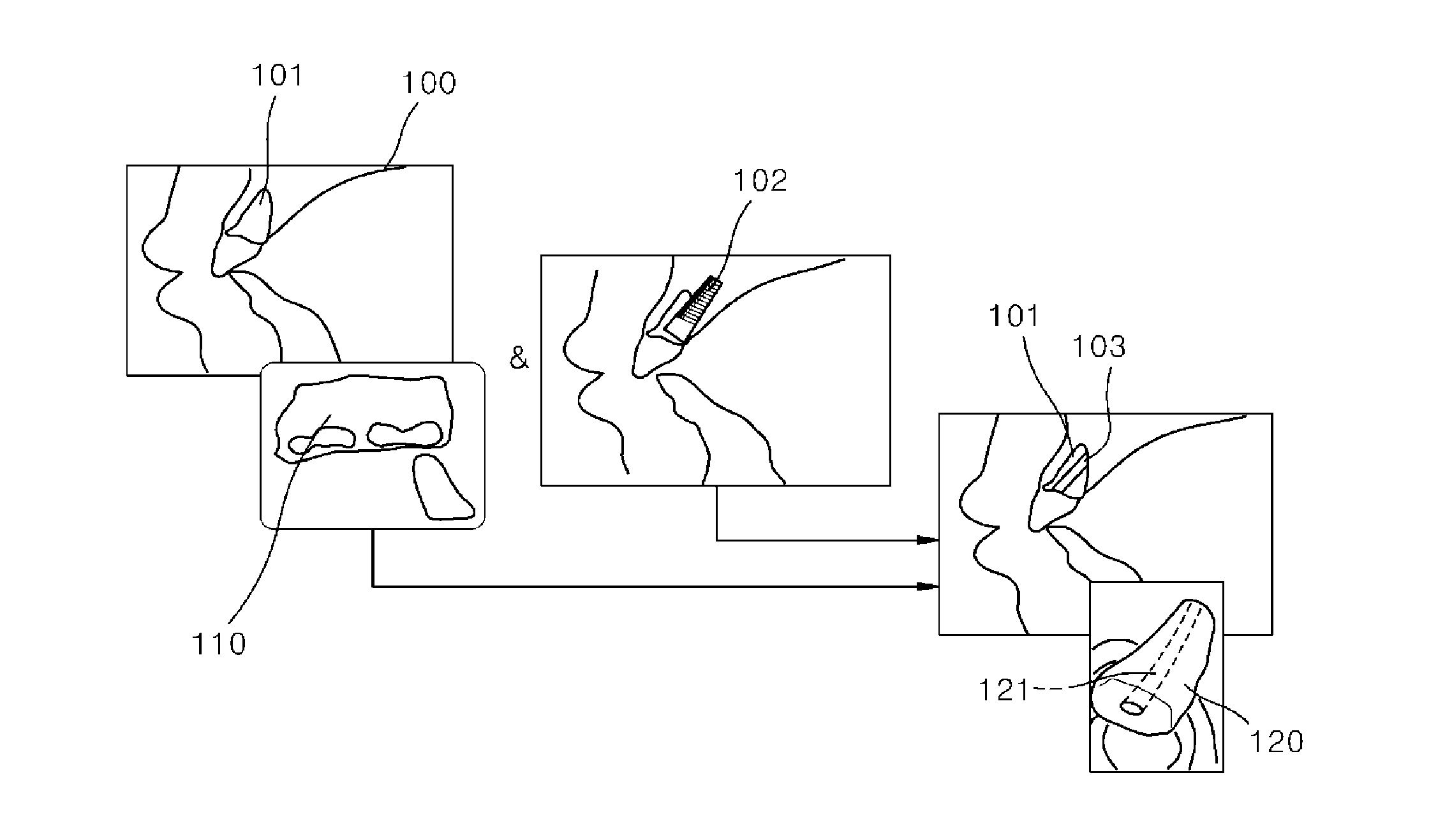
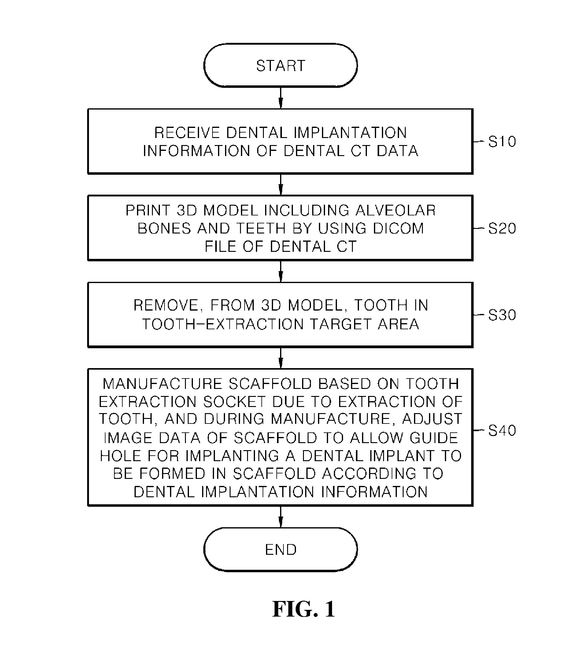
[0008]According to the present disclosure, there is provided a method of manufacturing a scaffold for treatment of a tooth extraction socket and implantation of a dental implant, the method including receiving dental implantation information of dental CT data which is previously input via a terminal of a manager; manufacturing, by using a three-dimensional (3D) printer, a 3D model including alveolar bones and teeth, which are distinguished therebetween, based on a medical image file that is a medical image file (DICOM file) of the dental CT data; performing virtual tooth-extraction by removing, from the manufactured 3D model, a region corresponding to a tooth in a tooth-extraction target area; and manufacturing, by using the 3D printer, a scaffold to be placed in an actual tooth extraction socket according to a shape of a tooth extraction socket that exists in the manufactured 3D model as a result of the virtual tooth-extraction, wherein, when the scaffold is manufactured, image dat...

second embodiment

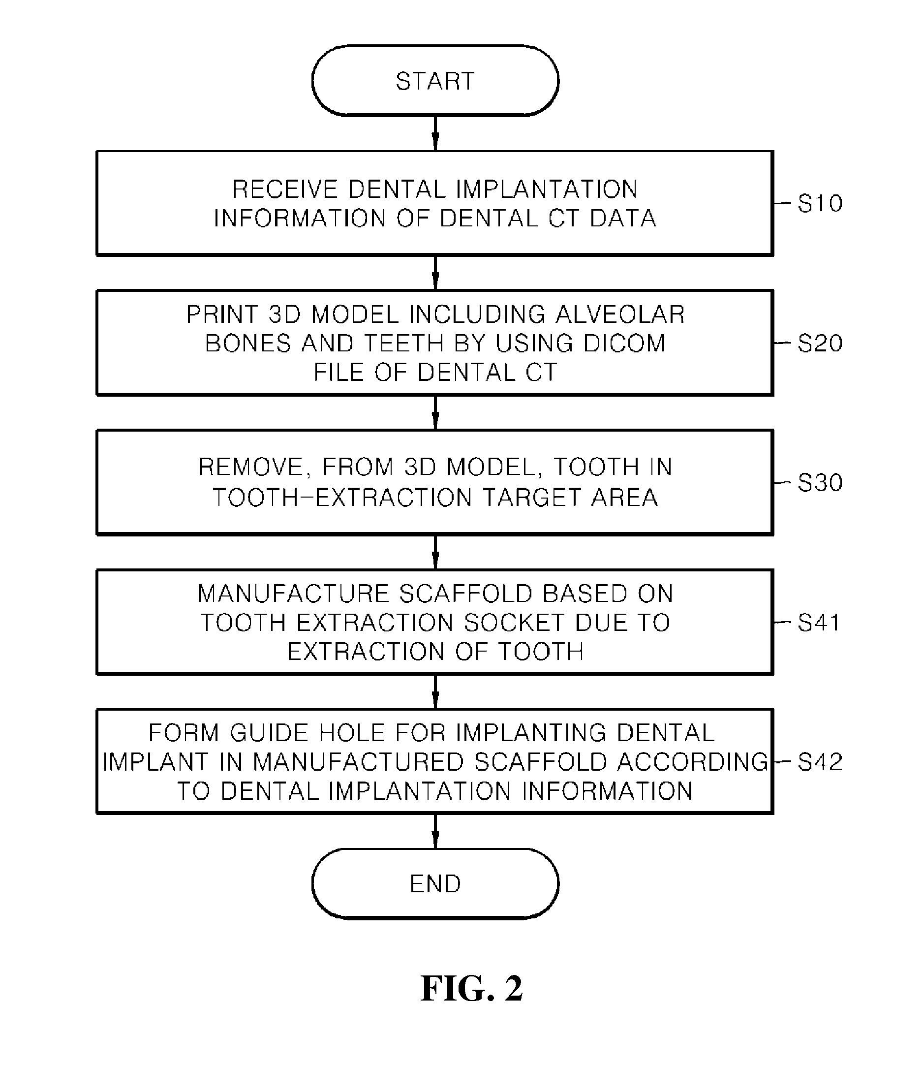
[0009]According to the present disclosure, there is provided a method of manufacturing a scaffold for treatment of a tooth extraction socket and implantation of a dental implant, the method including: receiving dental implantation information of dental CT data which is previously input via a terminal of a manager; manufacturing, by using a three-dimensional (3D) printer, a 3D model including alveolar bones and teeth, which are distinguished therebetween, based on a medical image file that is a medical image file (DICOM file) of the dental CT data; performing virtual tooth-extraction by removing, from the manufactured 3D model, a region corresponding to a tooth in a tooth-extraction target area; manufacturing, by using the 3D printer, a scaffold to be placed in an actual tooth extraction socket according to a shape of a tooth extraction socket that exists in the manufactured 3D model as a result of the virtual tooth-extraction; and forming a guide hole for implanting the dental impla...

third embodiment

[0010]According to the present disclosure, there is provided a method of manufacturing a scaffold for treatment of a tooth extraction socket and implantation of a dental implant, the method including receiving, from a computerized tomography (CT) imaging apparatus, a medical image file that is a medical image file (DICOM file) of dental CT data, and dental implantation information that is previously input via a terminal of a manager; generating image data of a scaffold to be placed in the tooth extraction socket formed when a tooth in a tooth-extraction target area is extracted, by using a region corresponding to the tooth in the tooth-extraction target area in the medical image file; amending the generated image data of the scaffold so as to allow a guide hole for implanting the dental implant to be formed in the scaffold, based on the dental implantation information;

[0011]and manufacturing a scaffold corresponding to the amended image data by using a three-dimensional (3D) printer...


PUM

PropertyMeasurementUnit
Angleaaaaaaaaaa

Abstract

Provided is a technique of allowing a dental implant to be stably placed after extraction, to be accurately placed in a tooth extraction socket, and to be stably placed in the tooth extraction socket according to implantation position and angle. A method of manufacturing a scaffold for treatment of a tooth extraction socket and implantation of a dental implant includes receiving dental implantation information of dental CT data which is previously input via a terminal of a manager; manufacturing, by using a three-dimensional (3D) printer, a 3D model comprising alveolar bones and teeth, which are distinguished therebetween, based on a medical image file that is a medical image file (DICOM file) of the dental CT data; performing virtual tooth-extraction by removing, from the manufactured 3D model, a region corresponding to a tooth in a tooth-extraction target area; and manufacturing, by using the 3D printer, a scaffold to be placed in an actual tooth extraction socket according to a shape of a tooth extraction socket that exists in the manufactured 3D model as a result of the virtual tooth-extraction, wherein, when the scaffold is manufactured, image data of the scaffold is amended to allow a guide hole for implanting the dental implant to be formed in the scaffold based on the dental implantation information.

Description

TECHNICAL FIELD[0001]The present disclosure relates to a dental implantation technique using manufacture of a scaffold to be inserted into a tooth extraction socket after tooth-extraction to help treatment, and more particularly, to a technique of fast and accurately manufacturing a scaffold to be immediately placed after tooth extraction in a tooth extraction process and simultaneously enabling dental implantation.BACKGROUND ART[0002]Although a tooth extraction socket formed after tooth extraction is spontaneously cured, the tooth extraction socket causes a loss of a height and width of alveolar bone during a curing process. The loss highly influences following implantation of a dental implant and a prognosis thereof. Accordingly, various bone grafts and barrier membranes using techniques have been used to maintain the form of alveolar bone. In particular, the techniques have been used to preserve the form of alveolar bone by immediately implanting a dental implant and grafting bon...

Claims


Application Information

Patent Timeline
IPC IPC(8): A61C8/00A61C1/08A61C13/00A61B6/03A61B6/14
CPCA61C8/008A61B6/032A61C1/084A61C13/0004A61C13/0019A61B6/14A61C8/0036A61B6/51B29C64/386B29C64/112B29C64/106
Inventor LEE, SANG HWA
Owner THE CATHOLIC UNIV OF KOREA IND ACADEMIC COOPERATION FOUND
Why Eureka
Try Eureka