Deep Image-to-Image Network Learning for Medical Image Analysis

a network learning and image technology, applied in image analysis, image enhancement, instruments, etc., can solve the problems of task-dependent approaches to various no systematic, universal approach to address all of these medical image analysis tasks,

Active Publication Date: 2017-07-13
SIEMENS CORP +1
View PDF0 Cites 81 Cited by
  • Summary
  • Abstract
  • Description
  • Claims
  • Application Information

AI Technical Summary

Problems solved by technology

Currently the technical approaches for various medical image analysis tasks are task-dependent.
There is no systematic, universal approach to address all of these medical image analysis tasks.

Method used


Image

  • Deep Image-to-Image Network Learning for Medical Image Analysis
  • Deep Image-to-Image Network Learning for Medical Image Analysis
  • Deep Image-to-Image Network Learning for Medical Image Analysis

Examples


Embodiment Construction

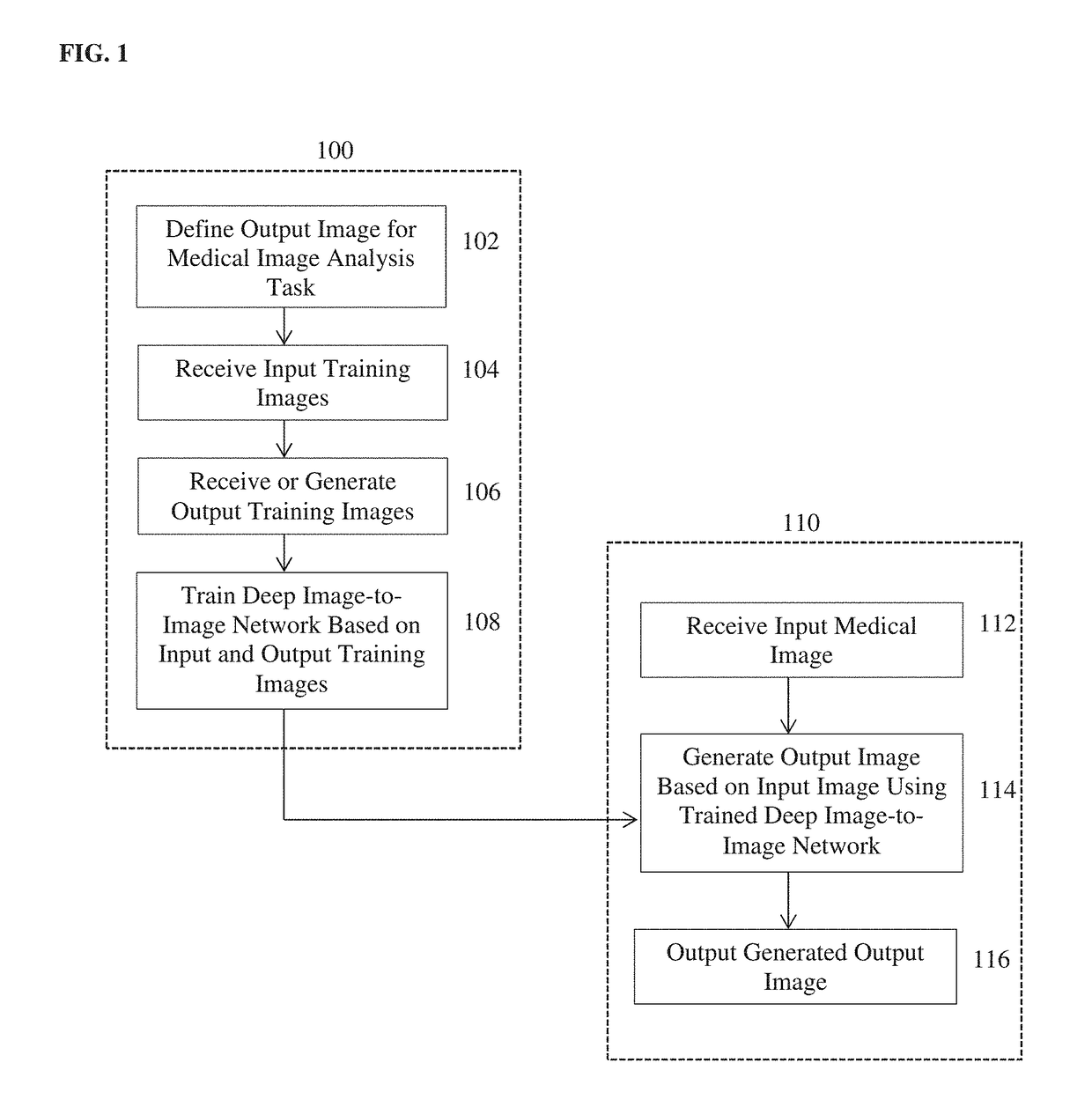
[0014]The present invention relates to a method and system for automatically performing medical image analysis tasks using deep image-to-image network (DI2IN) learning. Embodiments of the present invention are described herein to give a visual understanding of the medical image deep image-to-image network (DI2IN) learning method and the medical image analysis tasks. A digital image is often composed of digital representations of one or more objects (or shapes). The digital representation of an object is often described herein in terms of identifying and manipulating the objects. Such manipulations are virtual manipulations accomplished in the memory or other circuitry / hardware of a computer system. Accordingly, is to be understood that embodiments of the present invention may be performed within a computer system using data stored within the computer system.

[0015]Embodiments of the present invention utilize a deep image-to-image network (DI2IN) learning framework to unify many diffe...


PUM


Abstract

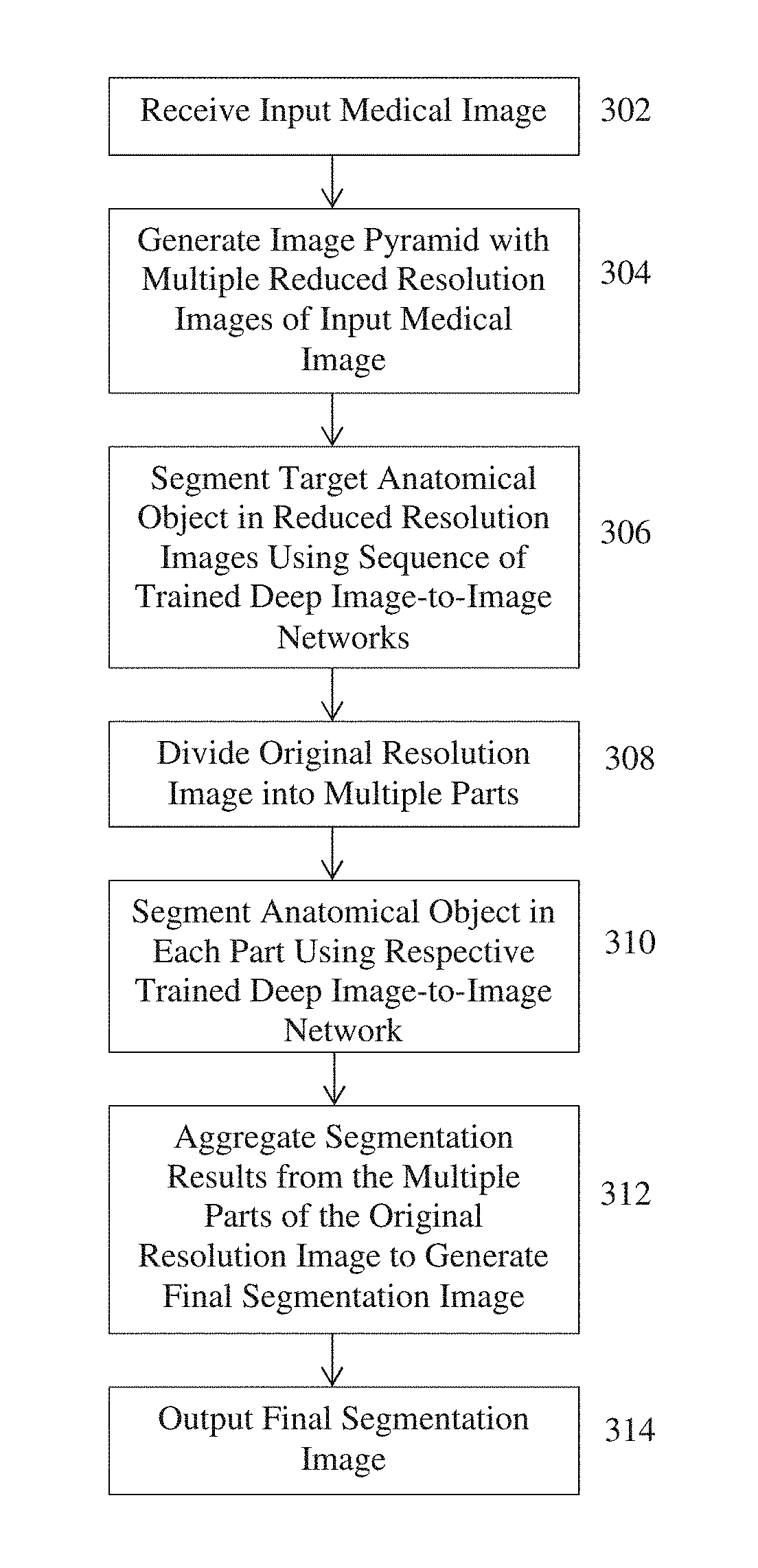
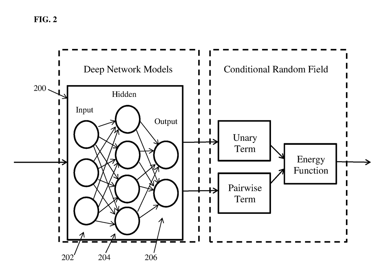
A method and apparatus for automatically performing medical image analysis tasks using deep image-to-image network (DI2IN) learning. An input medical image of a patient is received. An output image that provides a result of a target medical image analysis task on the input medical image is automatically generated using a trained deep image-to-image network (DI2IN). The trained DI2IN uses a conditional random field (CRF) energy function to estimate the output image based on the input medical image and uses a trained deep learning network to model unary and pairwise terms of the CRF energy function. The DI2IN may be trained on an image with multiple resolutions. The input image may be split into multiple parts and a separate DI2IN may be trained for each part. Furthermore, the multi-scale and multi-part schemes can be combined to train a multi-scale multi-part DI2IN.

Description

[0001]This application claims the benefit of U.S. Provisional Application No. 62 / 276,309, filed Jan. 8, 2016, the disclosure of which is herein incorporated by reference.BACKGROUND OF THE INVENTION[0002]The present invention relates to automated analysis of medical images, and more particularly, to automating various medical image analysis tasks using deep image-to-image network learning.[0003]Medical image analysis involves solving important tasks such as landmark detection, anatomy detection, anatomy segmentation, lesion detection, segmentation and characterization, cross-modality image registration, image denoising, cross-domain image synthesis, etc. Computer-based automation of these medical image analysis tasks brings significant benefits to medical imaging. For example, one such benefit of automating medical image analysis tasks is that it allows structured image reading and reporting for a streamlined workflow, thereby improving image reading outcomes in terms of accuracy, re...

Claims


Application Information

Patent Timeline
Patent Type & Authority Applications(United States)
IPC IPC(8): G06K9/66G06K9/46G06T7/11G06T7/00G06T7/174G06V30/194
CPCG06T2207/20016G06K9/6297G06T7/143G06K9/66G06T2207/20081G06T7/174G06T7/11G06K9/4609G06T2207/30004G06T7/0014G06T2207/10072G06T2207/10136G06T2207/30056G06T2207/30096G06V10/454G06V30/194G06F18/295
Inventor ZHOU, S. KEVINCOMANICIU, DORINGEORGESCU, BOGDANZHENG, YEFENGLIU, DAVIDXU, DAGUANG
Owner SIEMENS CORP
Why Eureka
Try Eureka