Image analysis for cervical neoplasia detection and diagnosis

a cervical neoplasia and image analysis technology, applied in the field of medical imaging, can solve the problems of lack of sophisticated laboratory equipment, lack of highly trained personnel and financial resources, and large number of cervical cancer cases, and achieve the effect of high probability of cervical neoplasia

Inactive Publication Date: 2011-11-10
CADES SCHUTTE A LIMITED LIABILITY LAW PARTNERSHIP
View PDF6 Cites 44 Cited by
  • Summary
  • Abstract
  • Description
  • Claims
  • Application Information

AI Technical Summary

Benefits of technology

[0030]Third, the present invention incorporates statistical features related to both acetowhitening and vessel structure. Previous approaches have used only acetowhitening features. The use of acetow...

Problems solved by technology

This considerable number of cervical cancer cases is mainly attributed to the lack of cervical cancer prevention programs in developing countries.
Even though cervical cancer prevention programs such as the Papanicolaou (Pap) smear have been effective in reducing the incidence and mortality of cervical cancer, developing countries often lack the sophisticated laboratory equipment, highly trained personnel and financial resources necessary to implement these programs (R. Sankaranarayanan, A. M. Budukh, and R. Rajkumar, “Effective screening programmes for cervical cancer in low- and middle-income developing countries,”Bulletin of the World Health Organization 79, pp.
Without a cost effective cervical cancer screening solution, cervical cancer remains a leading cause of cancer-related death among women in developing countries.
In these early studies, diagnostic image interpretation relied primarily on qualitative...

Method used


Image

  • Image analysis for cervical neoplasia detection and diagnosis
  • Image analysis for cervical neoplasia detection and diagnosis
  • Image analysis for cervical neoplasia detection and diagnosis

Examples


Embodiment Construction

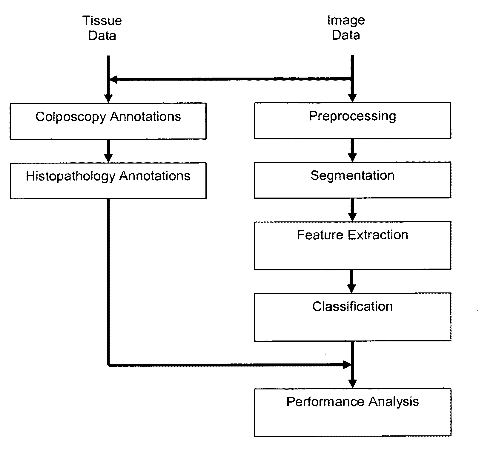
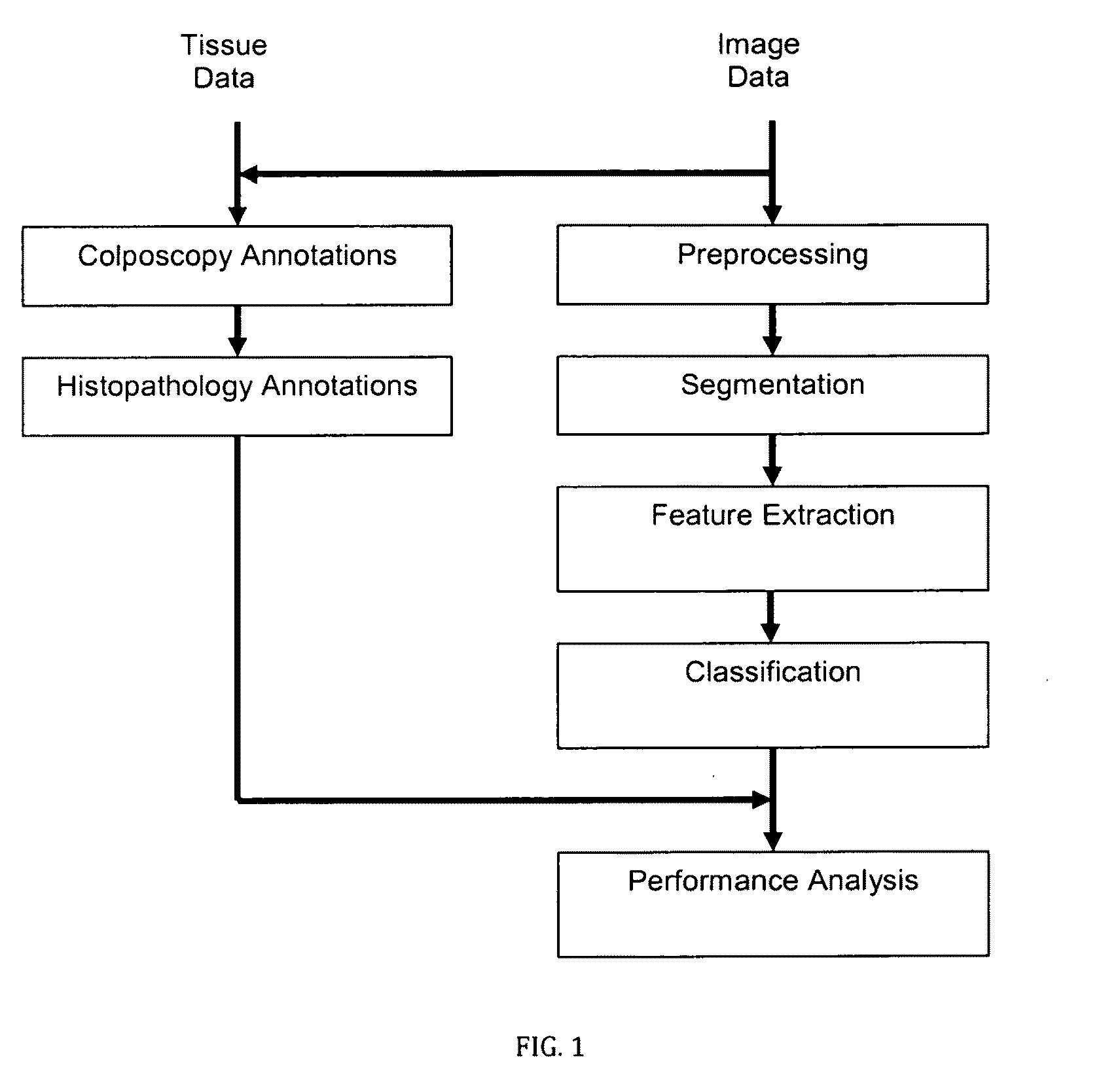
[0054]The presently preferred embodiment of the invention presented herein discloses an automated image analysis framework, schematically illustrated in FIG. 1, for cervical cancer detection and diagnosis. The framework uses high-resolution instrumentation to acquire high quality, clinical cervical image data, as well as tissue data in the form of colposcopy and histopatology annotations. The diagnostic image analysis framework is comprised of a series of image analysis steps in which image preprocessing, image segmentation, diagnostic feature extraction and diagnostic image assessment are applied in sequential order. In the final step, a performance analysis of the diagnostic image analysis output is applied.

Instrumentation and Data

Instrumentation

[0055]The image data used in the development of the cervical cancer detection and diagnosis framework were acquired with a digital imaging device designed specifically for the acquisition of cervical images and subsequent optical analysis ...


PUM


Abstract

The present invention is an automated image analysis framework for cervical cancerous lesion detection. The present invention uses domain-specific diagnostic features in a probabilistic manner using conditional random fields. In addition, the present invention discloses a novel window-based performance assessment scheme for two-dimensional image analysis, which addresses the intrinsic problem of image misalignment. As a domain-specific anatomical feature, image regions corresponding to different tissue types are extracted from cervical images taken before and after the application of acetic acid during a clinical exam. The unique optical properties of each tissue type and the diagnostic relationships between neighboring regions are incorporated in the conditional random field model. The output provides information about both the tissue severity and the location of cancerous tissue in an image.

Description

[0001]This application claims the priority of U.S. provisional patent application No. 61 / 343,757, filed on May 3, 2010.TECHNICAL FIELD[0002]The present invention generally relates to medical imaging, and more specifically to an automated image analysis framework for the detection and diagnosis of cervical cancerous lesions.BACKGROUND ART[0003]Although this invention is being disclosed in connection with cervical cancer, it is applicable to many other areas of medicine. Cervical cancer is preventable with early detection but still comprises approximately 12% of all cancer cases in women worldwide (World Health Organization, “Global Health Risks”, December 2009, incorporated herein by reference). This considerable number of cervical cancer cases is mainly attributed to the lack of cervical cancer prevention programs in developing countries. Even though cervical cancer prevention programs such as the Papanicolaou (Pap) smear have been effective in reducing the incidence and mortality o...

Claims


Application Information

Patent Timeline
IPC IPC(8): G06K9/00
CPCG06K9/6284G06K9/6297G06T7/0012G06T2207/10012A61B5/4331G06T2207/10024G06T2207/20076G06T2207/30096G06T2207/10016G06F18/2433G06F18/295
Inventor PARK, SUN YOUNGSARGENT, DUSTINGUSTAFSSON, ULF PETER
Owner CADES SCHUTTE A LIMITED LIABILITY LAW PARTNERSHIP
Why Eureka
Try Eureka