Method and system for automated brain tumor diagnosis using image classification

Inactive Publication Date: 2018-04-05
SIEMENS AG
View PDF1 Cites 17 Cited by
  • Summary
  • Abstract
  • Description
  • Claims
  • Application Information

AI Technical Summary

Benefits of technology

[0005]The present invention provides a method and system for automated classification of different types of tissue in medical images using machine learning based image classification. Embodiments of the present invention reconstruct image features of input endomicroscopy images using a learnt discriminative dictionary and classify the tissue in the endomicroscopy images based on the reconstructed image features using a

Problems solved by technology

Cancer is a major health problem throughout the world.
Unfortunately, intraoperative fast histopathology is often not sufficiently informative for pathologists to make an accurate diagnosis.
Biopsies are often non-diagnostic and yield inconclusive results for various reasons.
Such reasons include sampling errors, in which the biopsy may not originate from the most aggressive part of

Method used


Image

  • Method and system for automated brain tumor diagnosis using image classification
  • Method and system for automated brain tumor diagnosis using image classification
  • Method and system for automated brain tumor diagnosis using image classification

Examples


Embodiment Construction

[0014]The present invention relates to automated classification of different types of tissue in medical images using a machine learning based image classification. Embodiments of the present invention can be applied to endomicroscopy images of brain tumor tissue for automated brain tumor diagnosis. Embodiments of the present invention are described herein to give a visual understanding of the method for automated classification of tissue in medical images. A digital image is often composed of digital representations of one or more objects (or shapes). The digital representation of an object is often described herein in terms of identifying and manipulating the objects. Such manipulations are virtual manipulations accomplished in the memory or other circuitry / hardware of a computer system. Accordingly, is to be understood that embodiments of the present invention may be performed within a computer system using data stored within the computer system.

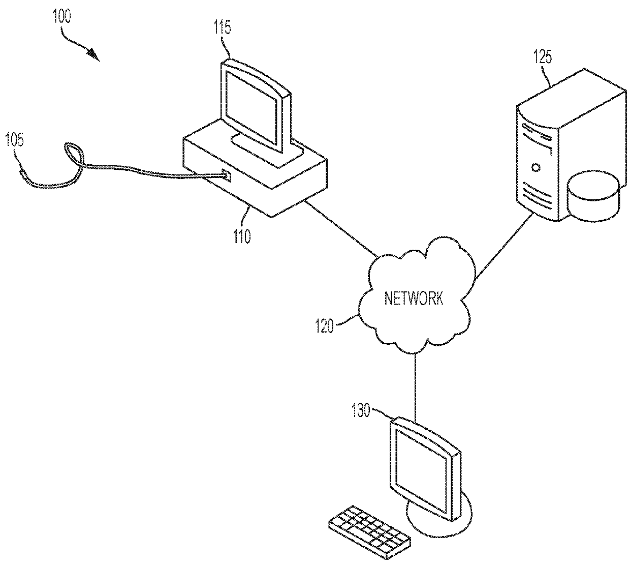
[0015]FIG. 1 illustrates an example...


PUM


Abstract

A method and system for classifying tissue endomicroscopy images are disclosed. Local feature descriptors are extracted from an endomicroscopy image. Each of the local feature descriptors is encoded using a learnt discriminative dictionary. The learnt discriminative dictionary includes class-specific sub-dictionaries and penalizes correlation between bases of sub-dictionaries associated with different classes. Tissue in the endomicroscopy image is classified using a trained machine learning based classifier based on the coded local feature descriptors encoded using a learnt discriminative dictionary.

Description

[0001]This application claims the benefit of U.S. Provisional Application No. 62 / 139,016, filed Mar. 27, 2015, the disclosure of which is herein incorporated by reference.TECHNICAL FIELD[0002]The present invention relates to classifying different types of tissue in medical image data using machine learning based image classification, and more particularly to automatic brain tumor diagnosis using machine learning based image classification.BACKGROUND OF THE INVENTION[0003]Cancer is a major health problem throughout the world. Early diagnosis of cancer is crucial to the success of cancer treatments. Traditionally, pathologists acquire histopathological images of biopsies sampled from patients, examine the histopathological images under microscopy, and make judgments as to a diagnosis based on their knowledge and experience. Unfortunately, intraoperative fast histopathology is often not sufficiently informative for pathologists to make an accurate diagnosis. Biopsies are often non-diag...

Claims


Application Information

Patent Timeline
IPC IPC(8): G06K9/00G06K9/62G06T7/00G06V10/772
CPCG06K9/00147G06K9/6255G06T7/0012A61B1/00009A61B5/4255G06V20/698G06V10/772A61B1/000094G06F18/28
Inventor WAN, SHAOHUASUN, SHANHUIBHATTACHARYA, SUBHABRATACHEN, TERRENCEKAMEN, ALI
Owner SIEMENS AG
Why Eureka
Try Eureka