
Quality control method and system for remote fundus screening, and storage device

Pending Publication Date: 2021-11-04
FUZHOU YIYING HEALTH TECH CO LTD
View PDF0 Cites 2 Cited by
  • Summary
  • Abstract
  • Description
  • Claims
  • Application Information

AI Technical Summary

Benefits of technology

The present invention is a system that improves the accuracy and reliability of fundus image interpretation and diagnosis. It uses pre-interpretation to determine if the fundus image meets the requirements for interpretation and consultation. This saves time and ensures a better user experience. The system also includes quality control processes and interactions with the interpretation center to further improve the accuracy of the diagnosis. The examinee does not have to go back for the fundus image, which avoids additional time and effort. The system also ensures that only authorized individuals can access and use the fundus image for diagnosis. Overall, this technology improves the efficiency and reliability of fundus image interpretation and diagnosis.

Problems solved by technology

DR screening can reduce the risk of blindness by 94.4%, but due to the lack of fundus reading or ophthalmology specialists in many primary medical institutions around the world, the DR screening rate is generally not high in many countries, and even less than 10% in China!
A common phenomenon that causes the examinee to go back and forth or repeat the examination for many times, which seriously affects the smooth progress of remote screening, causes unnecessary expenses or waste of time, and even affects the process or screening rate of fundus screening.
According to research conducted by Beijing Tongren Hospital, about 40% of the remote fundus images and medical information uploaded by basic terminal application agencies have quality problems.
In the process of image acquisition, due to the influence of human factors and eye factors such as shooting angle, cataracts, small pupils, etc., quality problems such as blurry and incomplete structure of the fundus image will appear.
The quality of the collected or uploaded fundus images determines the service quality of the remote screening system, which is an important problem that is difficult to solve in remote fundus screening.
In addition, those low-quality images that are difficult to interpret medically will directly reduce the reliability of subsequent analysis, cause interference with truly useful information, and affect the results and quality of subsequent services.

Method used

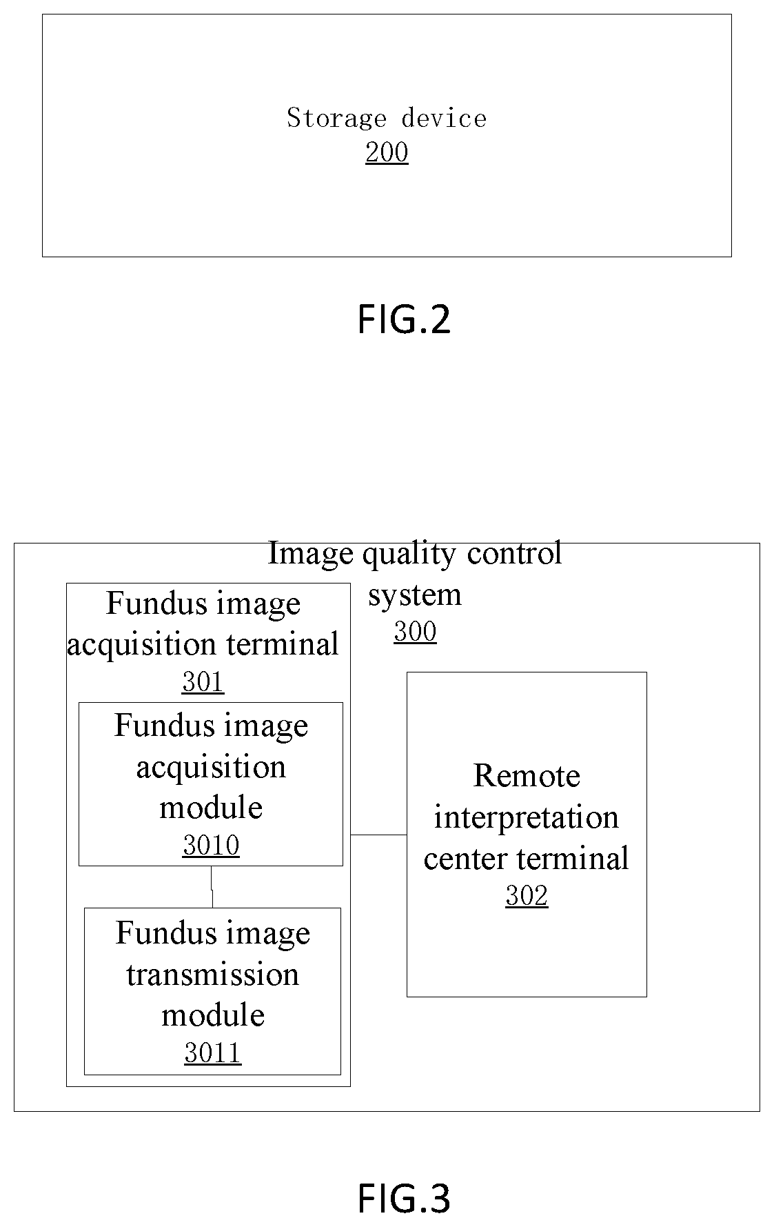

Image

  • Quality control method and system for remote fundus screening, and storage device
  • Quality control method and system for remote fundus screening, and storage device

Examples


Embodiment Construction

[0031]To describe the technical contents, the structural features, the achieved objective and effect in detail, the following will perform description in detail with reference to the specific embodiments and the accompanying drawings.

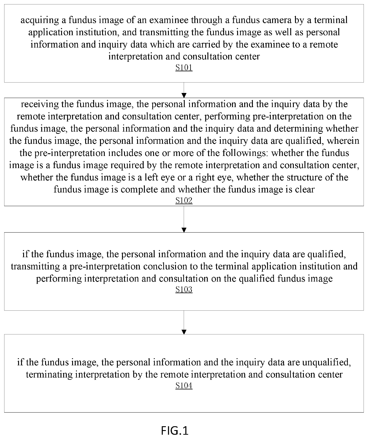
[0032]The key idea of the present invention is that the fundus images uploaded by multiple quality control systems such as the remote terminal application institution are checked by the operator of the basic or terminal application institution, the remote interpretation and consultation center or the pre-interpretation personnel of the remote interpretation and consultation center, the computer aided system, the relevant quality control system and the like, thereby ensuring that the fundus images finally for analysis and determination of diseases are 100% available.

[0033]Referring to FIG. 1, in the embodiment, an application scene of a quality control method for remote fundus screening is: a basic or terminal application institution, a fundus camera and...


PUM


Abstract

A quality control method for remote fundus screening includes acquiring a fundus image of an examinee through a fundus camera by terminal application institution and transmitting the fundus image as well as personal information and inquiry data which are carried by the examinee to a remote interpretation and consultation center. The remote interpretation consultation center determines whether the fundus image, personal information, and consultation data are qualified; if the fundus image, personal information, and consultation data are unqualified, the remote interpretation consultation center terminates the interpretation. Thus clear and usable qualified fundus image is obtained during follow-up or screening of a patient and the examinee is not needed to go back and forth or repeat the examination multiple times, unnecessary expenses or time waste is saved, and the progress or screening rate of fundus screening is not affected, guaranteeing for the effective and smooth progress of remote fundus medical screening.

Description

BACKGROUND OF THE INVENTION1. Field of the Invention[0001]The present invention relates to the field of fundus image processing, and in particular, to a quality control method and system for remote fundus screening, and a storage device.2. Description of the Prior Art[0002]Today, the fundus camera technology used for diabetic retinopathy (DR) screening is mature. Moreover, DR screening has prevention and treatment guidelines and diagnostic standards that are related to diabetes and can guide treatment. DR screening can reduce the risk of blindness by 94.4%, but due to the lack of fundus reading or ophthalmology specialists in many primary medical institutions around the world, the DR screening rate is generally not high in many countries, and even less than 10% in China! The experience of various countries has shown: “Telemedicine is an effective way to carry out DR screening and improve the screening rate and patient compliance”! Eyes are the only part of the human body where blood...

Claims


Application Information

Patent Timeline
IPC IPC(8): G06T7/00H04N7/18A61B3/12A61B3/14G16H30/20G16H50/20
CPCG06T7/0012H04N7/183A61B3/12G06T2207/30168G16H30/20G16H50/20G06T2207/30041A61B3/14G06T2207/10024G06T2207/20032G16H40/20G16H40/67G16H80/00
Inventor YU, LUNQIU, YING-QIANGLIN, JIA-WENCAO, XIN-RONGWANG, LI-NA
Owner FUZHOU YIYING HEALTH TECH CO LTD
Learn More