Method for operating a computed tomography apparatus having a diaphragm at the radiation detector

a computed tomography and radiation detector technology, applied in the direction of diaphragm/collimeter, instruments, application, etc., can solve the problems of finite number of slits of discrete width, high production cost of bearing bodies curved around two axes,

Inactive Publication Date: 2007-01-30
SIEMENS HEALTHCARE GMBH
View PDF13 Cites 45 Cited by
  • Summary
  • Abstract
  • Description
  • Claims
  • Application Information

AI Technical Summary

Benefits of technology

[0010]An object of the present invention is to provide a method for operating a computed tomography apparatus having a beam-gating diaphragm disposed near and preceding the radiation detector, wherein over-radiation of the patient at the beginning and at the end of a scan are avoided.

Problems solved by technology

Moreover, the production of a bearing body curved around two axes is expensive, because this must also be produced from x-ray-absorbing material, meaning from a material with a high atomic number.
A further disadvantage of the x-ray collimator known from U.S. Pat. No. 6,396,902 is that only a finite number of slits of discrete width can be applied or, respectively, introduced on or in the bearing body.

Method used


Image

  • Method for operating a computed tomography apparatus having a diaphragm at the radiation detector
  • Method for operating a computed tomography apparatus having a diaphragm at the radiation detector
  • Method for operating a computed tomography apparatus having a diaphragm at the radiation detector

Examples


Embodiment Construction

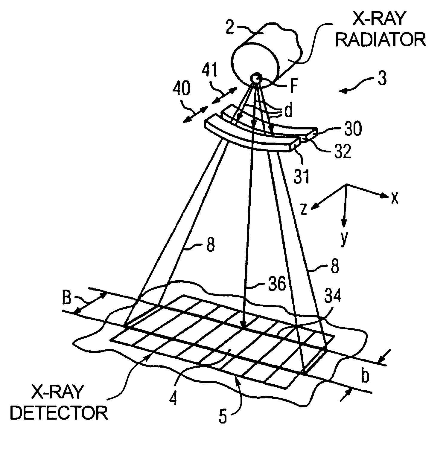
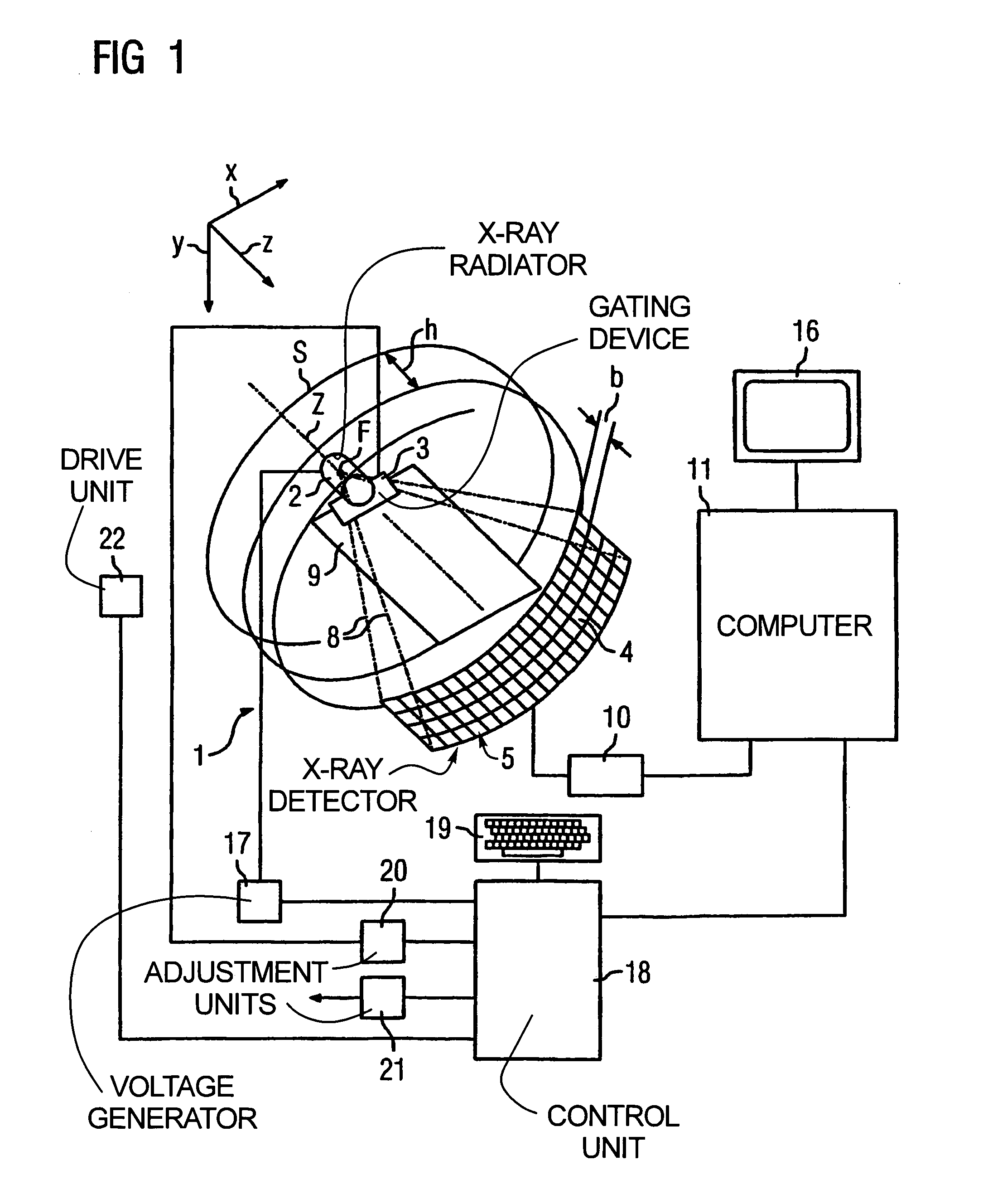
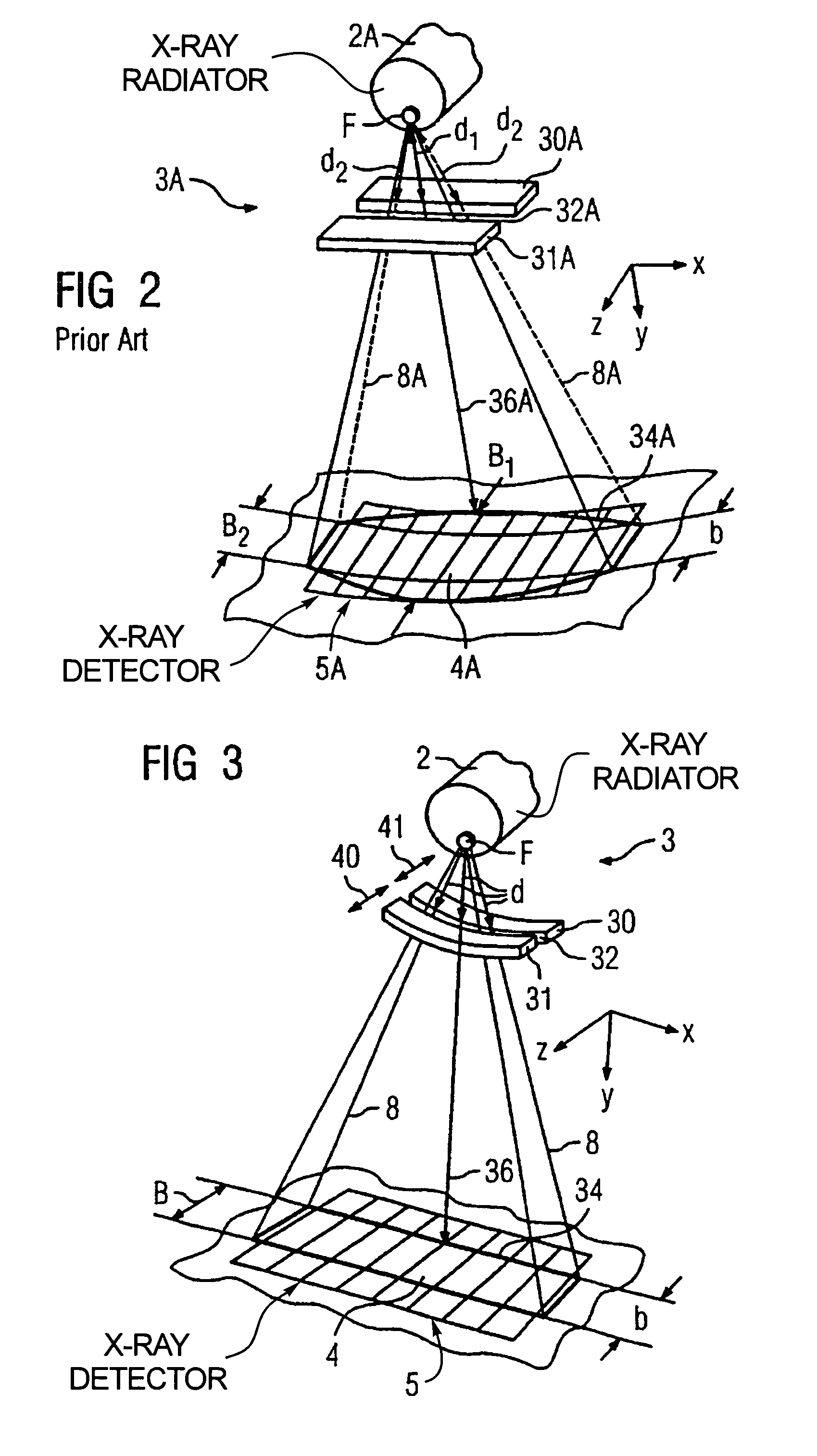
[0034]A CT apparatus of the third generation is shown in FIG. 1 in section. Its data acquisition arrangement includes an x-ray radiator 2 with a gating device 3 positioned in front of it, near the source, and an x-ray detector 5, fashioned as a laminar array of a number of rows and columns of detector elements (one of these is designated 4 in FIG. 1), with an optional beam diaphragm (not shown) positioned in front of the x-ray detector 5, close to the detector 5. For clarity, in FIG. 1 only four rows of detector elements 4 are shown; however, the x-ray detector 5 can have further rows of detector elements 4, optionally also with different widths b.

[0035]The x-ray radiator 2 with the gating device 3 on one side and the x-ray detector 5 with its beam diaphragm on the other side are mounted opposite one another on a rotary frame (gantry) (not shown), such that a pyramidal x-ray beam emitted by the x-ray radiator 2 in the operation of the CT apparatus 1 and gated by the adjustable gatin...


PUM


Abstract

In a method for operating a computed tomography apparatus, having an x-ray source rotatable around a system axis and a radiation detector with a detector-proximate beam-gating diaphragm, and a patient support, a spiral scan of a patient on the patient support is conducted by rotating the x-ray source around the system axis while moving the subject on the patient support substantially parallel to the system axis. The diaphragm have movable absorber elements that are curved, and are moved independently of each other toward and away from each other in a direction substantially parallel to the system axis during the spiral scan. The absorber elements are dynamically adjusted in an asymmetrical manner during the spiral scan to reduce overexposure of the examination subject to x-rays.

Description

[0001]This application is a continuation of PCT / DE 03 / 02890, Sep. 1, 2003.BACKGROUND OF THE INVENTION[0002]1. Field of the Invention[0003]The present invention concerns a method for operating a computed tomography apparatus with an x-ray radiator rotatable around a system axis, with an x-ray detector and with a radiator-side gating device that has two opposite absorber elements that can be adjusted in a straight line, in particular that can be adjusted with regard to their separation from one another, for variable delimitation of the ray beam. An examination subject is scanned during rotation of the x-ray radiator and relative translational movement between the x-ray radiator and the examination subject in the direction of the system axis.[0004]2. Description of the Prior Art[0005]In an examination of an examination subject or a patient in an x-ray diagnostic apparatus, the examination subject is moved into an x-ray beam emitted by an x-ray source, and the radiation actuated by the ...

Claims


Application Information

Patent Timeline
Patent Type & Authority Patents(United States)
IPC IPC(8): G21K1/04A61B6/03A61B6/06
CPCA61B6/032A61B6/06A61B6/4035G21K1/04A61B6/027
Inventor DISTLER, FRIEDRICHSEUFERT, MATTHIASSUSS, CHRISTOPH
Owner SIEMENS HEALTHCARE GMBH
Why Eureka
Try Eureka