Intraoral dental radiology positioning device for use with aiming ring

a positioning device and dental radiology technology, applied in the field of intraoral dental radiology positioning devices, can solve the problems of unnecessary radiation irradiation of tissues, patient overexposure, and inability to accurately target the x-ray, so as to prevent overexposure of patients

Active Publication Date: 2007-02-06
DIEDERICH JENNIFER A
View PDF11 Cites 18 Cited by
  • Summary
  • Abstract
  • Description
  • Claims
  • Application Information

AI Technical Summary

Benefits of technology

[0034]The collimation plate may include handles opposite of one another for the patient to grasp. The opening in the collimation plate is preferably substantially rectangular in shape, is orientated similarly to the film having a substantially rectangular shape, and the substantially central opening defined by the collimation plate is adapted to correspond dimensionally to the dimensions of the receptor. Preferably, the collimation plate is constructed of metal having a thickness of at least 0.075 in. to prevent overexposing the patient to needless additional radiation.

Problems solved by technology

A disadvantage of known devices is that the cross-sectional area of the beam used by the radiographic technique is typically larger than the surface area of the x-ray film.
When the cross-sectional area of the beam does not match the film size, the patient can be exposed to unnecessary radiation which irradiates tissues beyond the borders of the dental film.
Another disadvantage of current x-ray film positioning instruments is that there can be errors in aiming the x-ray.
These errors are frequently associated with a rectangular position-indicating device attached to an x-ray machine.
Also, there can be errors in orientation of the long axis of the substantially rectangular typical position indicating devices with the long axis of the film in the patient's mouth.
Aiming and orientation errors expose the patient to needless retakes of radiographs.
Current film positioning devices may have disadvantages when sterilizing because parts, such as cushioned areas, may be less accessible or receptive to steam, heat or chemical methods.
Difficulty in disinfecting or sterilizing is associated with a rectangular positioning indicating device attached to an x-ray machine
Another disadvantage of current x-ray film positioning instruments is that the patient can only bite on the bite plate to assist in positioning and holding a film positioning device in the mouth.
Typical devices may be difficult to grasp and manipulate in the patient's mouth making it problematic for the patient to assist in positioning the instrument.

Method used


Image

  • Intraoral dental radiology positioning device for use with aiming ring
  • Intraoral dental radiology positioning device for use with aiming ring
  • Intraoral dental radiology positioning device for use with aiming ring

Examples


Embodiment Construction

)

[0068]In describing the preferred embodiment of the present invention, reference will be made herein to FIGS. 1–29 of the drawings in which like numerals refer to like features of the invention. Features of the invention are not necessarily shown to scale in the drawings.

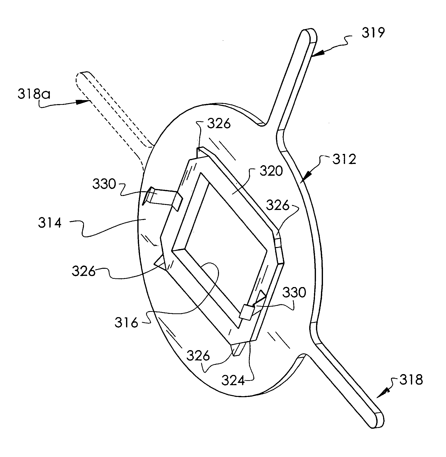
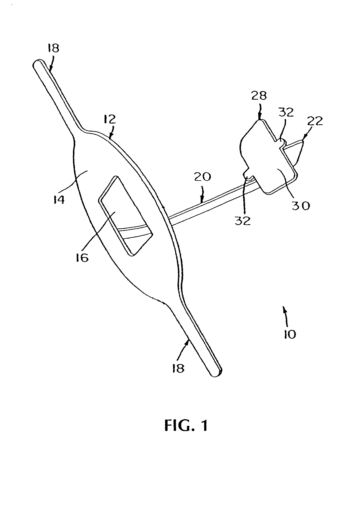
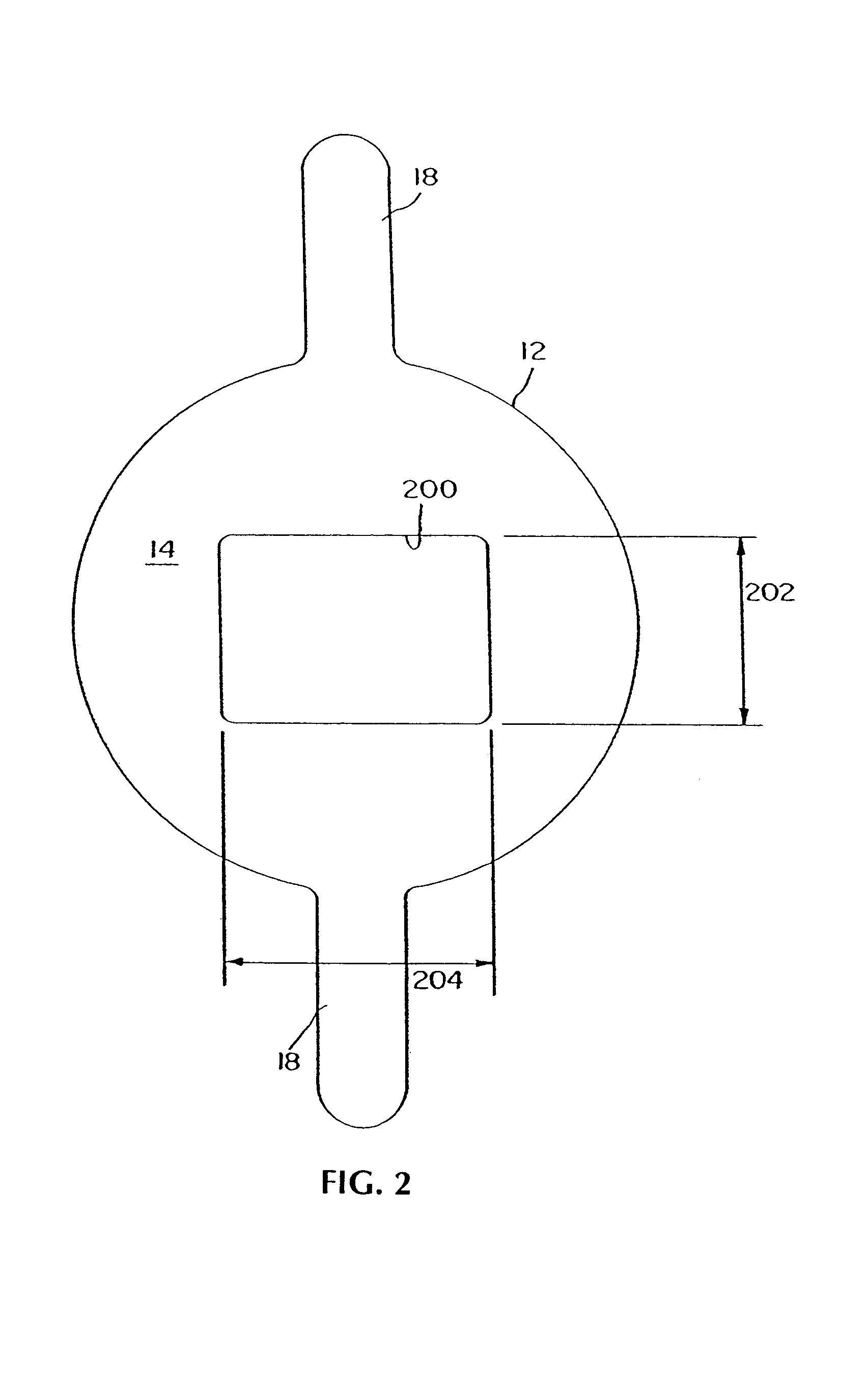
[0069]A preferred embodiment 10, shown in FIGS. 1–6 and 11, depicts the intraoral dental radiology receptor positioning device of the present invention. Referring to FIGS. 1–4, the receptor positioning device includes a substantially flat collimation plate 12, having a surface area 14. The surface area 14 defines a substantially central rectangular opening 16. The collimation plate 12 further includes opposing elongated handles 18 extending outward on opposite sides thereof. The receptor positioning instrument 10 further includes an elongated arm 20 and a film or electronic receptor holding member 28 having a back plate 30 and clips 32 for holding x ray film or electronic receptors. The elongated arm 20 is connecte...


PUM


Abstract

A receptor positioning device for taking dental bitewing radiographs of a patient's teeth comprises a receptor holding member adapted to receive a receptor for exposing x-radiation from the x-ray machine and a collimation plate having a substantially flat surface for aligning with an x-ray machine. The collimation plate has a rectangular opening in its surface for passage of radiation from the x-ray machine to expose a receptor in the receptor holding member and a pair of handles extending away from the plate for positioning the device. The collimation plate is constructed of metal having a thickness of at least 0.075 in., preferably at least 0.100 in., to reduce the passage of radiation from the x-ray machine to the patient except at the plate opening. The device also includes an arm between the collimation plate and the receptor holding member, the arm including a biting surface for engaging the patient's teeth to secure the device in the patient's mouth, and an aiming ring secured to the arm at a position distal from the receptor holding member, with the collimation plate being secured to the aiming ring. The aiming ring may also include notches and the collimation plate preferably includes clips for removably securing the plate to the aiming ring and tabs for locating the plate relative to the aiming ring notches.

Description

[0001]This application is related to an application by the same inventor filed on even date herewith as Ser. No. 10 / 899,249 entitled “Intraoral Dental Radiology Positioning Device.”BACKGROUND OF THE INVENTION[0002]1. Field of the Invention[0003]The present invention relates to devices for dental radiographic procedures or intraoral diagnostics, and more particularly, to intraoral dental radiology positioning devices relating to positioning x-ray film or receptors in a patient's mouth during radiographic procedures.[0004]2. Description of Related Art[0005]Intraoral x-ray diagnosis involves positioning an x-ray film within a patient's mouth next to the inner surface of the teeth or bone being studied. The film is then exposed to an x-ray beam generated outside the mouth and passing through the target. Known intraoral dental radiography typically employs a dental device having an alignment member including an x-ray film holding structure at one end, an aligning arm at another end, and ...

Claims


Application Information

Patent Timeline
Patent Type & Authority Patents(United States)
IPC IPC(8): A61B6/14
CPCG03B42/042A61B6/4435A61B6/145
Inventor DIEDERICH, JENNIFER A
Owner DIEDERICH JENNIFER A
Why Eureka
Try Eureka