Ionizing radiation imaging system and method with decreased radiation dose

a radiation dose and imaging system technology, applied in tomography, instruments, applications, etc., can solve the problems of induction of fatal cancer, inability to detect ionizing radiation, and many documented harmful effects of exposure to ionizing radiation,

Active Publication Date: 2007-04-10
PEDIATRIC IMAGING TECH
View PDF16 Cites 19 Cited by
  • Summary
  • Abstract
  • Description
  • Claims
  • Application Information

AI Technical Summary

Problems solved by technology

Exposure to ionizing radiation has many documented harmful effects, one of the most serious of which is the induction of fatal cancers.
All human suspects are susceptible to ionizing radiation in the doses provided by typical imaging technology.
However, children are reported to be at approximately ten times greater risk than an average middle-aged adult.
Using data from such survivors, Brenner et al. has predicted that the use of conventional x-ray CT technology on infants and children may cause the eventual cancer-related death of 1 out of every 1000 children examined using such CT technology.
This rate is considered by many to be unacceptably high.
Turning down the total power of the source (lowering the radiation dose) can produce lower quality images.
Adjustment of exposure parameters is limited by the existing technology (for example, sources and detectors) and can only decrease the total radiation dose to certain levels while still achieving desirable image quality.
Further, despite attempts to reduce radiation dose, a wide variability in image scanning techniques still exists that expose many objects to higher than necessary radiation doses.
In addition, multi-detector (multi-slice) image scanners are now being used and are inherently more complex, presenting an additional challenge with respect to dose reduction.

Method used


Image

  • Ionizing radiation imaging system and method with decreased radiation dose
  • Ionizing radiation imaging system and method with decreased radiation dose
  • Ionizing radiation imaging system and method with decreased radiation dose

Examples


Embodiment Construction

[0015]The present invention provides an ionizing radiation imaging system and method that reduces the radiation dose imparted on a object being imaged, while attaining a high quality image. By matching a peak emission of a source to a peak absorbance of a detector, high quality images can be attained using a minimized radiation dose to a object to be imaged.

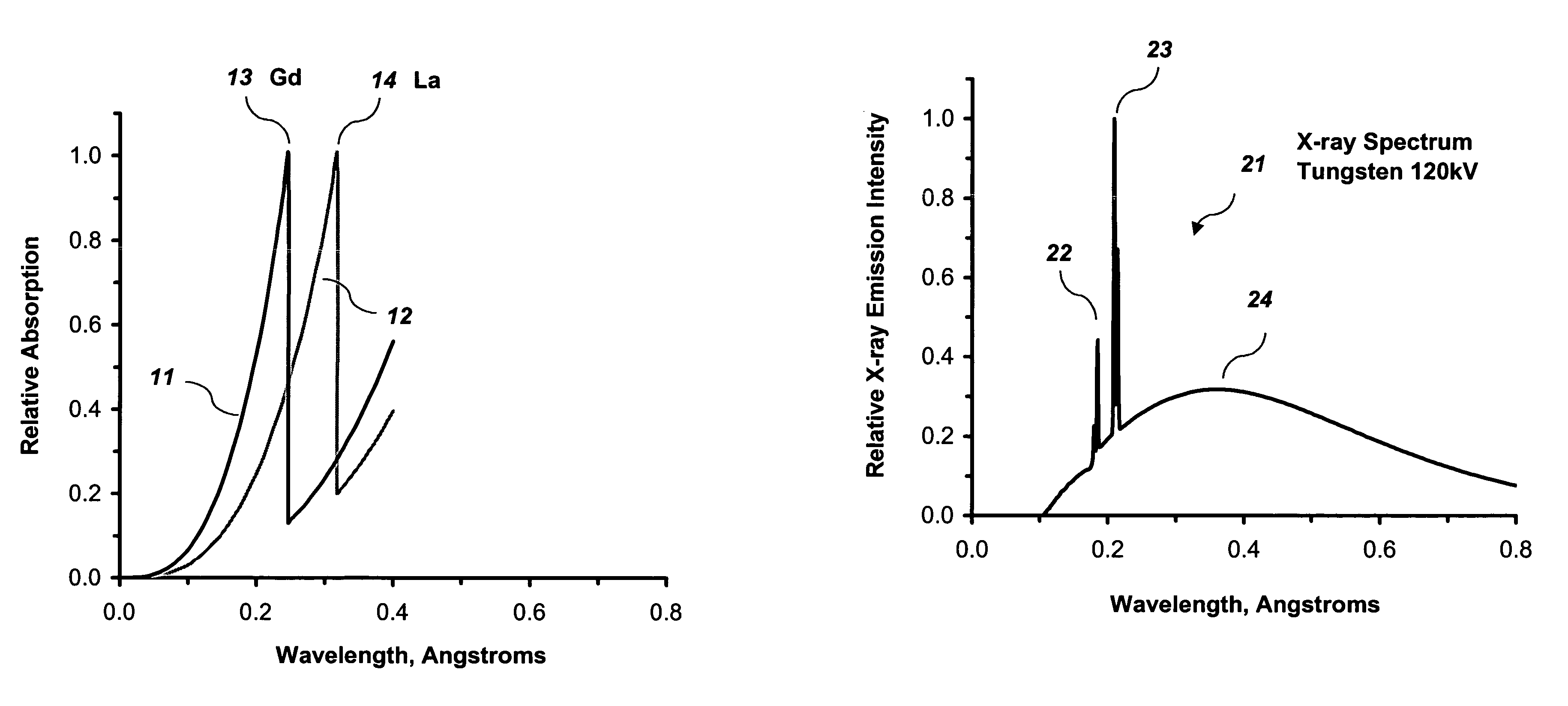
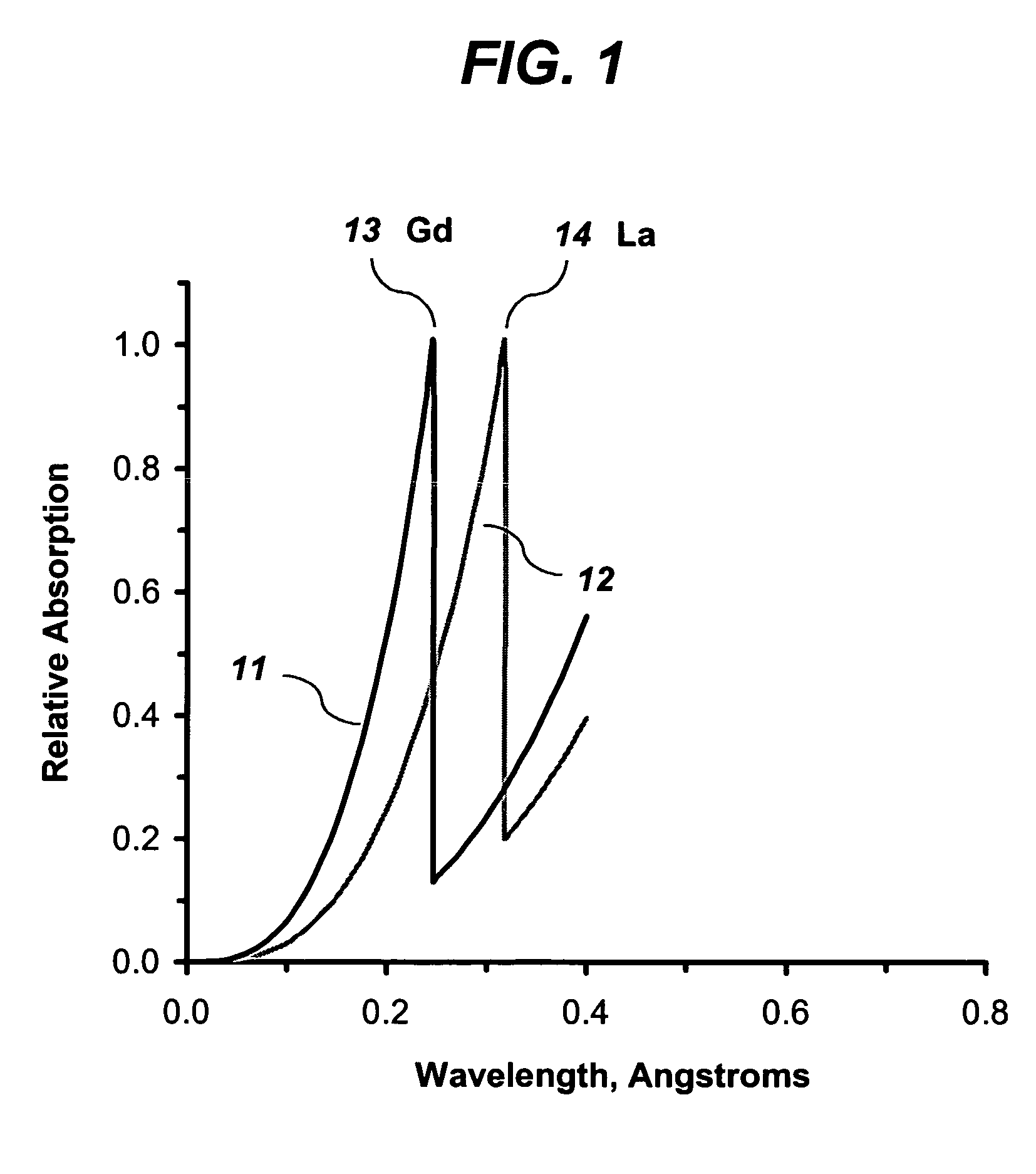
[0016]FIG. 1 illustrates a plot 11 of relative energy absorption versus energy wavelength for gadolinium and a plot 12 of relative energy absorption versus energy wavelength for lanthanum. Gadolinium and lanthanum are examples of atomic species that can be included in a detector according to the present invention. Plot 11 shows an absorption edge 13, or absorption maximum, for gadolinium at energy of wavelength of about 0.25 Angstroms (A). Plot 12 shows an absorption edge 14 for lanthanum at about 0.32 A. As can be seen in plots 11 and 12, maximum absorption of energy by an atomic species occurs at a wavelength that is at or just...


PUM

PropertyMeasurementUnit
wavelengthaaaaaaaaaa
wavelengthsaaaaaaaaaa

Abstract

A system and method for imaging a volume having a detector including a first atomic species with an absorption edge at a first wavelength. The system also includes a source having a second atomic species for emitting an ionizing energy. The second atomic species has a characteristic emission peak that substantially matches the absorption edge of the detector. A filter may be provided for blocking ionizing energy from the source in regions other than a region proximate the characteristic emission peak. Effective imaging with a lower radiation dose may be achieved with the system and method.

Description

RELATED APPLICATION DATA[0001]This application claims the benefit of priority of U.S. Provisional Patent Application Ser. No. 60 / 583,382, filed Jun. 28, 2004, and titled “Enhanced X-Ray-Detector-Based System and Method for CT”, which is incorporated by reference herein in its entirety.FIELD OF THE INVENTION[0002]The present invention generally relates to the field of imaging using ionizing radiation. In particular, the present invention is directed to an ionizing radiation imaging system and method with decreased radiation dose.BACKGROUND OF THE INVENTION[0003]Ionizing radiation, such as x-ray and gamma radiation, has been used for years in imaging systems, particularly in medical imaging. Exposure to ionizing radiation has many documented harmful effects, one of the most serious of which is the induction of fatal cancers. All human suspects are susceptible to ionizing radiation in the doses provided by typical imaging technology. However, children are reported to be at approximatel...

Claims


Application Information

Patent Timeline
Patent Type & Authority Patents(United States)
IPC IPC(8): G01N23/02A61B6/00A61B6/03
CPCA61B6/032A61B6/4042A61B6/542
Inventor BENZ, MARK G.BENZ, MATTHEW W.
Owner PEDIATRIC IMAGING TECH
Why Eureka
Try Eureka