Detector, three-dimensional direct positron imaging unit, and method to estimate the differential of the radiation dose provided to cancer cells and healthy tissues during hadrotherapy

a positron imaging and detector technology, applied in the field of medical imaging technique, can solve the problems of inability to resolve the position of tof-pet units, limitations to the resolution, contrast, brightness of clinical images, and poor resolution of today's commercial tof-pet units, so as to improve therefore the overall performance of the imaging unit. , the effect of improving the snr of the detector

Active Publication Date: 2019-06-11
THE TRUSTEES FOR PRINCETON UNIV
View PDF10 Cites 1 Cited by
  • Summary
  • Abstract
  • Description
  • Claims
  • Application Information

AI Technical Summary

Benefits of technology

[0012]In particular, it is an object of the present invention to improve the radial resolution of individual positron annihilation 5 events of known imaging techniques.
[0023]In one embodiment, the imaging unit further comprises means to force air, in particular air processed to reduce Radon concentration, inside the cavity where the subject is located. This solution improves SNR of the detector and therefore the overall performances of the imaging unit.

Problems solved by technology

Today's commercial TOF-PET units however have a very poor resolution—a few cm—for the determination of the position of the single annihilation event in the radial direction, due to the sub-optimal performance of the TOF measurement for the 511 keV γ-rays.
The poor resolution in the radial direction results in limitations to the resolution, contrast, and brightness of the clinical image.
As detailed above, the TOF-PET units commercially available today cannot resolve the position of individual positron annihilation with resolution close to the two fundamental scales of lengths of the PET described above.
The use of a tomographic procedure for the reconstruction of three-dimensional images is, by its own, a strong disadvantage of commercially available PET and TOF-PET units.
While the dose is of limited statistical consequence for adult cancer patients, it is a major cause of concern in pediatric oncology, with young patients in remission potentially subjected to up to 20-30 check-up procedures in the course of their lives.
Another, additional, limitation of PET and TOF-PET units is their limited efficiency as γ-ray detectors and their limited energy resolution.
The low efficiency in γ-ray detection stems from cost considerations: procurement of large surfaces of LSO and LYSO crystals is one of the driving costs, and this results in standard limitation of the thickness of crystals to no more than two interaction lengths.
A further limitation of PET and TOF-PET units is the low Signal-to-Noise Ratio (SNR).
The limited γ-ray detection efficiency results in the necessity of usage of large 18F activities, in turn resulting in very high rate of R events (RR), typically much higher than the rate of T events (RT) at normal condition of operation for PET and TOF-PET exams.
However, to date no method has been devised to monitor online the dose delivered to cancer cells relative to that delivered to surrounding healthy tissues.

Method used


Image

  • Detector, three-dimensional direct positron imaging unit, and method to estimate the differential of the radiation dose provided to cancer cells and healthy tissues during hadrotherapy
  • Detector, three-dimensional direct positron imaging unit, and method to estimate the differential of the radiation dose provided to cancer cells and healthy tissues during hadrotherapy
  • Detector, three-dimensional direct positron imaging unit, and method to estimate the differential of the radiation dose provided to cancer cells and healthy tissues during hadrotherapy

Examples


Embodiment Construction

[0031]While the invention is susceptible to various modifications and alternative constructions, some of the illustrated embodiments are shown in the drawings and will be described below in detail.

[0032]It must be understood, however, that there is no intention to limit the invention to the specific illustrated embodiments, but, on the contrary, the invention intends to cover all the modifications, alternative constructions and equivalents that fall within the scope of the invention as defined in the claims.

[0033]The use of “such as”, “etc.”, “or” indicates non-exclusive alternatives without limitations, unless otherwise indicated.

[0034]The use of “includes” means “includes, but is not limited to”, unless otherwise indicated.

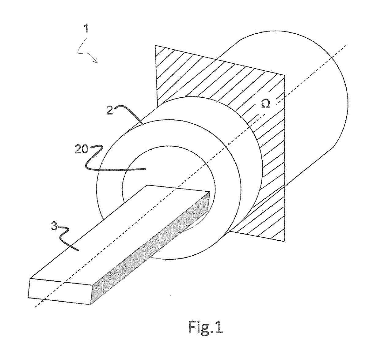
[0035]FIG. 1 illustrates an imaging unit 1 comprising a hollow body 2 with an internal cavity 20 allowing a table 3 to slide inside out it in order to bring a subject under a detector 4 housed inside the hollow body 2.

[0036]Detector 4 is adapted to detect emissi...


PUM


Abstract

Disclosed is a detector for a positron imaging unit, comprises a hollow body with an inner cylindrical wall and an outer wall spaced apart from the inner cylindrical wall. The hollow body includes a scintillating material, suitable to emit photons once hit by a 511 keV γ-ray, and one or more pairs of photo-detecting units (e.g. comprising PMTs or SiPM) for detecting photons emitted by the scintillating material; each photo-detecting unit of a pair being placed at opposite ends of the inner cylindrical wall along a radial direction. The scintillating material has scintillation decay time τ lower than 10 ns, an atomic number greater than 10, and a high scintillation yield greater than 8,000 photons / MeV, and comprises a mixture of xenon and argon. An imaging unit including the detector and a method to estimate the differential of the dose of radiation provided in a subject to cancer cells and to surrounding tissues in the course of hadrotherapy is also disclosed.

Description

CROSS-REFERENCE TO RELATED APPLICATIONS[0001]This application claims benefit of Italian Patent Application No. 102015000024439, filed Jun. 16, 2015, which is hereby incorporated in its entirety by reference.BACKGROUND OF THE INVENTION[0002]Positron emission tomography (PET) is a medical imaging technique that is commonly used to produce three-dimensional images of subjects' bodies inner organs and parts. This technique hinges on detection of the two 511 keV γ-rays emitted as a consequence of annihilation of an electron present in the subjects' tissues with a positron emitted by radionuclides inoculated to subjects in form of specific tracers (e.g., 18F-loaded fluorodeoxyglucose 18F-FDG). The clinical analysis of interest determines the choice for the tracers, which are selected to target specific cells or proteins based as it is made possible by their affinity for the metabolic processes. Information resulting from the determination of the position of interactions of the two 511 keV...

Claims


Application Information

Patent Timeline
Patent Type & Authority Patents(United States)
IPC IPC(8): A61B6/03G01T1/204A61B6/04G01T1/02G01T1/24G01T1/20G01T1/29A61N5/10
CPCA61B6/037A61B6/0421A61N5/1071G01T1/023G01T1/026G01T1/2018G01T1/249G01T1/2985G01T1/204A61N2005/1094A61N2005/109A61N2005/1087G01T1/20183G01T1/2019
Inventor GALBIATI, CRISTIANO
Owner THE TRUSTEES FOR PRINCETON UNIV
Why Eureka
Try Eureka