
Method and apparatus for displaying endoscopic images

a technology of endoscope and viewing set, which is applied in the field of system and method of displaying images, can solve the problems of tying the user to the viewing set of the endoscope, and limiting the way information about the internal structure can be conveyed. , to achieve the effect of displaying the endoscopic image from the viewing set of the endoscope, and not always convenient or even possibl

Active Publication Date: 2007-06-19
KARL STORZ IMAGING INC
View PDF51 Cites 85 Cited by
  • Summary
  • Abstract
  • Description
  • Claims
  • Application Information

AI Technical Summary

Problems solved by technology

However, they still only provide a view from the viewing point and in the viewing direction of the endoscope.
Because the endoscope, the user, and the internal structure being examined exist in an actual three-dimensional world, tying the user to the viewing set of the endoscope limits the way information about the internal structure can be conveyed.
This is not always convenient or even possible.
However, although these systems provide a three-dimensional model with a variable viewing set, they can only display the endoscopic image from the viewing set of the endoscope.
Because of this limitation, which is common for all existing endoscopic display systems, the true nature of the viewed area is often not conveyed adequately to the user.

Method used


Image

  • Method and apparatus for displaying endoscopic images
  • Method and apparatus for displaying endoscopic images
  • Method and apparatus for displaying endoscopic images

Examples


Embodiment Construction

[0016]The following detailed description illustrates the invention by way of example, not by way of limitation of the principles of the invention. This description will enable one skilled in the art to make and use the invention, and describes several embodiments, adaptations, variations, alternatives and uses of the invention, including what we presently believe is the best mode of carrying out the invention.

[0017]The preferred embodiment of the invention is a software program running on a computer. The computer communicates electronically with an endoscope and a display device such as a monitor. The computer includes a graphics processing unit such as those manufactured by NVidia Corporation. The graphics processing unit is specifically designed to quickly perform the types of graphics related calculations required by the present invention. Other devices may be connected to the computer as appropriate for a given application.

[0018]The program uses the graphical display programming...


PUM


Abstract

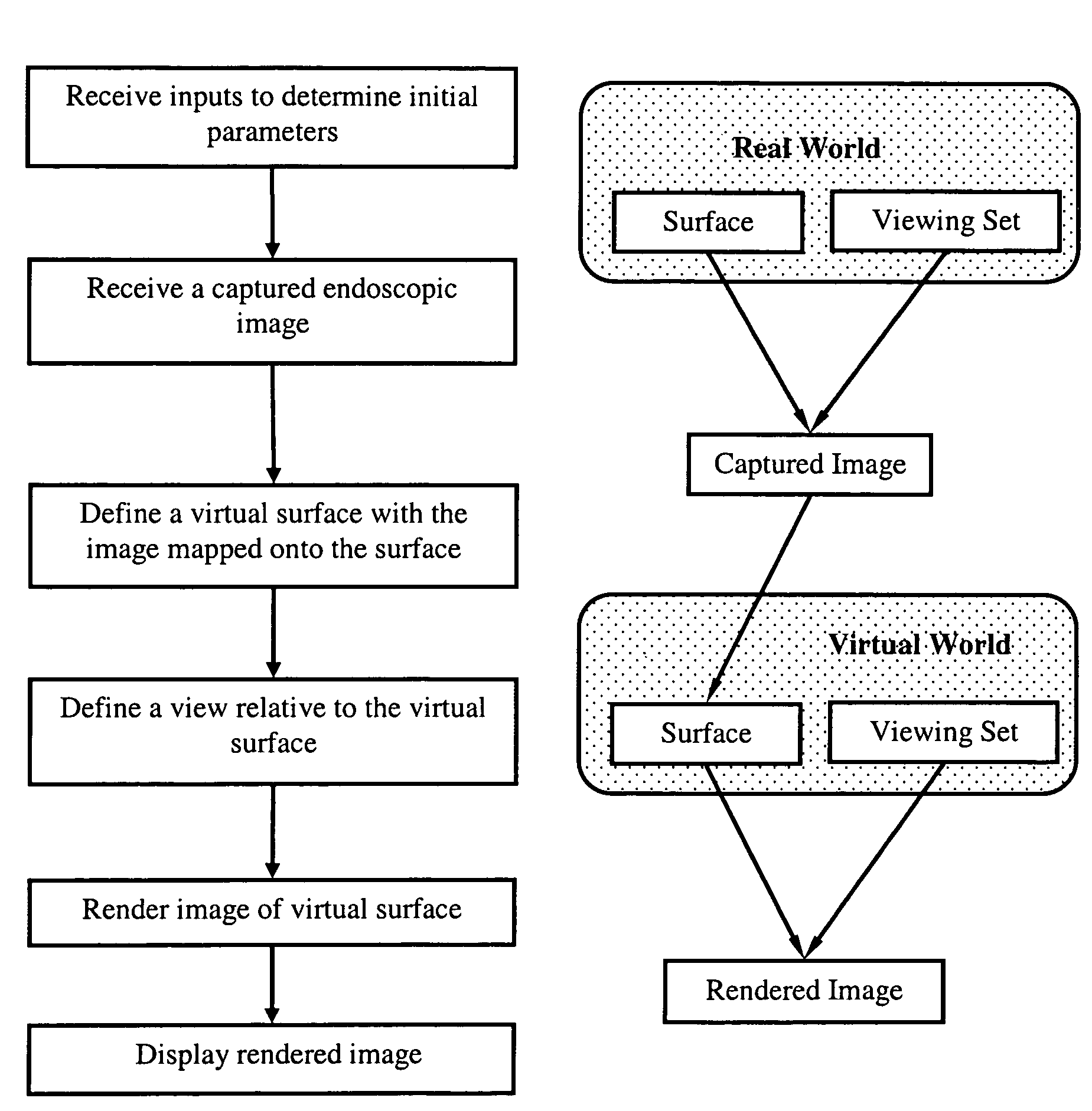
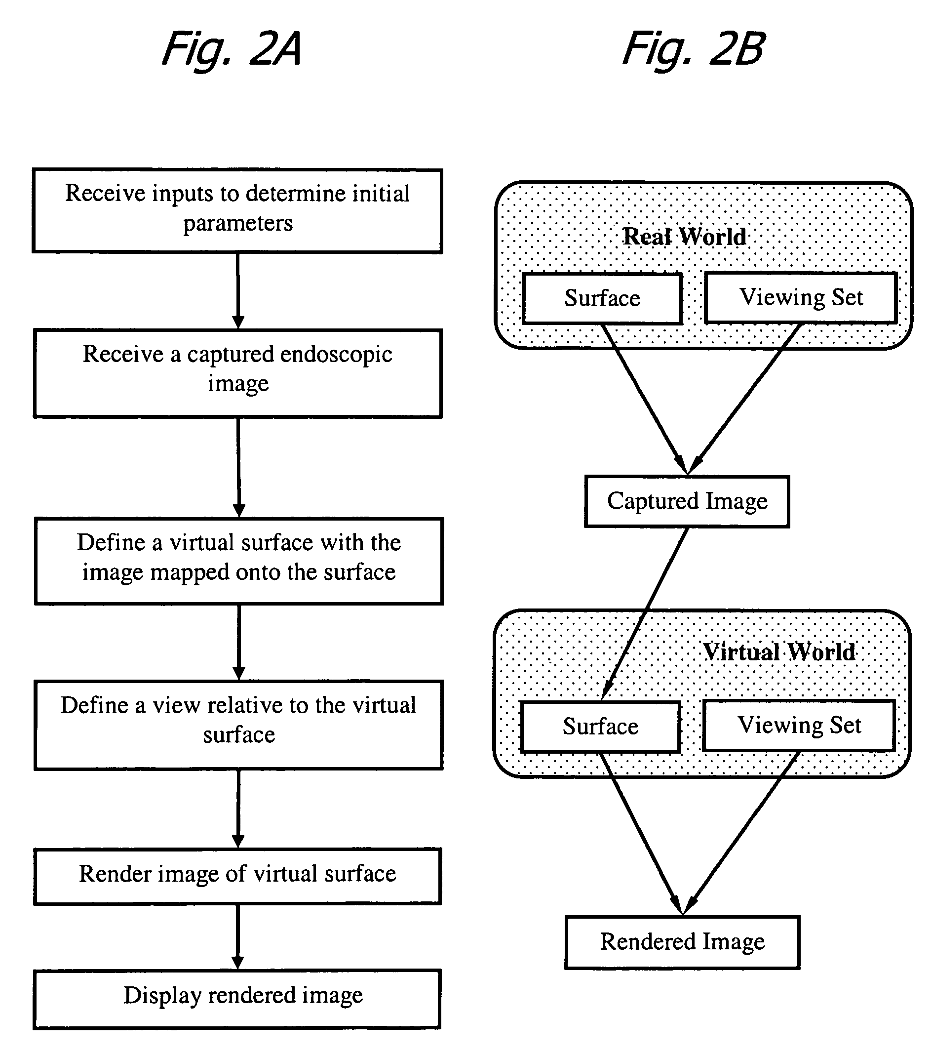
A method is disclosed for displaying endoscopic images that includes three-dimensional surfaces, variable viewing points, directions, and orientations. A computer receives a captured endoscopic image. A virtual surface is defined in the computer with the captured image textured onto the virtual surface. A virtual viewing point, virtual viewing direction, and virtual viewing orientation are defined relative to the virtual surface. A rendered image of the virtual surface is then created. The rendered image is displayed to the user. Video is displayed on the virtual surface by updating the image texture with each new frame.

Description

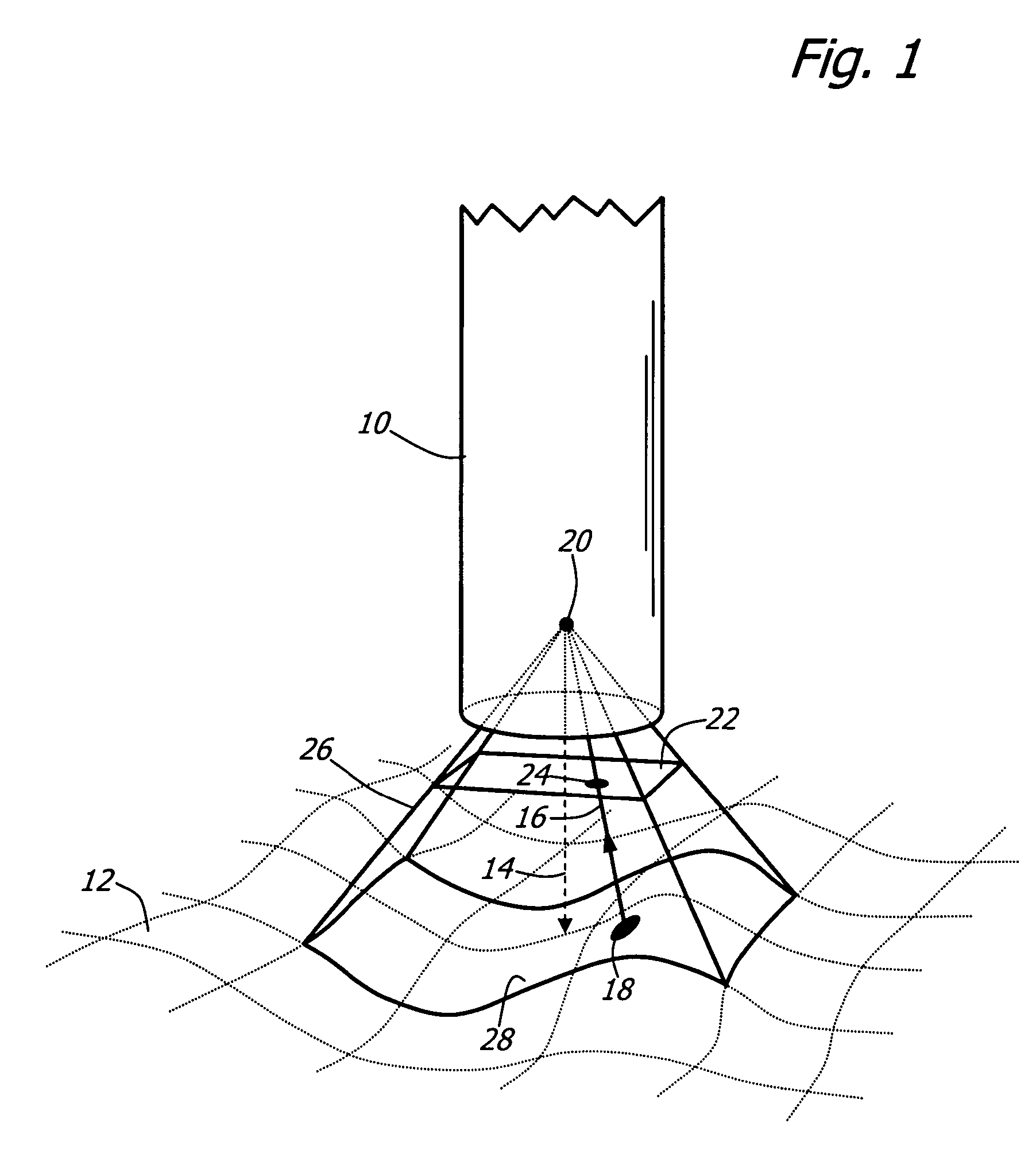
FIELD OF THE INVENTION[0001]The present invention relates to systems and methods for displaying images and specifically to the display of endoscopic images for medical and industrial applications.BACKGROUND OF THE INVENTION[0002]Endoscopes are elongated devices used to visualize the insides of cavities. Originally, endoscopes were equipped with eyepieces for direct observation. Today many endoscopes are equipped with electronic cameras, such as CCD or CMOS image sensors. These sensors are used to capture images from the area viewed with the endoscope. Endoscopic imaging is the process of capturing images from internal structures and transmitting them to an external viewer.[0003]FIG. 1 illustrates specific definitions related to image capture. An imaging device, in this case an endoscope 10, is pointed towards a surface to be viewed 12 in a viewing direction 14. A principal light ray 16 travels from a point 18 on the surface 12, to the endoscope 10, through the endoscope optics (not ...

Claims


Application Information

Patent Timeline
Patent Type & Authority Patents(United States)
IPC IPC(8): A61B1/04G06T15/20A61B1/00
CPCA61B1/00009A61B1/00045G06T3/0018
Inventor HALE, ERIC LAWRENCESCHARA, NATHAN JONHOEG, HANS DAVID
Owner KARL STORZ IMAGING INC
Learn More