Blood optode

a technology of optode and blood, which is applied in the field of medical methods and equipment, can solve the problems of blood examination in large volume, inability to carry out transmission measurement, and association of backscattered light, and achieves the effect of simple detector setup and evaluation

Active Publication Date: 2007-07-31
NIRLUS ENG
View PDF4 Cites 131 Cited by
  • Summary
  • Abstract
  • Description
  • Claims
  • Application Information

AI Technical Summary

Problems solved by technology

Blood examination in large, so called “central”, i.e. heart-near vessels, is impossible with the hereto standard methods for determining oxygenation due to the so called “centralization”, (i.e. inadequate blood circulation of the periphery) of emergency patients.
The difficulty of a measurement on large blood vessels is that their central position in the body renders a transmission measurement impossible, whereas, remission mainly takes place by diffuse backscattering of photons.
The essential problem is the association of backscattered light with a specific location of the scattering.
The extensive data analysis on the detector side is equally complicated.
It is common to all the aforementioned methods that the contribution to the measuring signal of blood from the interior of a large blood vessel cannot be considered in isolation or, in the case of WO 02 / 08740 A2, can only be established with considerable effort and expenditure.

Method used


Image

  • Blood optode
  • Blood optode
  • Blood optode

Examples


Embodiment Construction

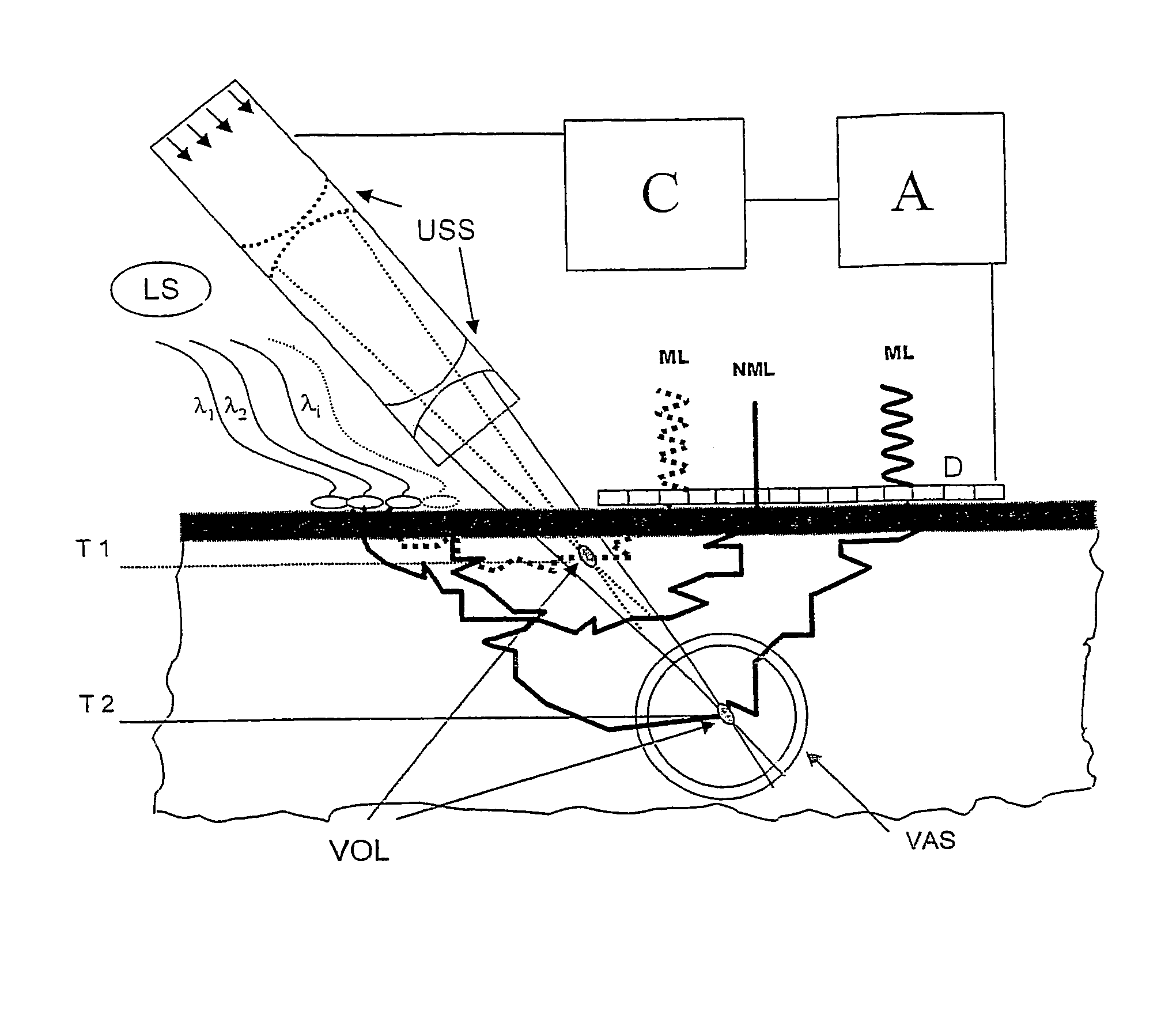
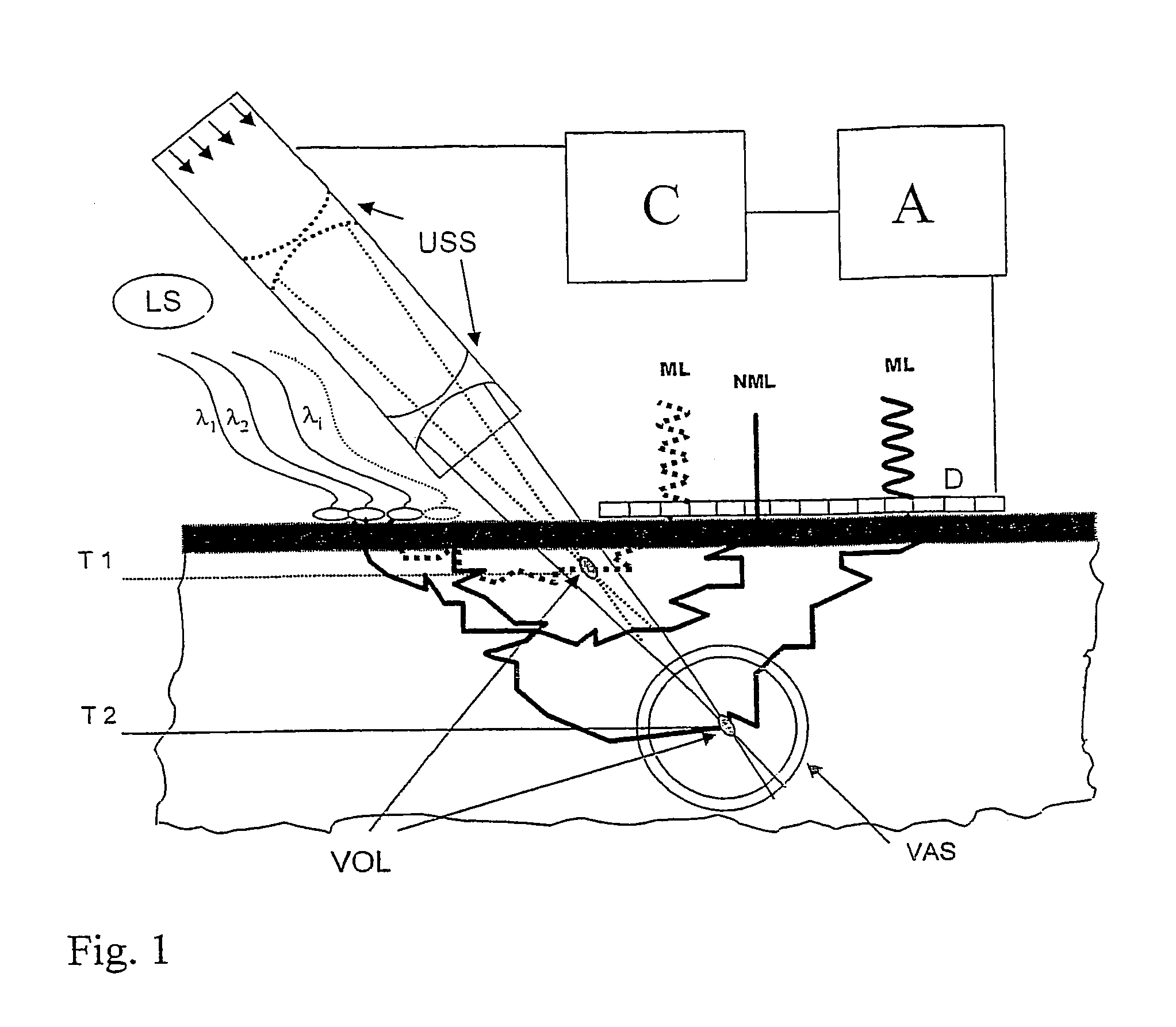
[0019]In FIG. 1, a light source LS emits light, preferably monochromatic light, with one or more discreet wavelengths, e.g. laser light, into the tissue. The light wavelengths λ1 to λi are chosen in such a way that preferably scattering takes place on selected blood constituents, particularly oxygen-rich and oxygen-poor hemoglobin. A significant component of the irradiated light passes out again at a plurality of exit points following numerous scattering processes. A matrix detector D, which comprises flat, juxtaposed, photosensitive pixels which generates an electric signal proportional to the light intensity, is placed on the skin surface in such a way that the detector covers the exit points adjacent to the irradiation point. The detector dimensions must correspond with the sought light penetration depth (see above), i.e. with the depth of the blood vessel examined. An evaluating unit A connected to the detector summates the detector signals and measures the backscattered light i...


PUM

PropertyMeasurementUnit
wavelengthaaaaaaaaaa
thicknessesaaaaaaaaaa
pressureaaaaaaaaaa

Abstract

A method for measuring in non-invasive manner the concentration of blood constituents by which backscattered light is measured under the action of ultrasonic radiation focused towards the inside of a central blood vessel. A light source and detection unit are arranged to detect the backscattered light on the skin surface above the blood vessel. The target tissue is illuminated by two discrete optical wavelengths. An average light intensity distribution is detected over the length of a pulse. The distribution is Fournier transformed, and the largest Fournier components and spectral position are determined in relation to the frequency of the ultrasonic radiation. The component concentration in the blood vessel is calculated taking into account the volume of the ultrasonic focus contributing to the signal and blood flow rate.

Description

PRIOR APPLICATIONS[0001]This U.S. §371 National Phase patent application bases priority on International Application No. PCT / DE2004 / 000470, filed on Mar. 10, 2004, which in turn bases priority on German Application No. DE 103 11 408.4, filed on Mar. 13, 2003.BACKGROUND OF THE INVENTION[0002]1. Field of the Invention[0003]The invention relates to a medical method and apparatus for measuring in non-invasive manner the concentration of blood constituents, particularly the hemoglobin concentration or oxygenation of the blood in large blood vessels.[0004]2. Description of the Prior Art[0005]Living tissue is largely transparent to electromagnetic radiation in the red and infrared range (wavelength 550 nm<λ<1000 nm). This so called “biological window” is limited towards longer wavelengths by strong absorption bands of water and towards shorter wavelengths by those of hemoglobin. It is in principle possible in this range to “see into” the tissue in depths ranging from a few mm to a fe...

Claims


Application Information

Patent Timeline
Patent Type & Authority Patents(United States)
IPC IPC(8): A61B5/00G01N21/49
CPCA61B5/0059A61B5/0095G01N21/49A61B5/7257
Inventor HERRMANN, VERA
Owner NIRLUS ENG
Why Eureka
Try Eureka