
Marker device and method of deploying a cavity marker using a surgical biopsy device

a biopsy device and cavity marker technology, applied in the field of applications for delivering and deploying markers, can solve the problems of inability to obtain an adequate sample of suspect tissue, failure to deploy the marker from the biopsy probe,

Inactive Publication Date: 2008-12-16
DEVICOR MEDICAL PROD
View PDF62 Cites 135 Cited by
  • Summary
  • Abstract
  • Description
  • Claims
  • Application Information

AI Technical Summary

Problems solved by technology

Moreover, large enough samples are obtainable, avoiding dry taps, or the inability to obtain an adequately sized sample of the suspect tissue, a common problem associated with other breast biopsy systems.
Actuating the marker placement device in some instances may fail to deploy the marker from the biopsy probe, however.

Method used


Image

  • Marker device and method of deploying a cavity marker using a surgical biopsy device
  • Marker device and method of deploying a cavity marker using a surgical biopsy device
  • Marker device and method of deploying a cavity marker using a surgical biopsy device

Examples


Embodiment Construction

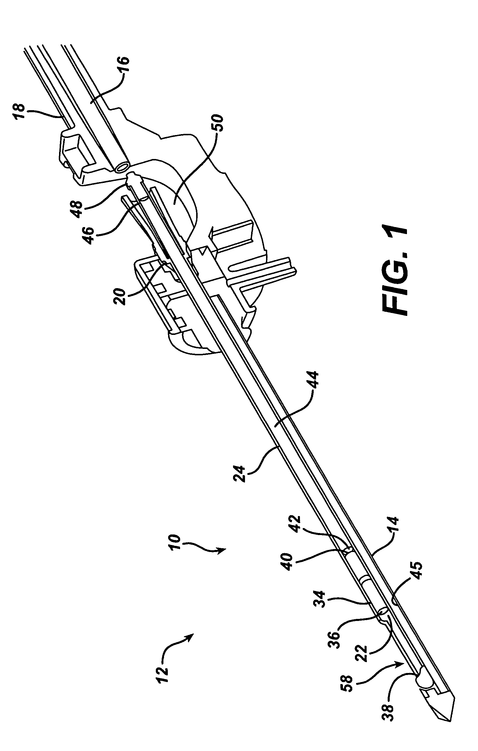
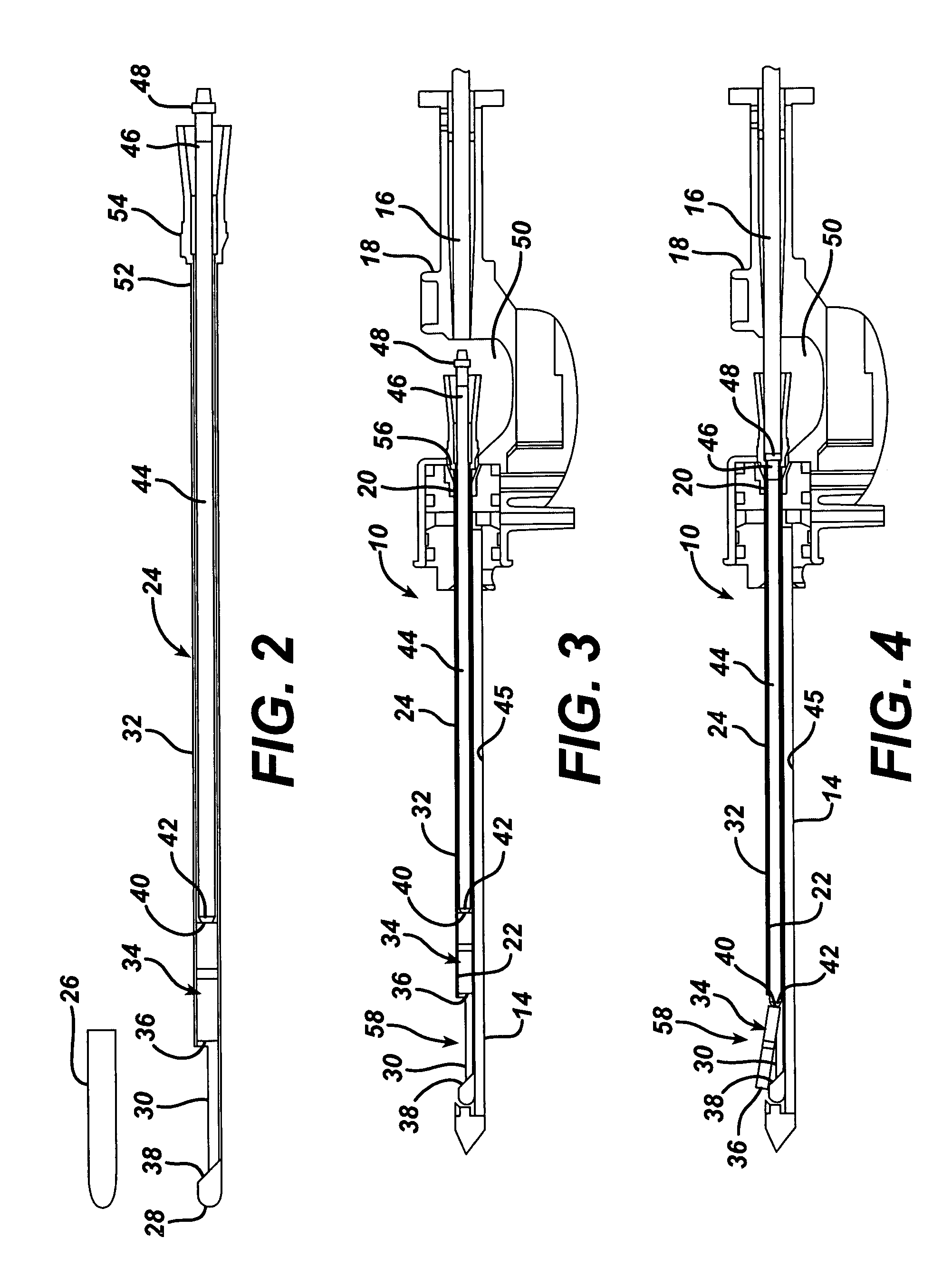
[0028]Turning to the Drawings, wherein like numerals refer to like components throughout the several views, in FIG. 1, a breast biopsy handle 10 of a biopsy system 12, a minimally invasive device, is used under local anesthetic and ultrasound guidance to collect multiple biopsy samples with a single insertion of a probe 14 into the breast of a patient. After which, a cutter 16 is retracted proximally in a housing 18 of the biopsy handle 10 to expose a distally opening entry cone 20 of a cutter lumen 22 of the probe 12. The surgeon may then insert and seat a biopsy marker introduction assembly 24, which is shown separately in FIG. 2.

[0029]Also shown in FIG. 2 is a Mylar sealing cap 26 that has been removed just prior to use from a distal end 28 of the introduction assembly 24 to expose a laterally disposed deployment opening 30 in an introducer tube 32. A marker 34 is positioned inside of the introducer tube 32 proximal to the deployment opening 30 that has sufficient length to allow...


PUM


Abstract

A biopsy marker introduction assembly is configured for introduction through a specimen retrieval recess into a probe cutter lumen of a handheld breast biopsy handle. Advancing the cutter causes an extending deployment rod to distally translate within an introducer tube of the introduction assembly, pushing a biopsy marker out of a distal end of the probe at a surgical biopsy site. Thereby, the surgeon is able to position the probe and effect deployment of the marker even if using the other hand to position an imaging device such as an ultrasonic transceiver.

Description

FIELD OF THE INVENTION[0001]The present invention relates, in general, to an applier for delivering and deploying a marker for implantation in tissue of a surgical patient, and more particularly, to such an applier for delivering and deploying an implantable biopsy marker for defining specific locations in human tissue during a biopsy procedure, especially in a human breast.BACKGROUND OF THE INVENTION[0002]One in nine American women will develop breast cancer in their lifetime. It is the leading cause of cancer deaths in women 40-55 years of age and the second leading cause of cancer deaths in women overall. Breast cancer will be diagnosed in approximately one in eight women in their lifetime, and one in 30 will die of this disease. Breast cancer does occur in males, but is much less common. Biopsy requests stem from a screening process generally performed via a physical examination (palpable) and / or mammogram (non-palpable). A biopsy is indicated if suspicious tissue is detected. F...

Claims


Application Information

Patent Timeline
Patent Type & Authority Patents(United States)
IPC IPC(8): A61B10/00A61B10/02A61B19/00
CPCA61B19/54A61B10/0275A61B2019/5408A61B2019/5487A61B90/39A61B2090/3908A61B2090/3987
Inventor BECKMAN, ANDREW T.APPLEGATE, RICK D.BOEHM, MICHAEL E.GARRISON, WILLIAM A.LADD, DOUGLAS N.
Owner DEVICOR MEDICAL PROD
Learn More