Laparoscopic and endoscopic trainer including a digital camera

a training equipment and digital camera technology, applied in the field of surgical training equipment, can solve the problems of inconvenient use of training environment, inability to translate hand eye coordination skills useful in conventional surgery into endoscopic surgery, and small video camera that can be disposed at an internal surgical field, etc., to achieve the effect of enhancing endoscopic skills training

Active Publication Date: 2009-09-29
TOLY CHRISTOPHER C
View PDF56 Cites 111 Cited by
  • Summary
  • Abstract
  • Description
  • Claims
  • Application Information

AI Technical Summary

Benefits of technology

[0011]The present invention is a surgical trainer for practicing videoendoscopic surgical techniques. The surgical trainer includes a relatively low-cost, portable digital camera disposed within the trainer, configured to provide a substantially real-time image of a practice volume defined by the trainer housing. Various embodiments are contemplated, including embodiments in which the position of the digital camera is fixed, and embodiments in which the digital camera is selectively positionable to obtain an image of different portions of the practice volume.
[0014]In one embodiment, the digital camera is movably positionable within the practice volume so that when a position of the anatomical structure is changed, the position of the digital camera can be changed to continue to provide a video feed imaging the anatomical structure. Movement of the digital camera enables a field of view obtained by the digital camera to be varied. Such movement enables the digital camera to obtain an image of at least a portion of the practice volume from a plurality of different angles. Preferably, movement of the digital camera will enable the proximity of the digital camera to be varied relative to at least a portion of the practice volume. Movable and positionable digital cameras are particularly beneficial for training involving the simulation of an actual endoscopic procedure, such as suturing, because endoscopic procedures often require the field of view provided by the endoscope, which is generally quite limited, to be shifted as the procedure progresses. As will be discussed in more detail below, it is particularly advantageous if the digital camera can be moved in a manner that enables the digital camera to move closer to (or farther from) a desired portion of the practice volume (i.e. to zoom in on or away from a particular portion of the practice volume). The ability simulates a technique often employed in videoendoscopy; moving a laparoscope or endoscope closer to (or away from) a particular portion of a surgical field, to zoom in (or out) relative to a specific portion of the surgical field. In another embodiment, the position of the digital camera is fixed, and the digital camera is configured to image a predefined portion of the practice volume. Trainers having digital cameras in fixed positions are particularly useful for skill based training exercises, such as basic instrument manipulation and two-dimensional recognition skills. If the field of view provided by a fixed digital camera is sufficiently broad, or if the simulated anatomical structure or other work piece can readily be repositioned relative to the digital camera, such an embodiment can also be used for simulating an actual endoscopic procedure.
[0015]In yet another embodiment, the digital camera is disposed in an upper portion of the practice volume, and light traveling along a light path from the anatomical structure to image acquisition elements in the digital camera moves in a generally upward direction. This configuration facilitates obtaining a “bird's eye view” of the anatomical structure, which most often will be disposed in a lower portion of the practice volume. Another embodiment includes at least one optical reflector used to direct light traveling along a light path from the anatomical structure to image acquisition elements in the digital camera, toward the digital camera. The use of one optical reflector provides a reverse image, while the use of two optical reflectors provides a normal (i.e., un-reversed) image. Of course, digital signal processing can be used to electronically reverse an image obtained using the digital camera. The use of properly positioned optical reflectors enables the digital camera to be disposed in a lower portion of the practice volume, and yet still obtain a bird's eye view of an anatomical structure that is also disposed in the lower portion of the practice volume.
[0017]Preferably, the support structure includes an elongate member having a proximal end disposed outside the practice volume, and a distal end disposed inside the practice volume. The digital camera is coupled with the distal end of the elongate member, such that a manipulation of the proximal end of the support structure changes a position of the digital camera within the practice volume. In at least one embodiment the proximal end of the elongate member includes a handle configured to simulate the handle of a conventional laparoscope. Embodiments that include an elongate member structure preferably also include a mounting bracket configured to support the elongate member. In some embodiments, the mounting bracket is configured to slidingly engage the elongate member, such that an amount of the elongate member disposed within the practice volume can be increased and decreased as desired. Such movement enables the digital camera to move closer to or away from portions of the practice volume, enabling the digital camera to zoom in or out. While some digital cameras include digital and / or optical zooms, requiring or enabling a user to manipulate the elongate member to achieve a desired zoom simulates the manipulation required of a conventional laparoscope or endoscope to achieve a similar zoom, increasing a realism of the training experience. In some embodiments the mounting bracket is configured to pivotally engage the elongate member, such that a position of the distal end of the elongate member within the practice volume can be adjusted. In some embodiments the mounting bracket is configured to pivotally engage the housing, such that a position of the distal end of the elongate member within the practice volume can be adjusted. Particularly preferred mounting brackets enable different ranges of motion to be achieved.
[0020]Generally, a size of the digital camera is significantly larger than a size of a distal end of a conventional laparoscope, such that the digital camera could not pass through an incision configured to receive a conventional laparoscope. This enables relatively inexpensive digital cameras, such as web cams, to be employed.
[0024]Yet another aspect of the present invention is a method for using a digital camera to enhance endoscopic skills training, by providing a substantially real-time image of the skill training exercise. This method includes the step of providing a surgical trainer defining a practice volume in which a digital camera is disposed. The digital camera is then used to obtain a video image of the practice volume that is displayed. The trainee remotely manipulates one or more objects within the practice volume using elongate tools, while viewing the manipulation on the display. The objects can be placed within the practice volume before or after the digital camera is energized. Once again, in addition to the immediate display of the digital images of the manipulations, such digital images can be stored for later review or transmitted in real-time or at a later time) over a network for viewing by others, and printouts of selected images can be made.

Problems solved by technology

While such systems work well in a surgical theater, their size, weight, limited mobility, and cost make such systems ill-suited for use in a training environment, where highly portable and lower cost devices are very desirable.
However, at the present time, small video cameras that can be disposed at an internal surgical field have not supplanted more conventional videoendoscopic systems that employ fiber optics to transmit internal images to an external camera, either for actual surgical use or in a training context, such as simulations or skill development exercises.
Hand eye coordination skills useful in conventional surgery do not translate well into endoscopic surgery.
In videoendoscopic surgery, the surgeon is not able to feel the tissue and / or organs associated with an operative site first hand, because the surgeon remotely manipulates the tissue and / or organs using elongate surgical tools from outside the surgical field.
The ability to work from a two-dimensional image of the surgical field, while remotely manipulating instruments, requires a significant amount of training.
Even basic surgical skills, such as suturing and knot tying, become challenging when performed endoscopically.
In a videoendoscopic environment, such basic surgical tasks require great skill and precision, which can only be achieved through training and practice.
However, such training methods are very expensive, since animals and cadavers are in limited supply and cannot be used repeatedly.
Also, animal specimens and human cadavers are not readily portable.
While such a trainer is effective for gaining skills related to remote manipulation of endoscopic instruments, since the trainee looks directly at the simulated anatomical structure, two-dimensional recognition skills cannot be learned and practiced.
It must be recognized, however, that box trainers, including those that employ mirrors to develop two-dimensional recognition skills, do not provide a very realistic simulation of a true videoendoscopic procedure.
While a conventional endoscopic camera could be introduced into a box trainer to provide a video image of the simulated anatomical structure at a practice site, conventional endoscopic cameras are not very portable, and are very expensive.
Such a training system, while being more realistic in simulating an actual surgical environment than a box trainer with a mirror, no longer offers the low cost and portability of a box trainer alone.

Method used


Image

  • Laparoscopic and endoscopic trainer including a digital camera
  • Laparoscopic and endoscopic trainer including a digital camera
  • Laparoscopic and endoscopic trainer including a digital camera

Examples


Embodiment Construction

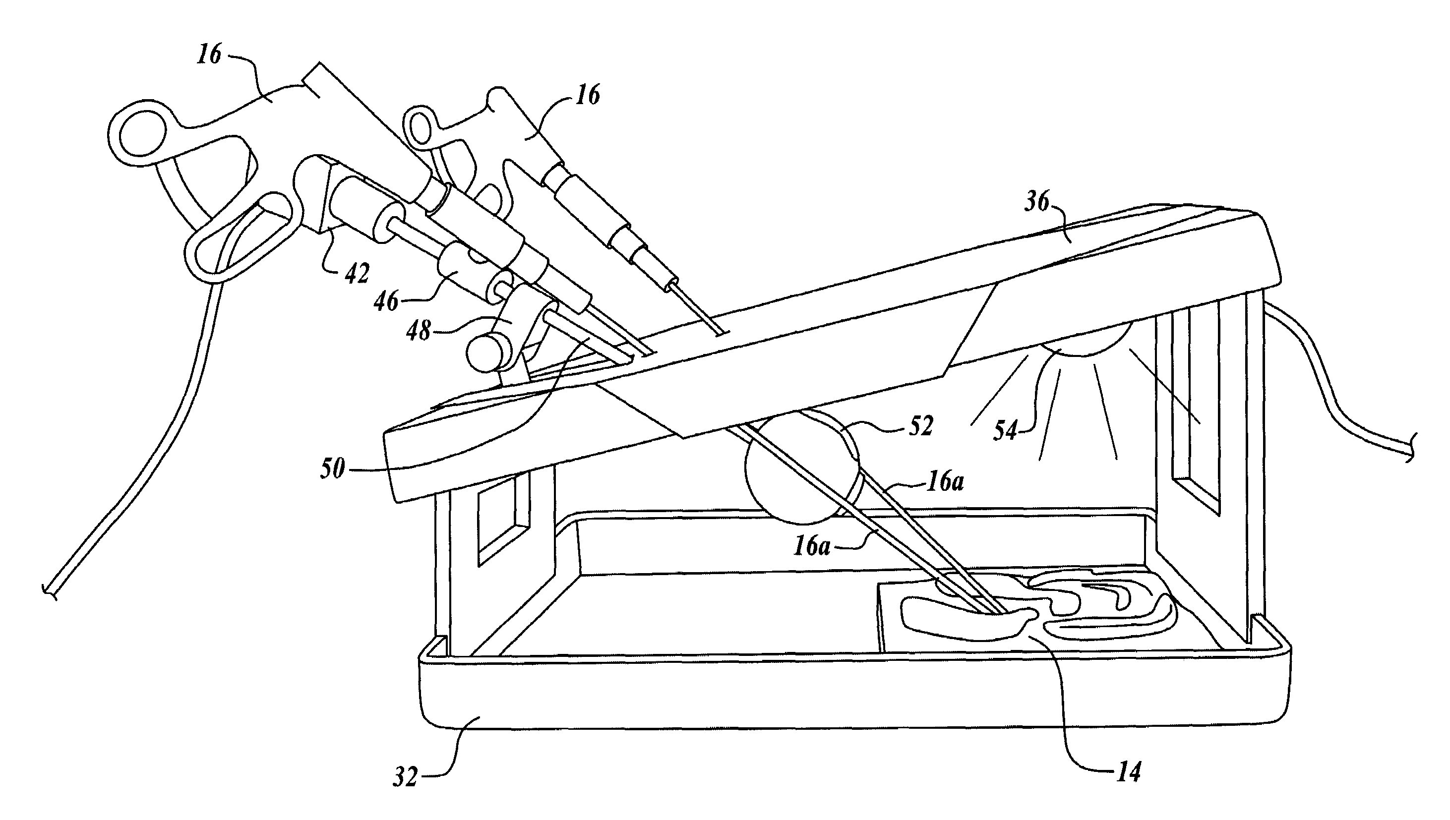
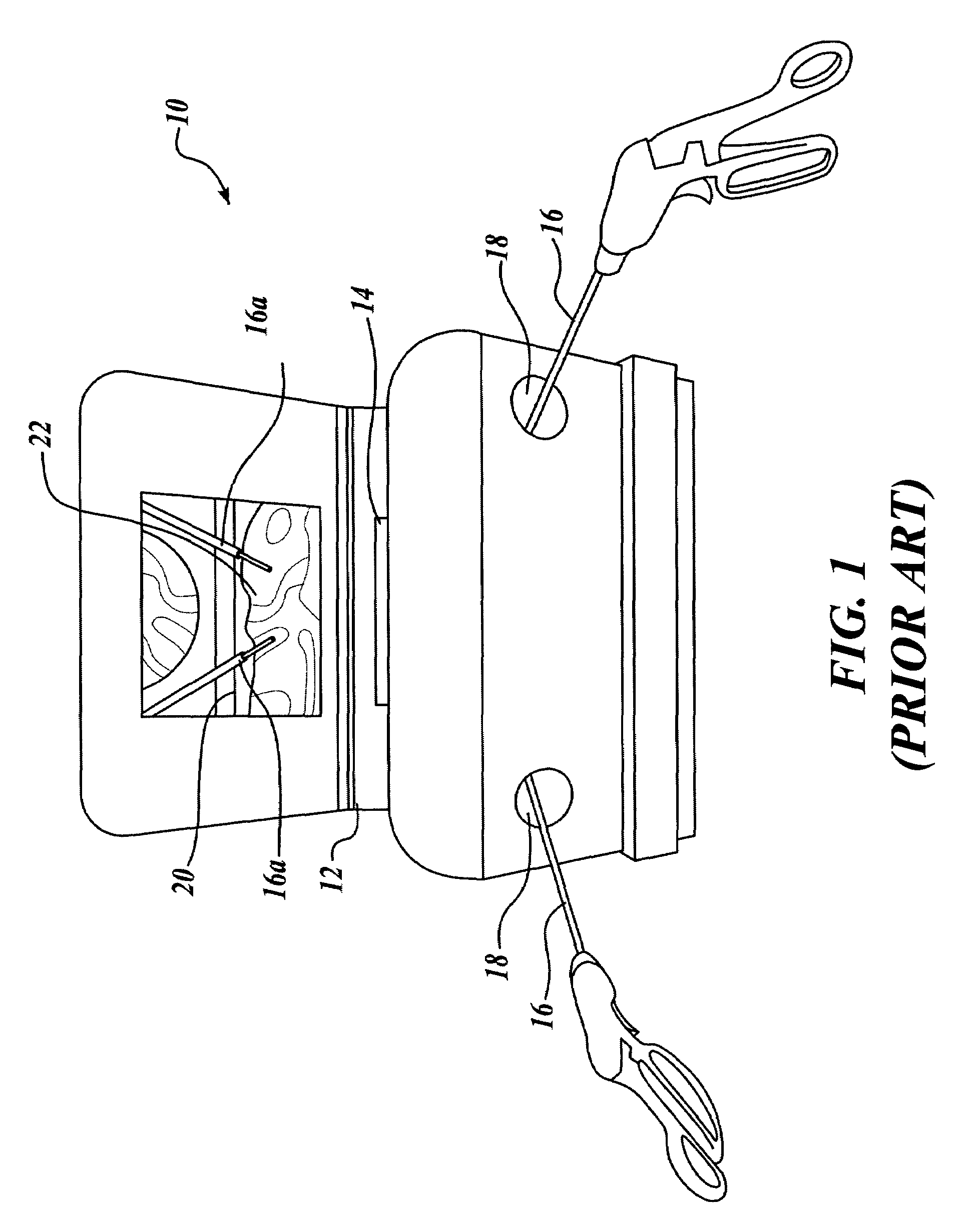
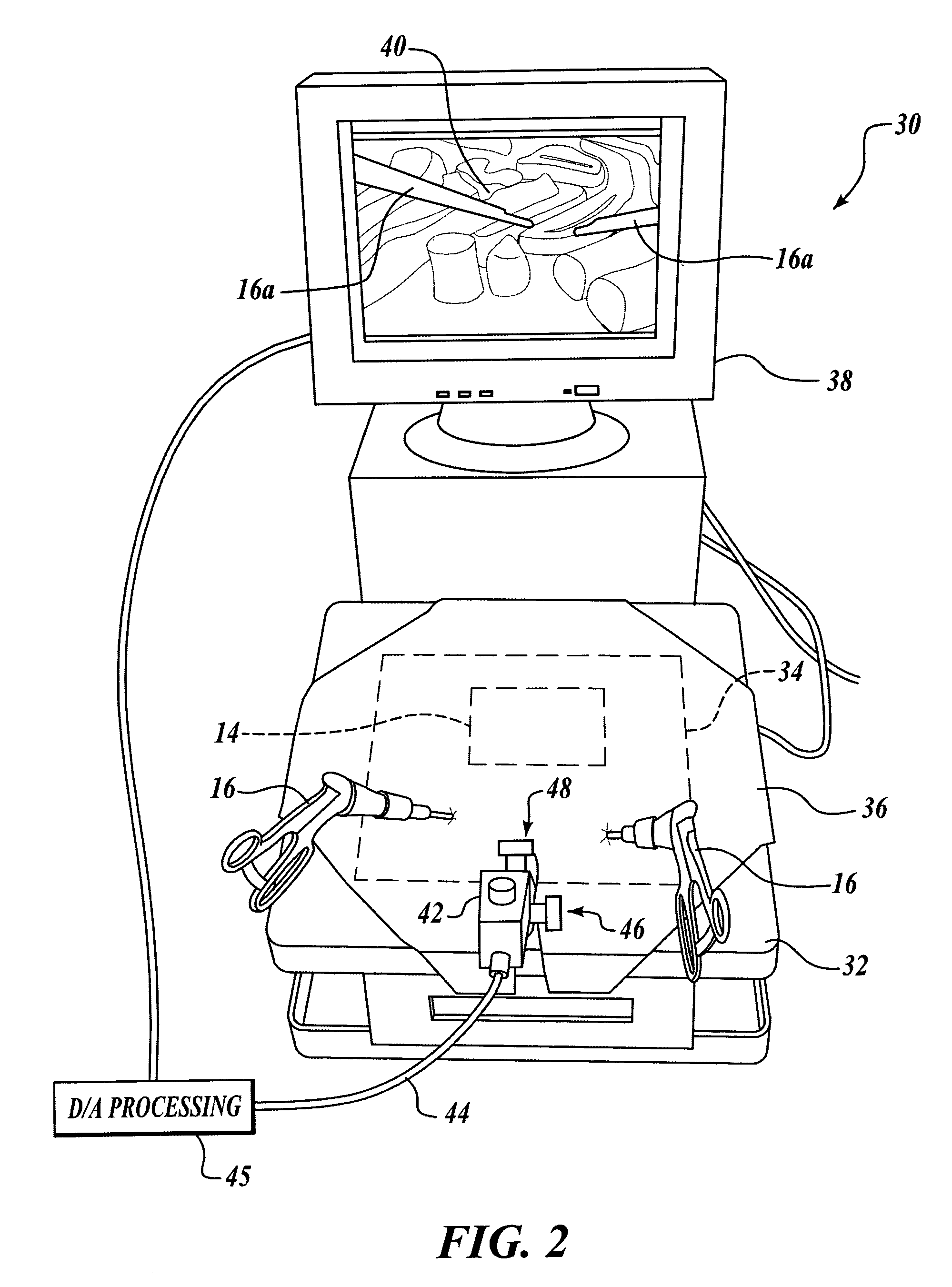
[0038]FIG. 1 schematically illustrates a prior art surgical trainer 10 that is configured for video endoscopic surgery training. Trainer 10 includes a housing 12. An anatomical structure 14 is disposed within housing 12, such that portions of housing 12 prevent a trainee from clearly viewing anatomical structure 14. Housing 12 includes a plurality of openings 18 into which surgical instrument 16 can be inserted. Preferably, surgical instruments 16 are endoscopic suturing instruments such as an ENDO STITCH endoscopic reciprocating suturing instrument manufactured by U.S. Surgical, Inc. Trainer 10 includes a reflector 20 in which an image 22 of anatomical structure 14 can be observed by a trainee. Note that distal ends 16a of surgical instruments 16 can be seen within image 22. Surgical trainer 10 thus provides a trainee with an opportunity to practice endoscopical surgical techniques such as suturing and knot tying, as well as gaining experience in two-dimensional pattern recognition...


PUM


Abstract

A videoendoscopic surgery training system includes a housing defining a practice volume in which a simulated anatomical structure is disposed. Openings in the housing enable surgical instruments inserted into the practice volume to access the anatomical structure. A digital video camera is disposed within the housing to image the anatomical structure on a display. The position of the digital video camera can be fixed within the housing, or the digital video camera can be positionable within the housing to capture images of different portions of the practice volume. In one embodiment the digital video camera is coupled to a boom, a proximal end of which extends outside the housing to enable positioning the digital video camera. The housing preferably includes a light source configured to illuminate the anatomical structure. One or more reflectors can be used to direct an image of the anatomical structure to the digital video camera.

Description

FIELD OF THE INVENTION [0001]The present invention generally relates to a surgical training apparatus, and more particularly to a surgical trainer that realistically simulates videoendoscopic surgery.BACKGROUND OF THE INVENTION [0002]In recent years many fully invasive surgical and operative medical procedures have been adapted to utilize videoendoscopic techniques to achieve minimally invasive procedures. Rather than requiring a relatively large incision to gain access to internal anatomical structures, videoendoscopic techniques require a plurality of much smaller incisions. Generally, one incision is made for a videoendoscopic camera, and two or more incisions are made to introduce surgical instruments. The diameters of the surgical instruments and the probe for the videoendoscopic camera are made as small as practical, to minimize the size of the incisions that are required. The endoscope is used to enable the surgeon to view, in real-time, the surgical field and the manipulatio...

Claims


Application Information

Patent Timeline
Patent Type & Authority Patents(United States)
IPC IPC(8): G09B23/28A61B1/00
CPCG09B23/285
Inventor TOLY, CHRISTOPHER C.
Owner TOLY CHRISTOPHER C
Why Eureka
Try Eureka