

Devices and methods for detecting amniotic fluid in vaginal secretions
a technology of amniotic fluid and vaginal secretions, which is applied in the direction of biomass after-treatment, analysis using chemical indicators, instruments, etc., can solve the problems of increasing both maternal and perinatal morbidity and mortality by about ten percent, and no “gold standard” is available, so as to minimize false negative and false positive results, the effect of precise setting up of the threshold of sensitivity
- Summary
- Abstract
- Description
- Claims
- Application Information
AI Technical Summary
Benefits of technology
Problems solved by technology
Method used
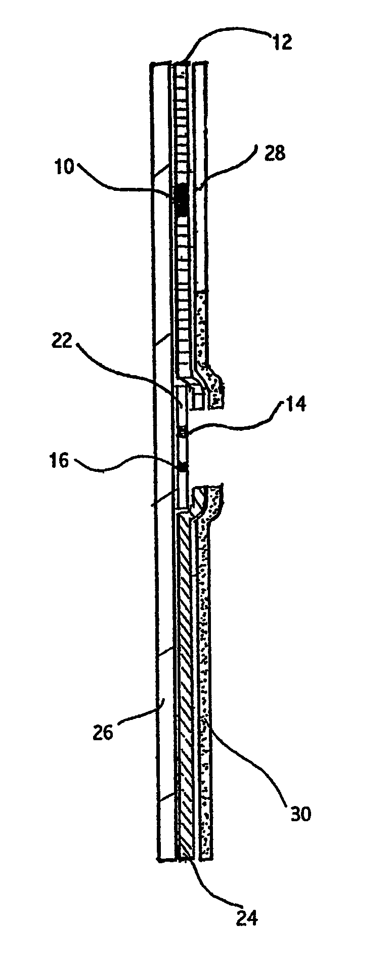
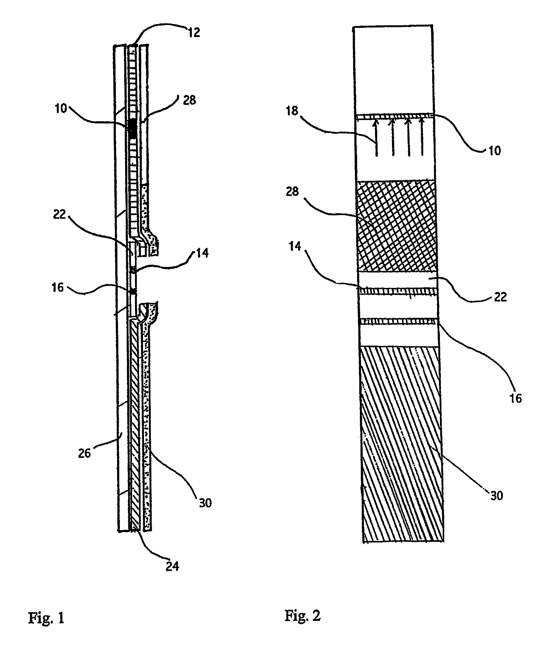
Image


Examples
example 1
Concentration of PAMG-1 in Blood and Amniotic Fluid
[0144]The PAMG-1 concentration was measured in the blood serum of non-pregnant women, pregnant women (37-40 weeks of gestation), and in amniotic fluid (39-40 weeks of gestation) by ELISA using monoclonal antibody pairs generated as described in Example 3.
[0145]Antibodies M1, M271, M152 and M392, were sorbed to polystyrene in 0.05M carbonate-bicarbonate buffer at pH 9.5 for 18 hours at 6° C. (100 μl of antibody solution in each well). Non-specific sorption to the polystyrene was removed with a 1% solution of bovine serum albumin (BSA) in phosphate buffer saline (PBS) at pH 7.0, 200 μl in each well, incubated for one hour at 37° C.
[0146]One hundred μl of PAMG-1 antigen was added to each well at concentrations of 50, 25, 12, 6, 3, 15, and 0.7 ng / ml. The samples of blood serum or amniotic fluid were diluted in a buffer of 0.01% BSA and 0.05% Twin 20 in PBS. The diluted samples were added to the wells and the wells were incubated for one...
example 2
Isolation of PAMG-1
[0152]D. Petrunin proposed the modified method of isolation of PAMG-1 in 1980 (Petrunin, D. D., Kozlyaeva, G. A., Tatarinov, Yu. S., Shevchenko, O. P., Bulletin of Experimental Biology and Medicine, No 5, p. 558, 1980 (in Russian)). The steps of the scheme are outlined in Table 4 below.
[0153]
TABLE 4Steps of isolation of PAMG-1Steps of IsolationPurity %Yield %Amniotic fluid 16-25 weeks pregnancy4100Precipitation by 0.5% lanthanum chloride2590Precipitation by ammonium sulphate at 50%3570saturationPrecipitation by lithium sulphate at 60% saturation6060Reverse Phase Chromatography Separation9030
[0154]PAMG-1 was separated from the amniotic fluid at 16 to 25 weeks of gestation. The fluid was obtained from women whose pregnancy was terminated due to medical considerations. We added 10% solution of lanthanum chloride at the volumetric ratio 20:1 (so that its final concentration was 0.5%) to the amniotic fluid and kept at 4° C. for 18 hours. Precipitate formed and was sepa...
example 3
Production of the Stable Hybrid Lines of Present Invention
[0155]Hybridoma Experiment 1. Lymphocytes from popliteal lymph nodes of 5 BALB / c mice were used. Mice were immunized by five injections of PAMG-1 in foot pads. Each injection consisted of 100 μg of PAMG-1 and Freund's Complete Adjuvant in a 1:1 ratio. After the cell fusion, the cells were seeded in 1152 wells. Total of 363 primary hybridomas were tested, 38 of them were PAMG-1-positive. Then the specificity of monoclonal antibodies was tested by studying their cross-reactive binding of proteins—alpha-2-microglobulin of fertility, human chorionic gonadotropin, trophoblastic beta-1-glycoprotein, human placental lactogen, alpha-fetoprotein, and human serum albumin. Fourteen primary hybridomas were selected whose monoclonal antibodies were not cross-reactive to other proteins. Then a method of limiting dilutions was used to clone twice the primary PAMG-1-specific hybridomas. Finally, five clones were selected that were apparently...
PUM
| Property | Measurement | Unit |
|---|---|---|
| concentration | aaaaa | aaaaa |
| concentration | aaaaa | aaaaa |
| concentrations | aaaaa | aaaaa |
Abstract
Description
Claims
Application Information