
Method and apparatus for providing mammographic image metrics to a clinician

a technology for providing mammography and metric data, applied in the field of providing mammography metric data to a clinician, can solve problems such as grave consequences for patients

Inactive Publication Date: 2010-08-24
GENERAL ELECTRIC CO
View PDF36 Cites 22 Cited by
  • Summary
  • Abstract
  • Description
  • Claims
  • Application Information

AI Technical Summary

Problems solved by technology

Accordingly, due to the highly subjective nature of the mammographic image review process, it is possible that a clinician may incorrectly diagnose a mammographic image, which can lead to grave consequences for a patient due to the misdiagnosis.

Method used


Image

  • Method and apparatus for providing mammographic image metrics to a clinician
  • Method and apparatus for providing mammographic image metrics to a clinician
  • Method and apparatus for providing mammographic image metrics to a clinician

Examples


Embodiment Construction

[0017]A detailed description of the invention is provided herein, with reference to the accompanying drawings.

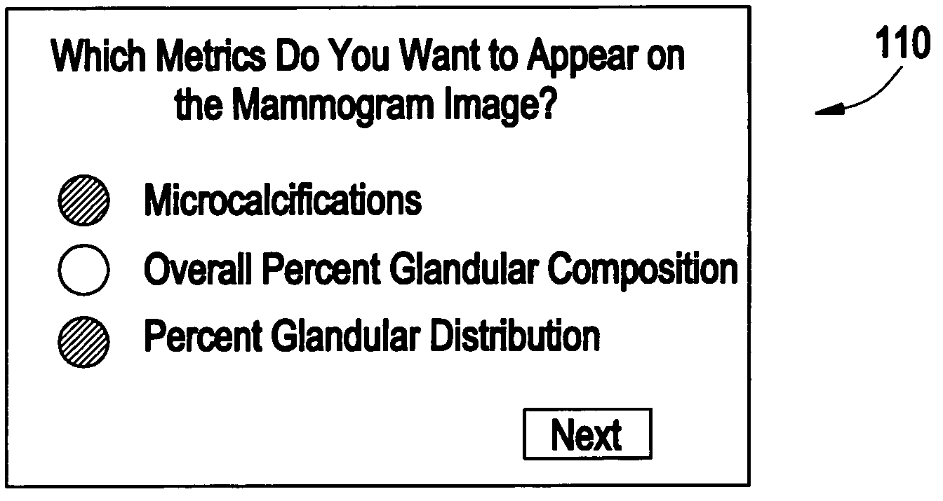
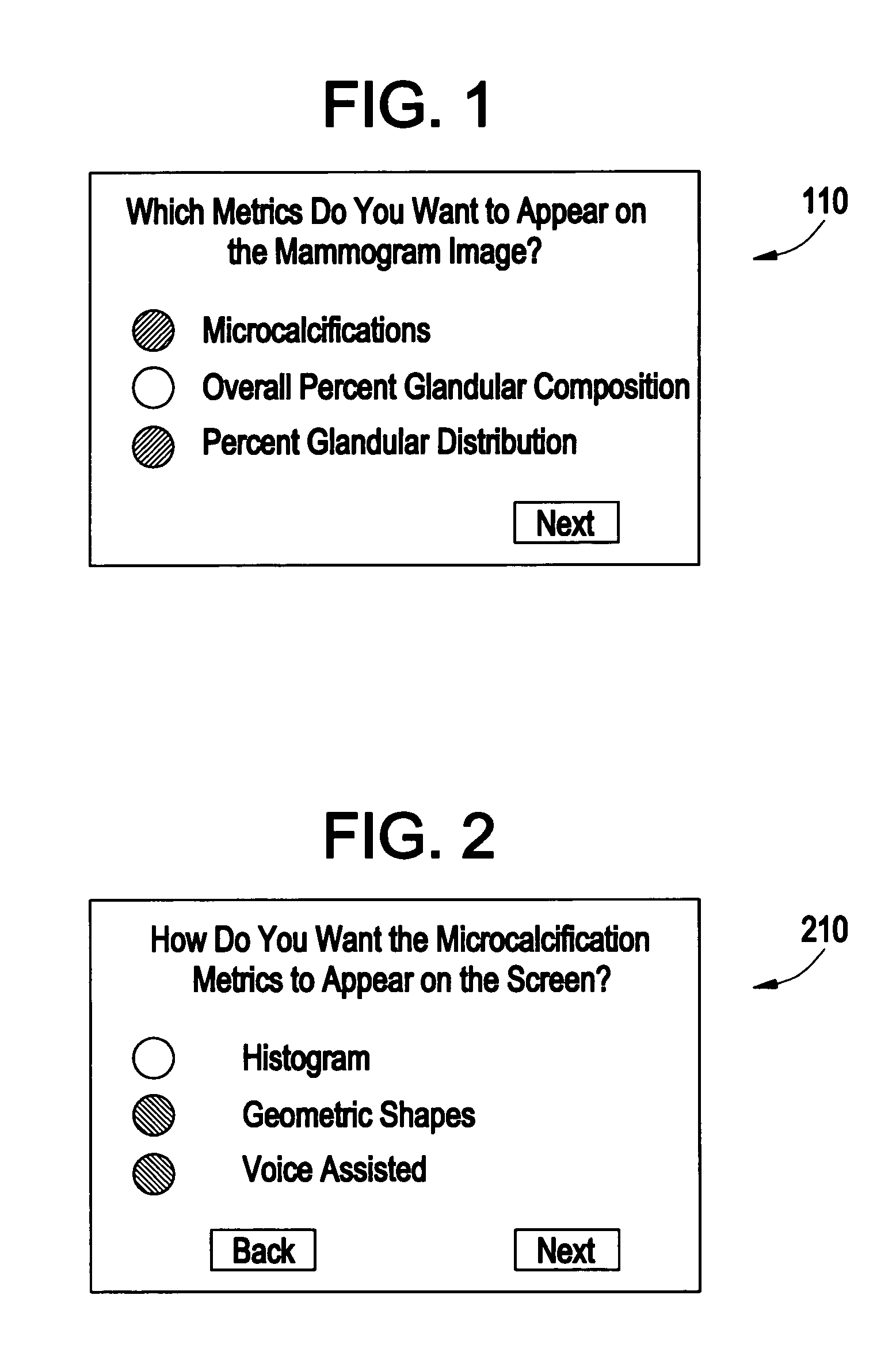
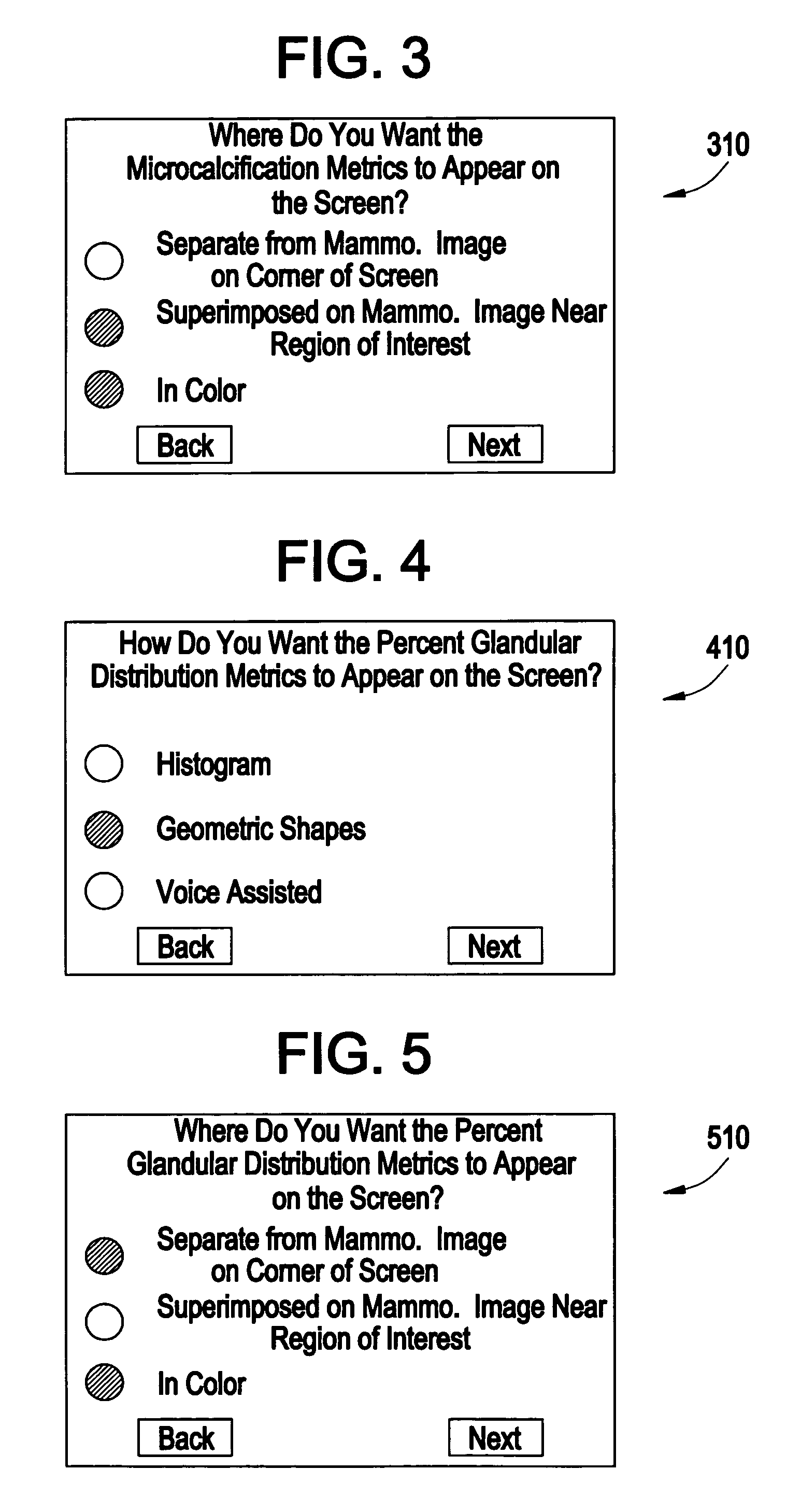
[0018]The present invention has been developed based on the premise that, given a mammographic image, it may be useful to present to a clinician certain quantitative metrics extracted from the image. For instance, it may be helpful to the clinician to have access to 1) the overall percent glandular composition or 2) the percentage glandular distribution, for instance. Further, after delineation of findings (microcalcifications, masses, or vessels, e.g.), either via computer-aided diagnosis (CAD) algorithms (automatic) or by hand-labeling (manual) or semi-automatic methods, it may be useful to the clinician to have at their disposal a summary of the quantitative measures of the findings.

[0019]For instance, a number of metrics may be useful for making decisions about the specific pathology associated with microcalcifications. Among others, metrics on the “clustering” of the mi...


PUM


Abstract

At least one metric for a mammographic image is computed by a workstation, and provided on a workstation display along with the mammographic image. A clinician can select one or more different metrics to be computed for an image, as well as where they are to be shown and the manner in which they are to be shown on the display. Speech (may also be an audible sound which is not speech) may also be used in the workstation to audibly provide metrics information to the clinician.

Description

STATEMENT REGARDING FEDERALLY SPONSORED RESEARCH & DEVELOPMENT[0001]The government may have rights in this invention pursuant to subcontract 22287 issued from the Office of Naval Research / Henry M. Jackson Foundation.BACKGROUND OF THE INVENTION[0002]The present invention relates generally to providing computer-calculated metrics on a mammographic image to a clinician on soft copy display (e.g., monitor of a work station) to be used by the clinician for screening or diagnostic evaluation purposes.[0003]Currently, clinicians, such as radiologists, perform diagnostic evaluation on mammographic images by looking for particular features within the mammographic images. That is, based on review of many different types of mammographic images, a clinician can determine whether or not a mammographic image has any potentially cancerous or suspicious regions.[0004]Accordingly, due to the highly subjective nature of the mammographic image review process, it is possible that a clinician may incorr...

Claims


Application Information

Patent Timeline
IPC IPC(8): G06K9/00A61B6/00G03B42/02G06F17/00G06Q50/00G06T1/00G06T7/00
CPCG06F19/321G06F19/3406G06F19/345G06T7/0012G16H30/20G16H40/63G16H50/20G16Z99/00
Inventor KAUFHOLD, JOHN P.CLAUS, BERNHARD E. H.EBERHARD, JEFFREY W.
Owner GENERAL ELECTRIC CO
Learn More