
Method for the generation of antigen-specific lymphocytes
a technology of antigen-specific lymphocytes and lymphocytes, applied in the field of gene delivery and immunology, can solve the problems of limited genetic background, unsuitable for therapeutic applications, and significant time, and achieve the effects of enhancing t cell function, enhancing b cell function, and enhancing immune cell function
- Summary
- Abstract
- Description
- Claims
- Application Information
AI Technical Summary
Benefits of technology
Problems solved by technology
Method used
Image

Examples
example 1
In Vitro Demonstration of Functional Expression of Antigen-Specific Tcrs Using Retroviral Vector
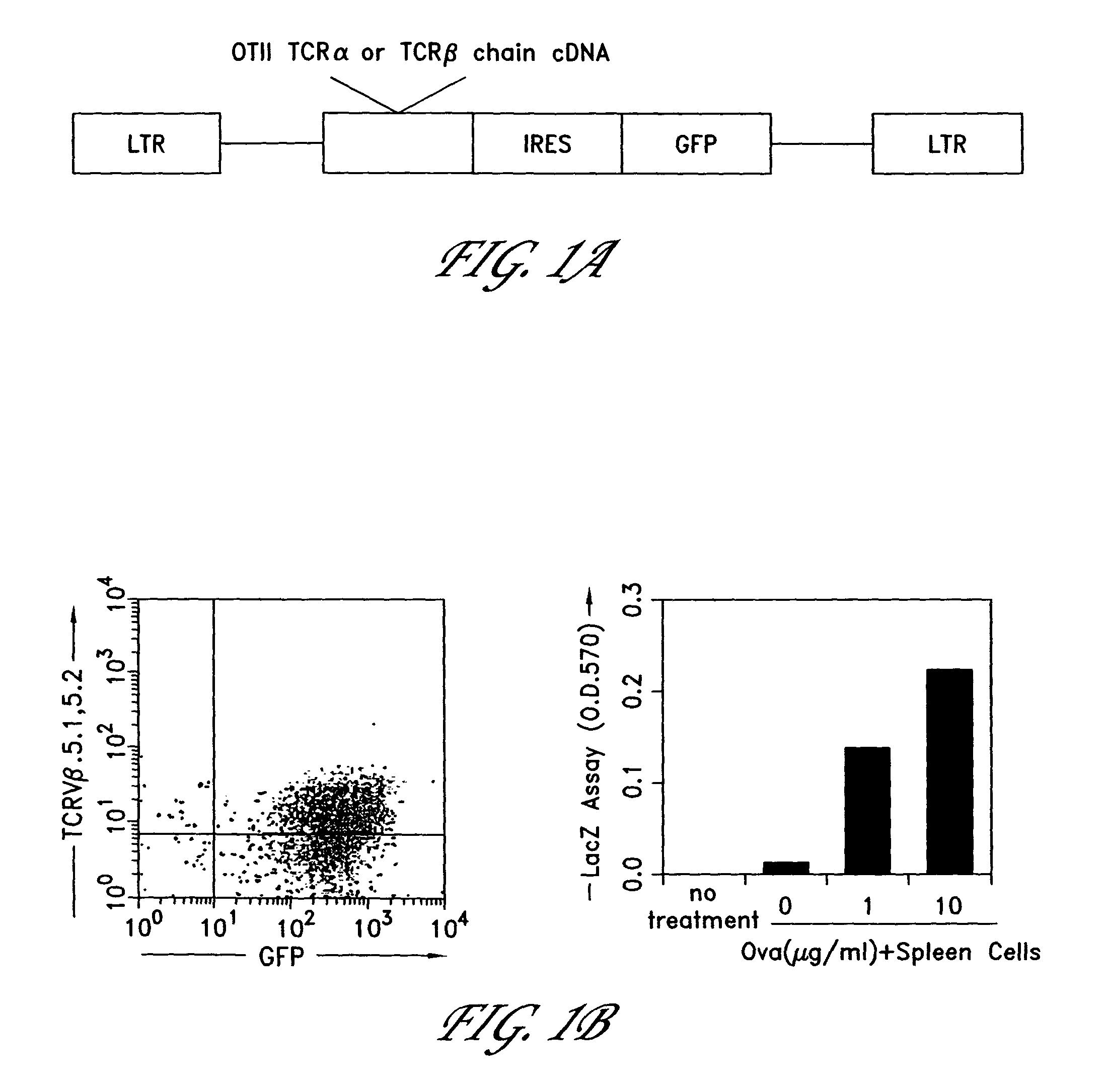
[0226]This example demonstrates the successful expression of a functional TCR in a hybridoma cell line. The bicistronic MIG retroviral expression vector was created by placing GFP downstream of the pCITE1 IRES (Novagen) and cloning it into MSCV 2.2 vector (Van Parijs et al. 1999, Immunity, Vol. 11, 281-288). This retroviral vector (shown in FIG. 1A) expresses both GFP, to mark infected cells, and a heterologous gene of interest. OTII T Cell Receptor (TCR) α or β chain cDNAs were cloned into this vector. The OTII TCR is a well-defined TCR derived from a CD4+ class II-restricted T cell clone that responds to a known antigen, residues 323-339 of chicken ovalbumin (OVAp). The OTII TCR was used as a model system in our experiments. A MIG-OTI-2A vector, having the same sequence as the MIG-OTII-2A vector except for the substitution of the OTI TCR seqeuence, may be used in other embodiments. The ...
example 2
Generation of Functional Antigen-Specific T Cells in Mice of Defined Genetic Background
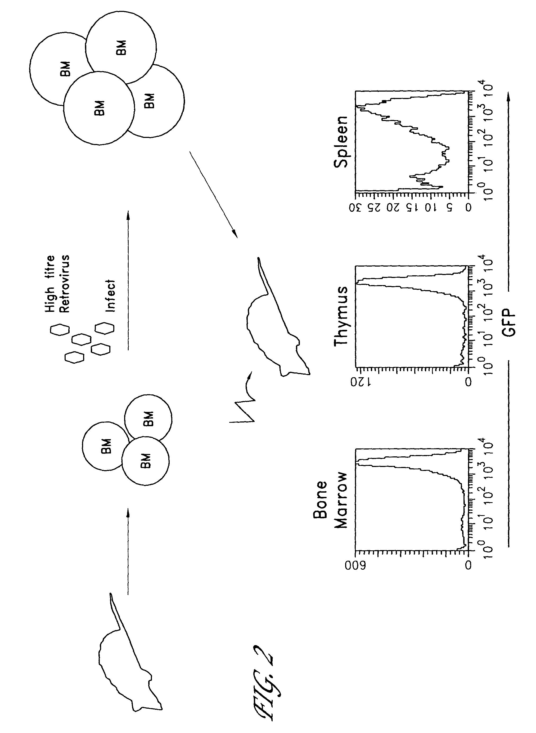
[0229]FIG. 2 shows schematically the methods of the invention applied to the generation of a transgenic mouse. Bone marrow cells were obtained from mice of the desired genetic background (in these experiments, wild type or IL-2 knockout RAG1-deficient mice) and infected them with retrovirus expressing the TCR gene, as described above. The infected BM cells were then transferred into a lethally irradiated RAG1 deficient host mouse and allowed to reconstitute functionally normal T cells.
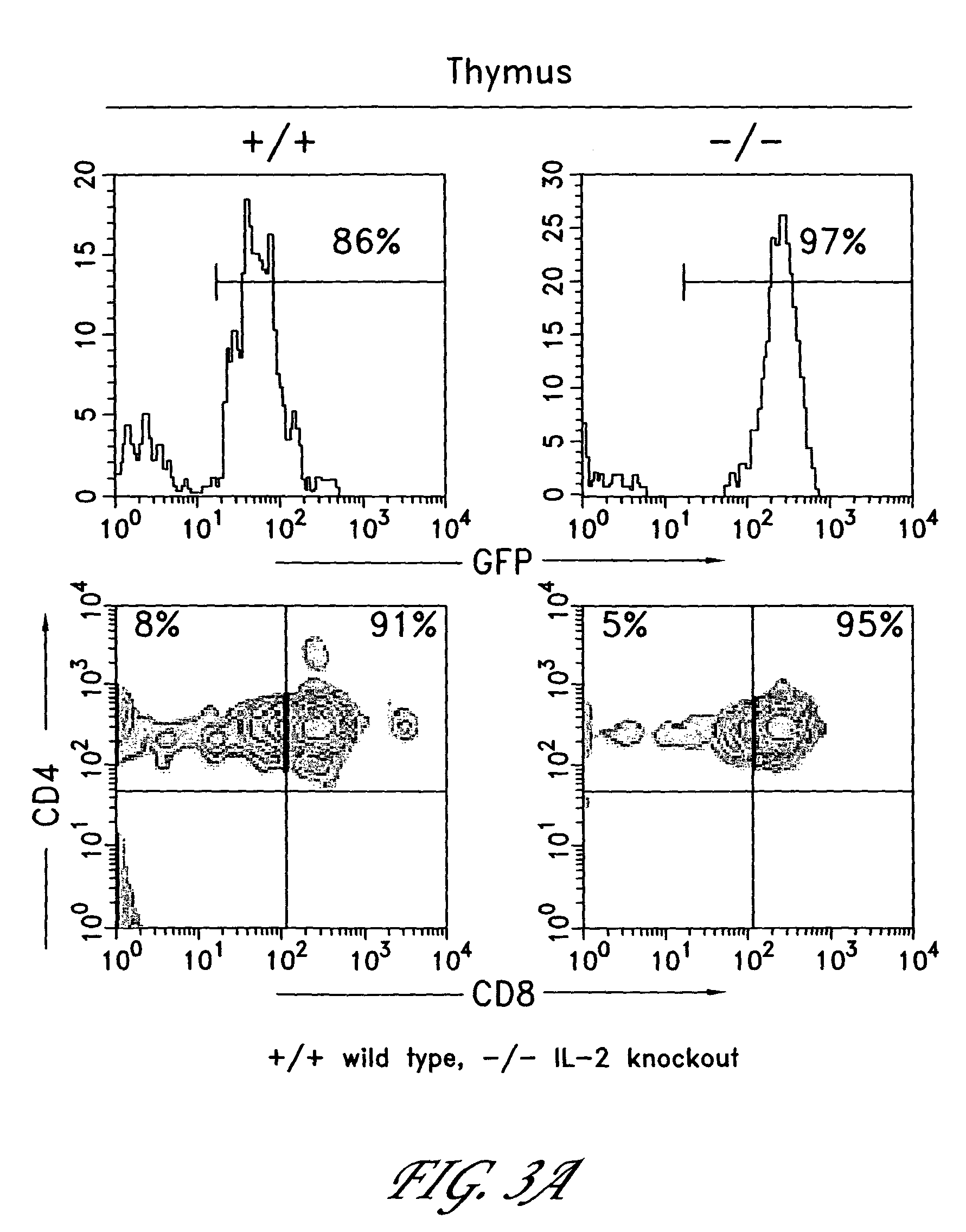
[0230]In both wild type (wt) and IL-2 knock-out (IL-2 ko) RAG1-deficient genetic backgrounds, expression of the OTII TCRα and β cDNAs in stem cells by the MIG retrovirus led to the development of phenotypically normal OT.II CD4+ T cells in the thymi of host mice. The cellularity of the thymi derived from mice expressing OTIIα and β chains was greatly increased compared to those from control mice that received bone ma...
example 3
Generation of Wild Type Mice Expressing Antigen-Specific TCRs
[0239]The ability to generate wild-type mice expressing antigen-specific TCRs was investigated. Bone marrow cells were obtained from wild-type B6 mice that had been previously treated with 5-fluorouracil as described above. Bone marrow cells were infected with the MIG retrovirus comprising sequences encoding the OTII TCRα and TCRβ subunits, as well as a GFP marker protein. The infected bone marrow cells were then transferred into an irradiated host animal and allowed to reconstitute functionally normal T cells.
[0240]As can be seen in FIG. 6A, approximately 65% of the cells extracted from the thymi of mice receiving infected BM cells expressed GFP. FIG. 6B shows that of the CD4+GFP+ thymocytes, about 21% expressed the OTII Vβ element. Further, the GFP positive thymocytes showed normal distribution of CD4 and CD8 markers (FIG. 6C).
[0241]In addition, infected BM cells were found to develop into mature CD4+ T cells expressing ...
PUM
| Property | Measurement | Unit |
|---|---|---|
| pH | aaaaa | aaaaa |
| temperature | aaaaa | aaaaa |
| density | aaaaa | aaaaa |
Abstract
Description
Claims
Application Information