
Medical image management system and medical image management method

a medical image management system and image management technology, applied in medical science, surgery, diagnostics, etc., can solve the problems of inability to obtain images, inability to know how surgeons are operated on, and inability to obtain conventional imaging devices, so as to reduce memory size, manage medical images easily, effective indices

Active Publication Date: 2012-11-20
SEVENTH DIMENSION DESIGN
View PDF3 Cites 3 Cited by
  • Summary
  • Abstract
  • Description
  • Claims
  • Application Information

AI Technical Summary

Benefits of technology

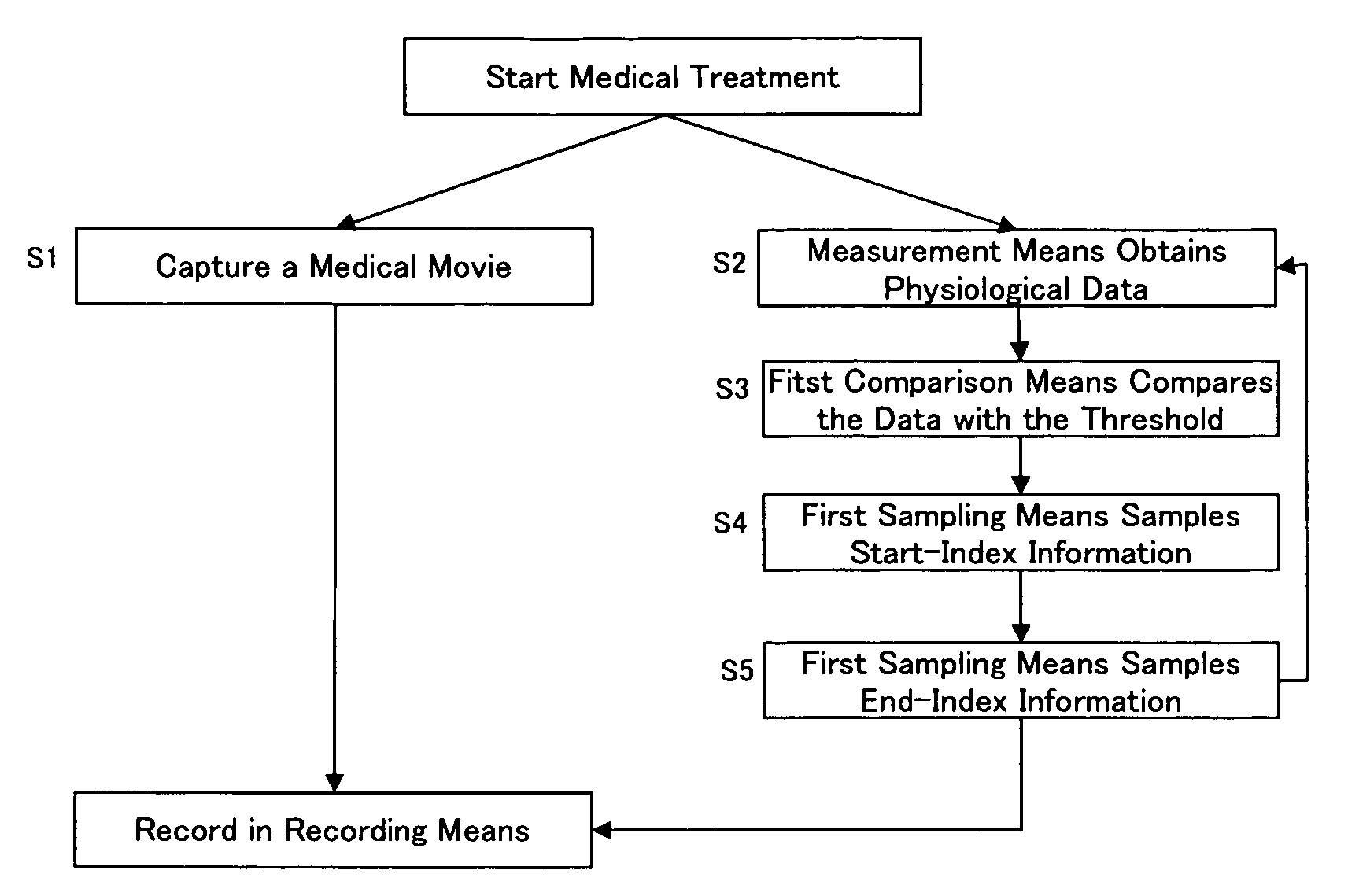
[0040]According to one embodiment of the present invention, the medical image management system adds indices to a medical movie according to changes in the physiological data of a surgeon who performs the medical treatment on a patient. Using the medical image management system, one can understand the changes in the surgeon's mental condition. For example, the indices added to the movie facilitate to pick up the stressful events for the surgeon. It is also possible to manage data about stressful events for an inexperienced surgeon. Such data can be used to give effective instructions to other surgeons. By using the device, it is further possible to know both important events and stressful events for the surgeon. This helps to provide safe medical treatment on patients and to improve the rate of successful treatment.
[0059]According to another embodiment of the medical image management method of the present invention, it is possible to effectively estimate the physiological condition of the surgeon and to add index information to the medical movie by using the data of surgeon's heart beat, blood pressure, sweat production, body temperature, electroencephalogram, grip strength, point of gaze, blink, pupil, eye movement, respiratory rate (including apneic period), pneumogram, number of swallowing, skin electric conductance, electric potential difference of muscle, neurotransmitter level, blood glucose level, blood flow rate, blood composition, amount of various hormones, chewing pressure, electrocardiogram, galvanic skin reflex, fingertip pulse wave, posture or position, tear production, tear composition, saliva production, saliva composition, gastric secretion, gastric fluid composition, facial expression (measurement in characteristic analysis), vocal change (measurement in characteristic analysis), lip reading (measurement in characteristic analysis), limb shivering, urine (protein level, sugar level, occult blood level).

Problems solved by technology

While the foot switch enables the surgeon to capture the desired still-images, if the surgeon forgets to operate the foot switch, the images cannot be obtained.
Moreover, unless the surgeon decides to capture still images, nothing will be obtained.
However, the surgeon concentrating his attention to a surgery tends to forget to operate a footswitch or the like to capture still images.
The conventional imaging devices are not helpful in knowing how surgeons react to a specific situation.
Particularly, it is difficult to direct his attention to capturing still images with the conventional method while performing medical treatment, especially during such a stressful event that it causes changes in the physiological data of the surgeon.
It is also significantly difficult to tell stressful situations for an inexperienced surgeon.
It is also difficult to tell what medical event makes him confused.
Thus, he does not understand what stressful situation he will meet in a surgery room until he is involved in actual medical treatment.
Therefore, it is difficult to use the physiological data to make an accurate decision about when the surgeons become nervous.

Method used


Image

  • Medical image management system and medical image management method
  • Medical image management system and medical image management method
  • Medical image management system and medical image management method

Examples


Embodiment Construction

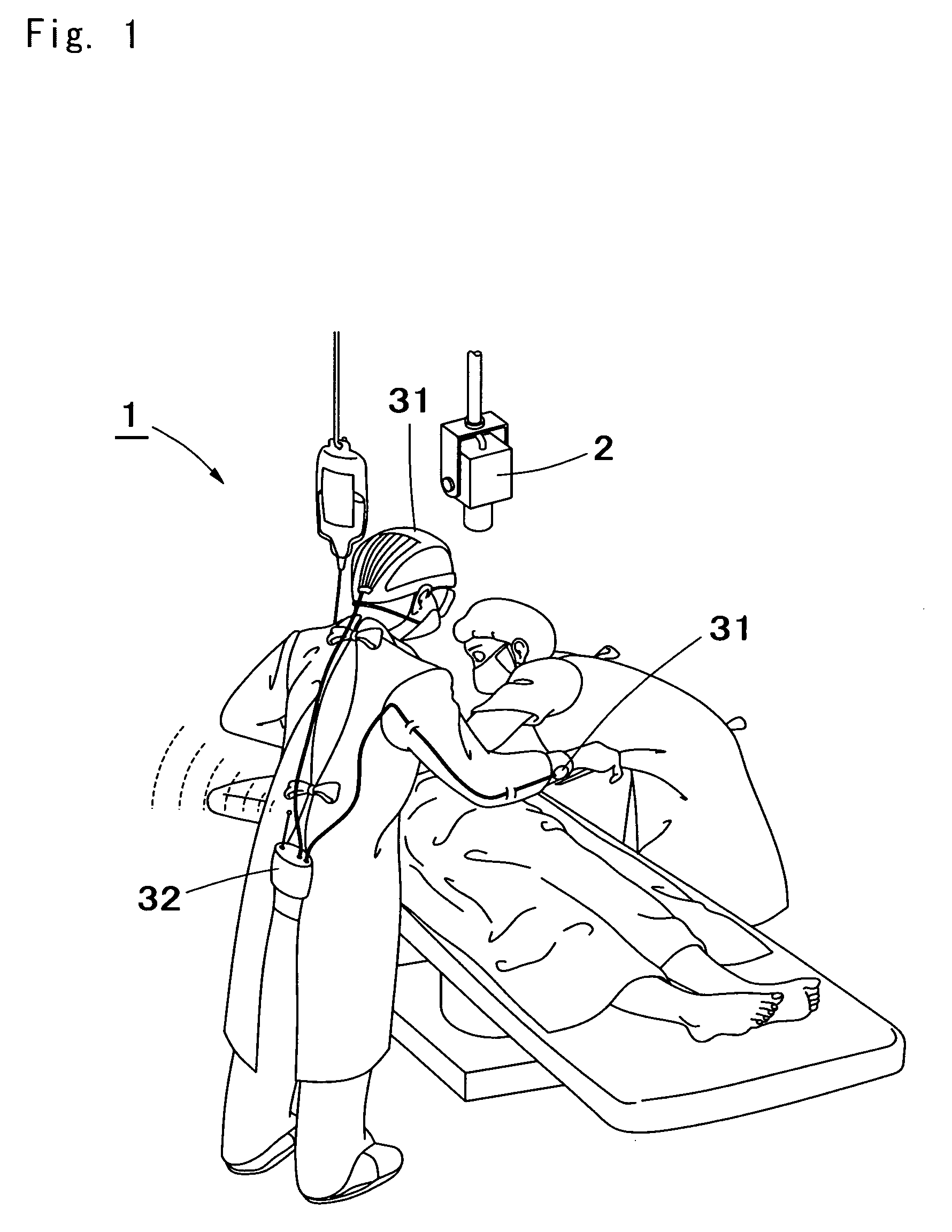
[0079]The term “surgeon” used herein refers to a person who performs medical treatment on a patient. Surgeons include any persons who are concerned in a surgery, such as persons who partake in a surgery (e.g. surgeons and anesthesists), persons who help the surgeon, nurses, and pharmacists, but not limited to these. In the description below, examples where one surgeon is involved are explained but the number of the surgeons is not particularly limited. If several surgeons are involved in a surgery, the physiological data of all of the surgeons is preferably obtained.

[0080]The term “physiological data” refers to data obtained by measuring at least one physiological parameter selected from a group consisting of surgeon's heart beat, blood pressure, sweat production, body temperature, electroencephalogram, grip strength, point of gaze, blink, pupil, eye movement, respiratory rate (including apneic period), pneumogram, number of swallowing, skin electric conductance, electric potential ...


PUM


Abstract

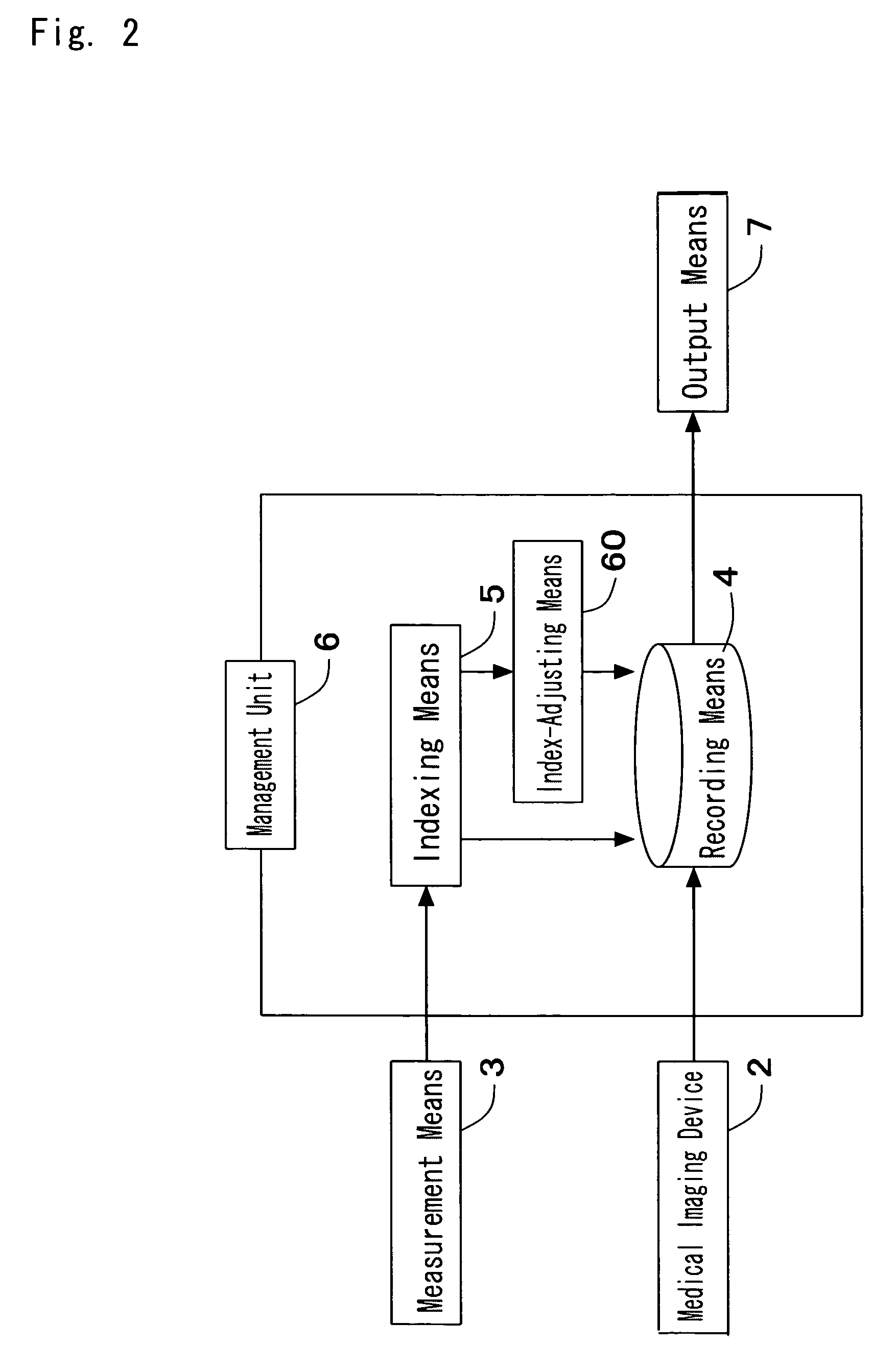
A medical image management system includes: a medical imaging device for imaging a patient to receive a medical treatment and creating a medical moving picture; and a recording unit for recording the medical moving picture imaged by the medical imaging device. The system further includes: a measurement unit for measuring biological information on a person who performs the medical treatment; and an indexing unit for adding an index to the medical moving picture recorded in the recording unit, according to the measurement result obtained by the measurement unit. By providing this system, it is possible to add an index to the medical moving picture recorded, according to the change of the biological information on the person who performs medical treatment on a patient.

Description

CROSS REFERENCE TO RELATED APPLICATIONS[0001]This application claims the right of priority under 35 U.S.C. §120, as authorized by 35 U.S.C. §365(c), to International Application No. PCT / JP2004 / 008991, filed on Jun. 25, 2004 by the same inventor (published under PCT Article 21(2) in Japanese and not English), which is incorporated herein by reference in it's entirety.BACKGROUND OF THE INVENTION[0002]1. Technical Field[0003]The present invention relates to a medical image management system and a medical image management method. The present invention particularly relates to a medical image management system and a medical image management method that can add indices to the recorded medical movies according to the physiological data of a surgeon who performs medical treatment on a patient.[0004]2. Description of the Related Art[0005]Conventionally, movies of a surgery are captured by surgical cameras such as endoscopes and microscopes for ophthalmic surgeries. The in-vivo images are disp...

Claims


Application Information

Patent Timeline
Patent Type & Authority Patents(United States)
IPC IPC(8): A61B5/04A61B5/00A61B19/00G06F17/30G16H10/60
CPCA61B5/00A61B19/52A61B19/5212A61B90/36A61B90/361
Inventor UEDA, ATSUHIRO
Owner SEVENTH DIMENSION DESIGN
Learn More