Devices, systems, and methods for pericardial access

a pericardial access and pericardial catheter technology, applied in the field of pericardial access devices, systems and methods, can solve the problems of inconsistent delivery, significant decrease in the ability of the heart to pump blood, and chronic heart failure, and achieve the effects of short operation time, cost saving, and decreased supply

Active Publication Date: 2010-06-24
CVDEVICES
View PDF46 Cites 34 Cited by
  • Summary
  • Abstract
  • Description
  • Claims
  • Application Information

AI Technical Summary

Benefits of technology

[0019]Surgical drainage (or pericardiectomy) is excessive for many patients. The best option is pericardiocentesis with the Seldinger technique, leaving a pigtail drainage catheter that should be kept in place until drainage is complete. Sagrista Sauleda J., Permanyer Miralda G., Soler Soler J. (2005). “[Diagnosis and management of acute pericardial syndromes].” Rev Esp Cardiol 58(7): 830-41. This less-invasive technique resulted in a short operative time and decreased supply, surgeon, and anesthetic costs. When comparing procedure costs of a pericardial window versus an echo-guided pericardiocentesis with catheter drainage at our institution, there was a cost savings of approximately $1,800 / case in favor of catheter drainage. In an era of accelerating medical costs, these savings are of considerable importance. Buchanan C. L., Sullivan V. V., Lampman R., Kulkarni M. G. (2003). “Pericardiocentesis with extended catheter drainage: an effective therapy.” Ann Thorac Surg 76(3): 817-82.
[0020]Clearly, there is a clinical need for a mini-invasive, safe and effective approach to treatment of pericardial effusion and tamponade. The present application takes advantage of a safe and effective pericardial access approach previously disclosed in combination with a special catheter used specifically for fluid drainage, fluid diagnosis, resuscitation and therapy delivery to treat the underlying cause of the effusion.
[0021]Thus, there is need for an efficient, easy to use, and relatively inexpensive device, system and technique that can be used to access the heart for local delivery of therapeutic and diagnostic substances, as well as of CRT leads and other types of leads. There is also a need for an efficient, easy to use, and relatively inexpensive device, system and technique that can be used to access a space containing fluid within a tissue to remove the fluid and to optionally deliver a substance if necessary.

Problems solved by technology

If left untreated, ischemic heart disease can lead to chronic heart failure, which can be defined as a significant decrease in the heart's ability to pump blood.
However, endoluminal drug delivery has several shortcomings, such as: (1) inconsistent delivery, (2) low efficiency of localization, and (3) relatively rapid washout into the circulation.
Such patients generally have an abnormality in conduction that causes the right and left ventricles to beat (i.e., begin systole) at slightly different times, which further decreases the heart's already-limited function.
Like the local delivery of drugs to the heart, the placement of CRT leads on the heart can be challenging, particularly when the target placement site is the left ventricle.
Problems with these methods of lead placement can include placement at an improper location (including inadvertent placement at or near scar tissue, which does not respond to the electrical signals), dissection or perforation of the coronary sinus or cardiac vein during placement, extended fluoroscopic exposure (and the associated radiation risks) during placement, dislodgement of the lead after placement, and long and unpredictable times required for placement (ranging from about 30 minutes to several hours).
The additional clearance tends to decrease the likelihood that the myocardium will be inadvertently punctured when the pericardial sac is pierced.
Although this technique works well in the normal heart, there are major limitations in diseased or dilated hearts—the very hearts for which drug delivery and CRT lead placement are most needed.
When the heart is enlarged, the pericardial space is significantly smaller and the risk of puncturing the right ventricle or other cardiac structures is increased.
Additionally, because the pericardium is a very stiff membrane, the suction on the pericardium provides little deformation of the pericardium and, therefore, very little clearance of the pericardium from the heart.
Therefore, it is not surprising that cardiac perforation can quickly result in tamponade, which can be lethal.
Ultimately, compression of the heart by a pressurized pericardial effusion results in markedly elevated venous pressures and impaired cardiac output producing shock which, if untreated, it can be rapidly fatal.
End-stage renal disease is a major public health problem.
Renal failure is a common cause of pericardial disease, producing large pericardial effusions in up to 20% of patients.
Purulent (bacterial) pericarditis in adults is rare, but always fatal if untreated.
Management of pericardial effusions continues to be a challenge.
There is no uniform consensus regarding the best way to treat this difficult clinical entity.
This procedure portends significant operative and anesthetic risks because these patients often have multiple comorbidities.
Less invasive techniques such as blind needle pericardiocentesis have high complication and recurrence rates.
However, practices in community hospitals have rarely been studied in detail.
Medical management is usually ineffective and should be used only while arrangements are made for pericardial drainage.
Surgical drainage (or pericardiectomy) is excessive for many patients.
This less-invasive technique resulted in a short operative time and decreased supply, surgeon, and anesthetic costs.

Method used


Image

  • Devices, systems, and methods for pericardial access
  • Devices, systems, and methods for pericardial access
  • Devices, systems, and methods for pericardial access

Examples


Embodiment Construction

[0096]It will be appreciated by those of skill in the art that the following detailed description of the disclosed embodiments is merely exemplary in nature and is not intended to limit the scope of the appended claims.

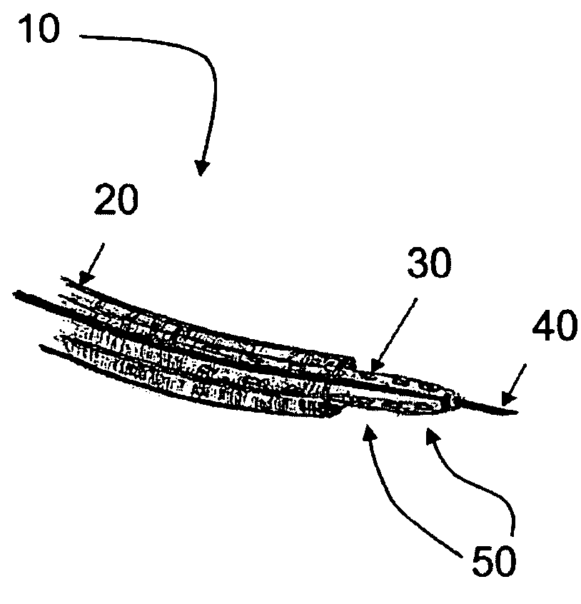
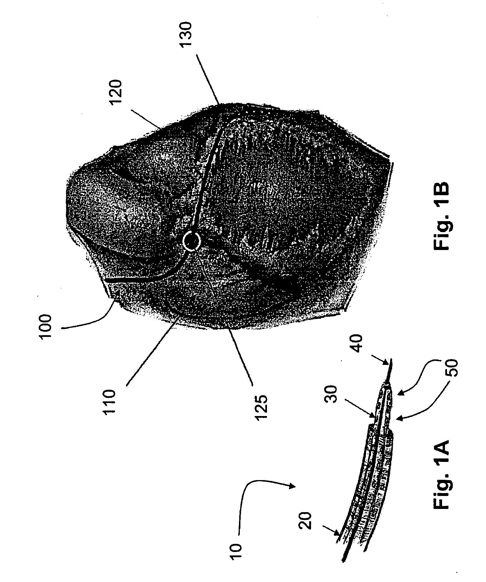
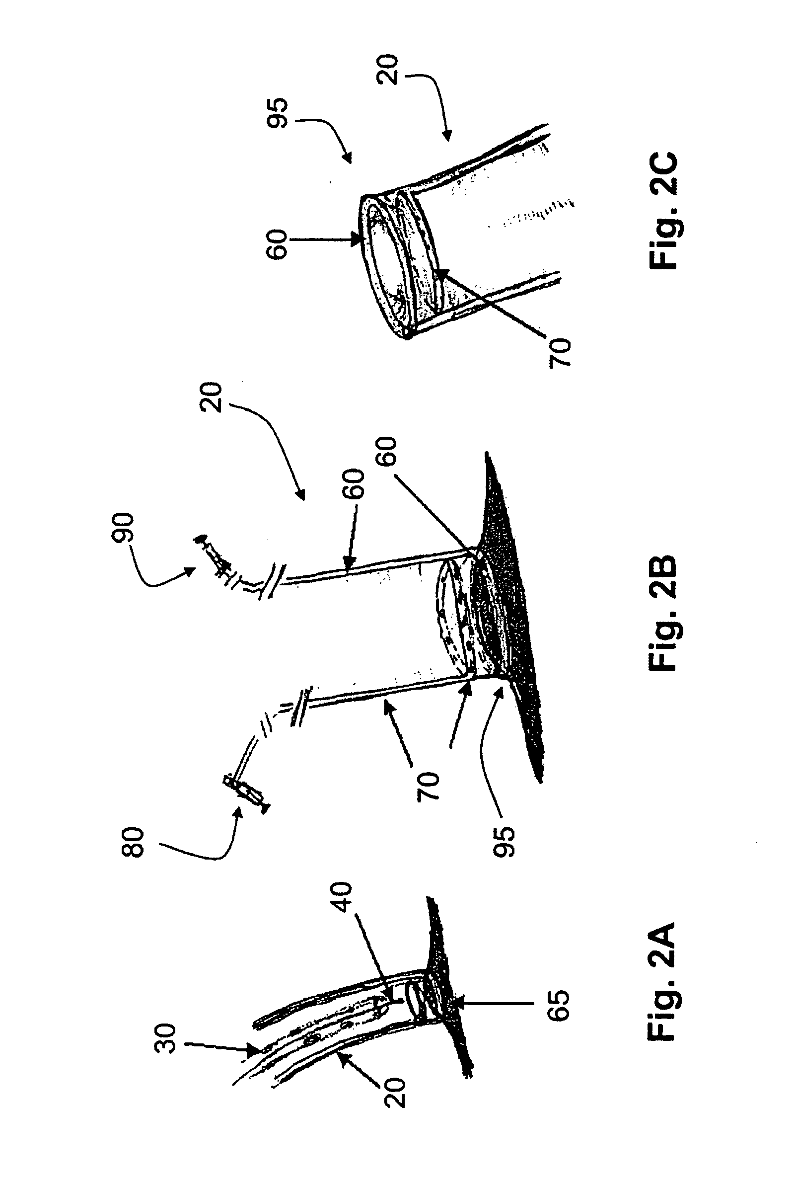
[0097]The disclosed embodiments include devices, systems, and methods useful for accessing various tissues of the heart from inside the heart. For example, various embodiments provide for percutaneous, intravascular access into the pericardial space through an atrial wall or the wall of an atrial appendage. In at least some embodiments, the heart wall is aspirated and retracted from the pericardial sac to increase the pericardial space between the heart and the sac and thereby facilitate access into the space.

[0098]Unlike the relatively stiff pericardial sac, the atrial wall and atrial appendage are rather soft and deformable. Hence, suction of the atrial wall or atrial appendage can provide significantly more clearance of the cardiac structure from the pericardium as...


PUM


Abstract

Devices, systems, and methods for accessing tissue, including the internal and external tissues of the heart, are disclosed. At least some of the embodiments disclosed herein provide access to the external surface of the heart through the pericardial space for localized delivery of leads to the heart tissue. At least some of the embodiments disclosed herein provide access to the pericardial space for removal of accumulated fluid. In addition, various disclosed embodiments provide devices, systems, and methods for engaging a tissue, for removing fluid from a space within a body, and for the delivery of substances to a targeted site for therapeutic purposes.

Description

[0001]This International Patent Application claims priority to, and in at least some designated countries should be considered a continuation-in-part application of, International Patent Application No. PCT / US2008 / 053061, filed Feb. 5, 2008, which claims priority to, and in at least some designated countries should be considered a continuation-in-part application of, International Application Serial No. PCT / US2007 / 015207, filed Jun. 29, 2007, which claims priority to U.S. Provisional Patent Application Ser. No. 60 / 914,452, filed Apr. 27, 2007, and U.S. Provisional Patent Application Serial No. 60 / 817,421, filed Jun. 30, 2006. The present application also claims priority to U.S. Provisional Patent Application Serial No. 60 / 914,452, filed Apr. 27, 2007. Each of these applications are incorporated herein by reference.BACKGROUND[0002]Ischemic heart disease, or coronary heart disease, kills more Americans per year than any other single cause. In 2004, one in every five deaths in the Unit...

Claims


Application Information

Patent Timeline
Patent Type & Authority Applications(United States)
IPC IPC(8): A61B17/34A61M1/00A61B17/00A61M25/01A61M60/191A61M60/289A61M60/468A61M60/508A61M60/865
CPCA61B2017/00606A61M2230/005A61B2017/306A61B2017/308A61B2018/00392A61M25/00A61M25/003A61M25/0074A61M25/0084A61M2025/0036A61M2025/0039A61M2025/004A61M2025/0089A61M2025/0096A61M2025/0681A61M2210/125A61N1/0587A61B17/0057A61B2017/00247A61B2017/003A61B2017/00584A61B2017/00592A61B2017/00601A61M1/1068A61M2210/122A61M1/107A61B90/37A61M1/1086A61M1/122A61M1/1008A61M2205/3331A61M25/06A61B2017/00628A61M2205/32A61M2205/33A61M2205/3303A61M60/865A61M60/289A61M60/508A61M60/191A61M60/468A61M2025/015A61M25/0054A61M25/0136A61M25/0141A61M25/0147
Inventor KASSAB, GHASSAN S.NAVIA, SR., JOSE A.
Owner CVDEVICES
Why Eureka
Try Eureka