Light energy sealing, cutting and sensing surgical device

a surgical device and light energy technology, applied in the field of surgical forceps, can solve the problems of few surgical instruments having the capability to treat and monitor tissue treatment without the use of additional surgical instruments, and leave foreign body material inside a patient, and achieve the effect of better indication of seal quality

Active Publication Date: 2016-06-28
TYCO HEALTHCARE GRP LP
View PDF247 Cites 199 Cited by
  • Summary
  • Abstract
  • Description
  • Claims
  • Application Information

AI Technical Summary

Benefits of technology

[0013]As described in more detail below with reference to the accompanying figures, the present disclosure generally relates to surgical light energy devices that include an end effector assembly that can fuse (e.g., seal) and / or separate (e.g., cut) tissue. The present disclosure also provides one or more devices that sense and / or monitor tissue properties at various stages of treatment to determine when the treatment is complete, efficacy of a tissue seal and / or to measure jaw pressure (e.g., a potential requirement for a quality seal). Optical sensing provides better indication of seal quality than current methods, such as, electrical impedance measurements. Additionally, tissue separation may be accomplished with the same light energy device used for tissue sealing eliminating the need for a separate mechanical blade that is traditionally used for tissue separation in jaw members. The present disclosure also provides one or more methods for providing feedback to the user, generator and / or control algorithm with regard to temperature at or proximate a surgical site, jaw closure pressure, jaw positioning, and other various feedback information.

Problems solved by technology

However, these traditional procedures often leave foreign body material inside a patient.
Other treatment methods are known in the art, however, very few surgical instruments have the capability to treat and monitor tissue treatment without the use of additional surgical instruments.

Method used


Image

  • Light energy sealing, cutting and sensing surgical device
  • Light energy sealing, cutting and sensing surgical device
  • Light energy sealing, cutting and sensing surgical device

Examples


Embodiment Construction

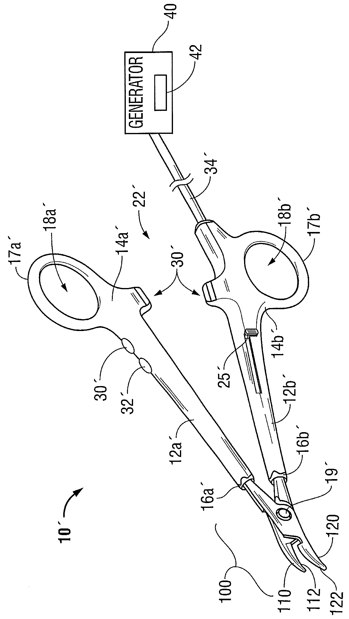
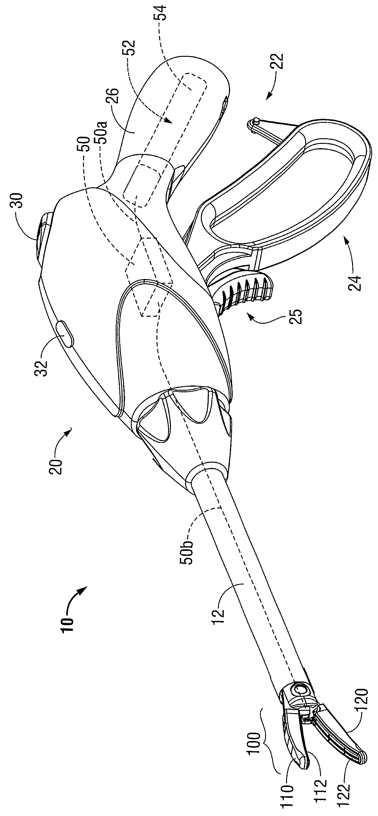
[0053]Referring now to FIGS. 1A and 1B, an endoscopic surgery forceps 10 and an open forceps 10′ are shown, respectively. For the purposes herein, either an endoscopic instrument or an open surgery instrument may be utilized with any of the embodiments of end effector assemblies described herein. It should be noted that different electrical, optical and mechanical connections and other considerations may apply to each particular type of instrument. However, the novel aspects, with respect to the end effector assembly and the operating characteristics thereof, remain generally consistent with respect to both the endoscopic or open surgery designs. It also should be noted that any of the embodiments described below may be configured to also include traditional vessel sealing capabilities.

[0054]The present disclosure provides for an apparatus, system and method for sealing tissue using light energy. Light (e.g., from about 200 nm to about 11,000 nm) is used to heat the tissue due to ab...


PUM


Abstract

The present disclosure is directed towards a medical instrument. The medical instrument includes a housing and an end effector assembly operably connected to the housing. The end effector assembly includes first and second jaw members each having a tissue contacting surface, at least one of the first and second jaw members movable between a first, spaced-apart position and a second proximate position, wherein in the second position, the jaw members cooperate to define a cavity configured to receive tissue between the jaw members. The end effector also includes at least one light-emitting element coupled to at least one of the first and second jaw members, the at least one light-emitting element adapted to deliver light energy to tissue grasped between the first and second jaw members to treat the tissue.

Description

BACKGROUND[0001]1.Technical Field[0002]The present disclosure relates to surgical forceps having components to treat and / or monitor tissue being treated. More particularly, the present disclosure relates to open or endoscopic surgical forceps that utilize light energy to treat (e.g, seal, cut, etc.) and / or to sense tissue properties.[0003]2. Description of Related Art[0004]In many surgical procedures, body vessels, e.g., blood vessels, ducts, adhesions, fallopian tubes, or the like are sealed to defunctionalize or close the vessels. Traditionally, staples, clips or sutures have been used to close a body vessel. However, these traditional procedures often leave foreign body material inside a patient. In an effort to reduce foreign body material left within the patient and to more effectively seal the body vessel, energy techniques that seal by heating tissue have been employed.[0005]Endoscopic or open forceps are particularly useful for sealing since forceps utilize mechanical action...

Claims


Application Information

Patent Timeline
Patent Type & Authority Patents(United States)
IPC IPC(8): A61B18/20A61B18/22A61B18/00A61B17/29A61B18/18
CPCA61B18/22A61B17/29A61B18/20A61B2018/0063A61B2018/00184A61B2018/00601A61B2018/1807A61B2018/2272A61B2018/20361A61B2018/2253A61B2018/2055A61B18/1445A61B18/18A61B2017/00057A61B2018/00642A61B2018/2277A61B2018/2294A61B18/28
Inventor NAU, JR., WILLIAM H.KELLER, CRAIG A.KERR, DUANE E.
Owner TYCO HEALTHCARE GRP LP
Why Eureka
Try Eureka