
Detection method of nucleic acid and kit and using thereof

a detection method and nucleic acid technology, applied in the field of detection methods of nucleic acid, can solve the problem of few acceptable results, and achieve the effect of simple nature and high sensitivity

Active Publication Date: 2016-08-02
BEIJING ENTRY EXIT INSPECTION & QUARANTINE BUREAU +1
View PDF5 Cites 0 Cited by
  • Summary
  • Abstract
  • Description
  • Claims
  • Application Information

AI Technical Summary

Benefits of technology

[0007]Aiming to overcome the defects of the prior art, the present invention provides a simple, rapid, naked-eye visible nucleic acid detection method with significant color change, a kit depending on the method, and the application of the method and the kit in the inspection and quarantine.
[0009]The inventors of the present invention found that the use of asymmetric PCR instead of the conventional PCR can obtain ssDNA-rich analyte nucleic acid and increase efficiency of hybridization between the probe and the analyte nucleic acid, further enhancing AuNP SPR effect in the probe and the color change as well.
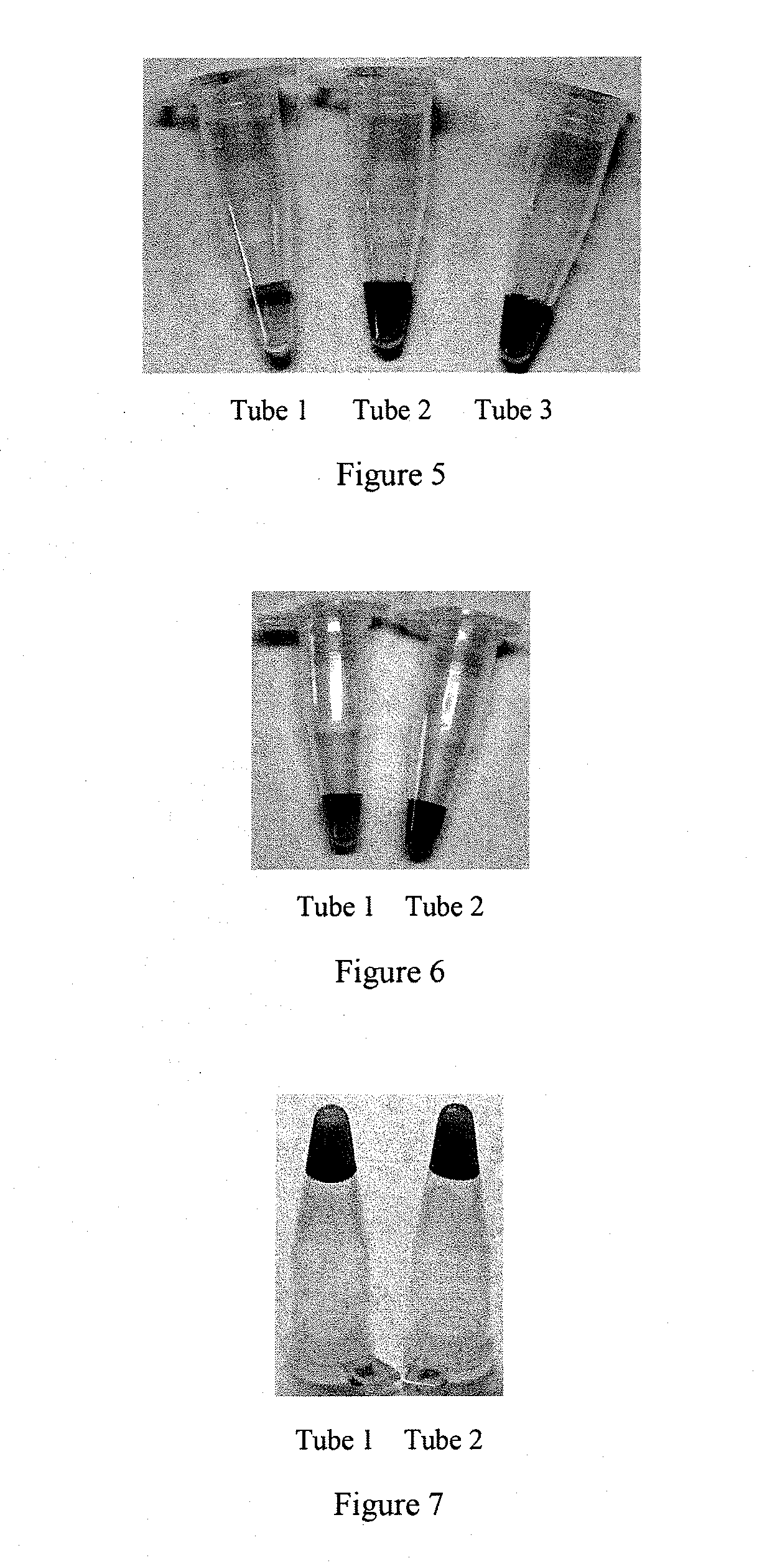
[0013]It is carried out as following when detecting genomic DNA fragment of Bacillus anthracis using the method according to the present invention: first, use the analyte nucleic acid as a template conducting asymmetric PCR, then mix the asymmetric PCR product with the probe hybridization solution containing the specific DNA fragment of genome of the Bacillus anthracis; and the color will change significantly within 2 minutes, but no color change occurs if the fragment does not exist. It is indicated that the present invention is a quick and easy, sensitive and specific, equipment-free and naked eyes visible nucleic acid detection method. A case indicating high sensitivity and simple nature of this method is that for 50 μL asymmetric PCR reaction with 0.01 ng of template nucleic acid, on the condition of taking 5 μL of asymmetric PCR product and mixing with 5 μL of probe solution at a probe concentration of 1.5 μM, significant color change can develop. The method can also be applied to detection of DNA fragment of mouse β-actin and Vibrio cholerae, indicating a certain degree of universality of this method.

Problems solved by technology

Although many ways can be used to enhance the binding, few is acceptable due to complexity of the ways themselves, let alone groping and optimization of the detection systems themselves each time upon determination.

Method used


Image

  • Detection method of nucleic acid and kit and using thereof
  • Detection method of nucleic acid and kit and using thereof

Examples


preparation example 1

[0046]The present preparation is for synthesis of AuNP with a particle diameter of approximately 13 nm.

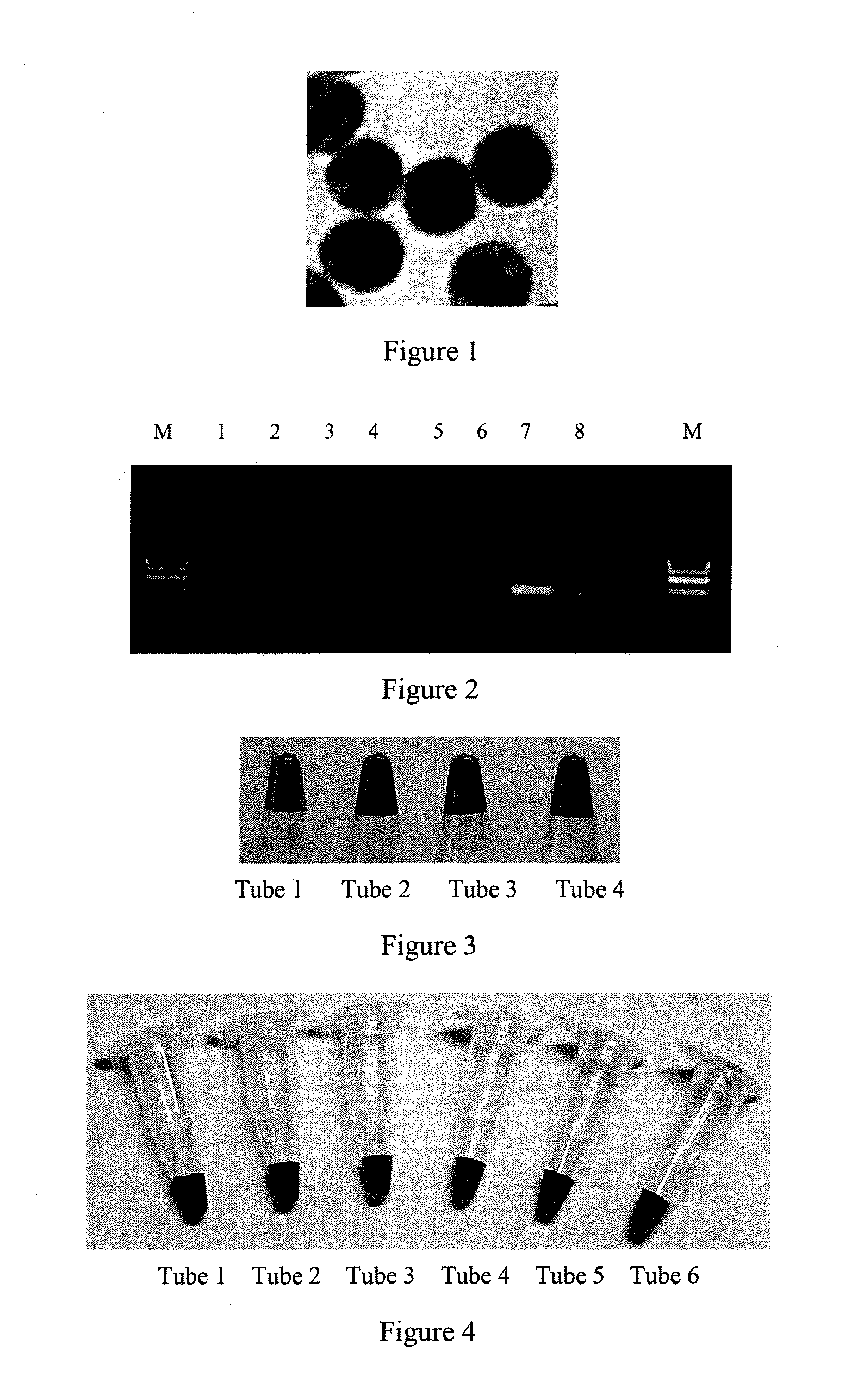
[0047]The AuNP is prepared using the classical chloroauric acid-sodium citrate reduction method. Soak all glass containers in aqua regia, rinse with nanoscale water, dry. In a 50 mL Erlenmeyer flask add 40 mL of nanoscale water and 0.4 mL of 1 g / mL HAuCl4 (chloroauric acid). Stir vigorously using a magnetic stirrer, heat to boiling. Then quickly add all at once 1.2 mL of 1 g / mL sodium citrate, allow the solution gradually change from light yellow to dark red, continue to heat for 15 minutes, then stop heating, continue to stir allowing to cool to room temperature. Keep the solution closed and stored at 4° C. free from light, use UV-visible spectrophotometer to measure the maximum absorption peak at 520 nm, the concentration is 3 nM. Drip 10 μL of the analyte onto the copper mesh, drain dry under vacuum, allow undergoing the TEM. The results are shown in FIG. 1, the AuNP diameter is...

preparation example 2

[0048]According to cDNA sequence of mouse β-actin, choose the suitable hybrid sites and design the limiting primer P1 the non-limiting primer P2, oligonucleotide Oligo1 and Oligo2 for subsequent molecular hybridization, the sequences are as follows:

[0049]

P1:(SEQ ID NO: 6)5′-GAT GCC ACA GGA TTC CAT A-3′;P2:(SEQ ID NO: 7)5′-CTT CTC TTT GAT GTC ACG CA-3′;Oligo1:(SEQ ID NO: 8)5′-TGC GTG ACA TCA AAG AGA AG-3′;Oligo2:(SEQ ID NO: 9)5′-GAT GCC ACA GGA TTC CAT A-3′;

[0050]Wherein, C6 of the 5′ end of the Oligo1 and Oligo2 for coupling are mercapto-modified. What mentionable is that the oligonucleotide probes used in the examples of the present invention are commercially available.

preparation example 3

[0051]The present preparation example is for the preparation of a liquid that contains the probe. Dissolve the 5OD mercapto modified oligonucleotides dry powder in 80 μL of the disulfide lysate (170 mM phosphate buffer, pH 8.0) obtaining a solution of oligonucleotide after complete solution. Divide the solution into four tubes, each containing 20 μL. Dissolve DTT (dithiothreitol) in disulfide lysate obtaining freshly prepared 0.1M DTT solution. Add 80 μL of the DTT solution to 20 μL of the oligonucleotide solution obtaining 100 μL DNA DTT reduction solution. Wrap it with aluminum foil and place at room temperature for 1 hour, vortex for 5 seconds every half hour. At the same time, with at least 10 mL above nano-grade water prewash the Nap-5 column. Feed the 100 μL DNA DTT reduction solution to the Nap-5 purification column, add 400 μL of Milli-Q water to wash the column, and finally add 500 μL of Milli-Q water to elute obtaining a mercapto group-containing oligonucleotide analyte. C...


PUM

PropertyMeasurementUnit
diameteraaaaaaaaaa
diameteraaaaaaaaaa
diameteraaaaaaaaaa

Abstract

A detection method of nucleic acid is provided. The method includes: providing nucleic acid to be tested, making the nucleic acid to be tested react under asymmetric PCR conditions with a pair of primers for target nucleic acid amplification, DNA polymerase, adenine deoxyribonucleotide, guanine deoxyribonucleotide, cytosine deoxyribonucleotide and thymidine deoxyribonucleotide in a PCR buffer solution, mixing the reaction product and liquid that contains probe molecules, and judging whether the nucleic acid to be tested contains the target nucleic acid by observing the obtained mixture color or color change. A kit is also provided that can be used in the nucleic acid detection by the said method. Application of the method and the kit in inspection and quarantine is also provided. The method is a quick and easy, sensitive and a specific detection method of nucleic acid with direct observation using naked eyes. The method does not need additional equipment.

Description

CROSS REFERENCE TO RELATED APPLICATION[0001]This application is a 35 U.S.C. 371 national stage application of PCT / CN2011 / 072682, which was filed Apr. 12, 2011 and is incorporated by reference as if fully set forth.TECHNICAL FIELD[0002]The present invention relates to a detection method of nucleic acid, a kit for detection of nucleic acid applying the method, and use of the kit for inspection and quarantine.BACKGROUND ART[0003]Molecular diagnosis is a leading-edge technology in modern biological analysis and detection of specific nucleic acid sequence is an important part of biological analysis. Conventional molecular identification methods, such as fluorescent quantitative PCR (Qi Y. Appl Environ Microbiol, 2001, 67 (8): 3720-3727), RFLP (restriction fragment length polymorphism, S R Klee. J. Applied Microbiology. 2006, 100, 1364-5072), gene chip and sequencing, are complex and require large, expensive equipment and long time. They are very inaccessible for quick detection of some p...

Claims


Application Information

Patent Timeline
Patent Type & Authority Patents(United States)
IPC IPC(8): C12Q1/68
CPCC12Q1/6816C12Q1/686C12Q1/689C12Q2565/107
Inventor LIANG, XINGJIEDENG, HUAMA, XIAOWEILI, CHAN
Owner BEIJING ENTRY EXIT INSPECTION & QUARANTINE BUREAU
Learn More