

System and method for intracranial implantation of therapeutic or diagnostic agents
a diagnostic agent and intracranial implantation technology, applied in the field of system and method for intracranial implantation of therapeutic or diagnostic agents, can solve problems such as obviating the need, and achieve the effect of accurate positioning of the wafer into the tumor, simple and intuitive manipulation
- Summary
- Abstract
- Description
- Claims
- Application Information
AI Technical Summary
Benefits of technology
Problems solved by technology
Method used
Image

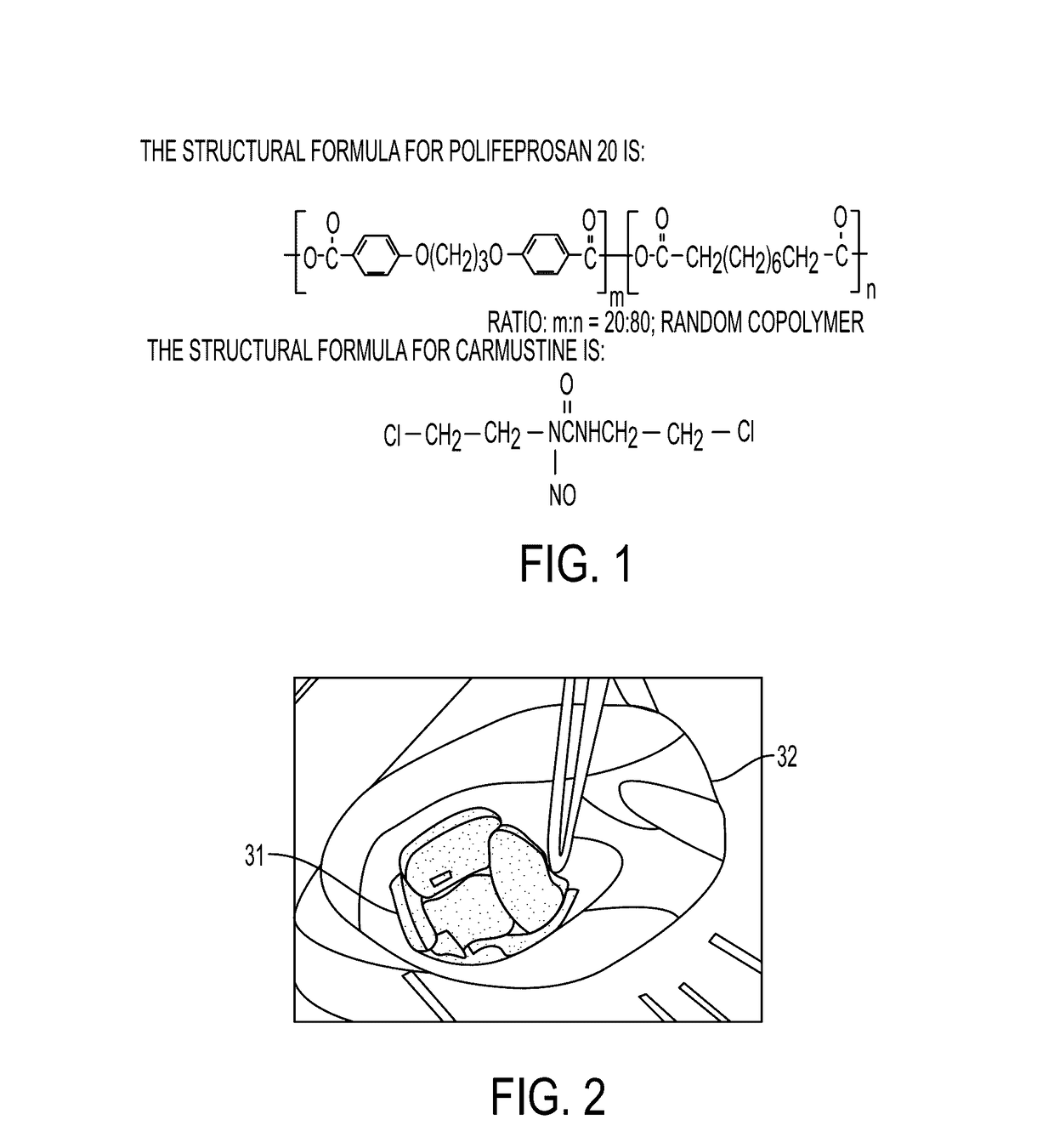
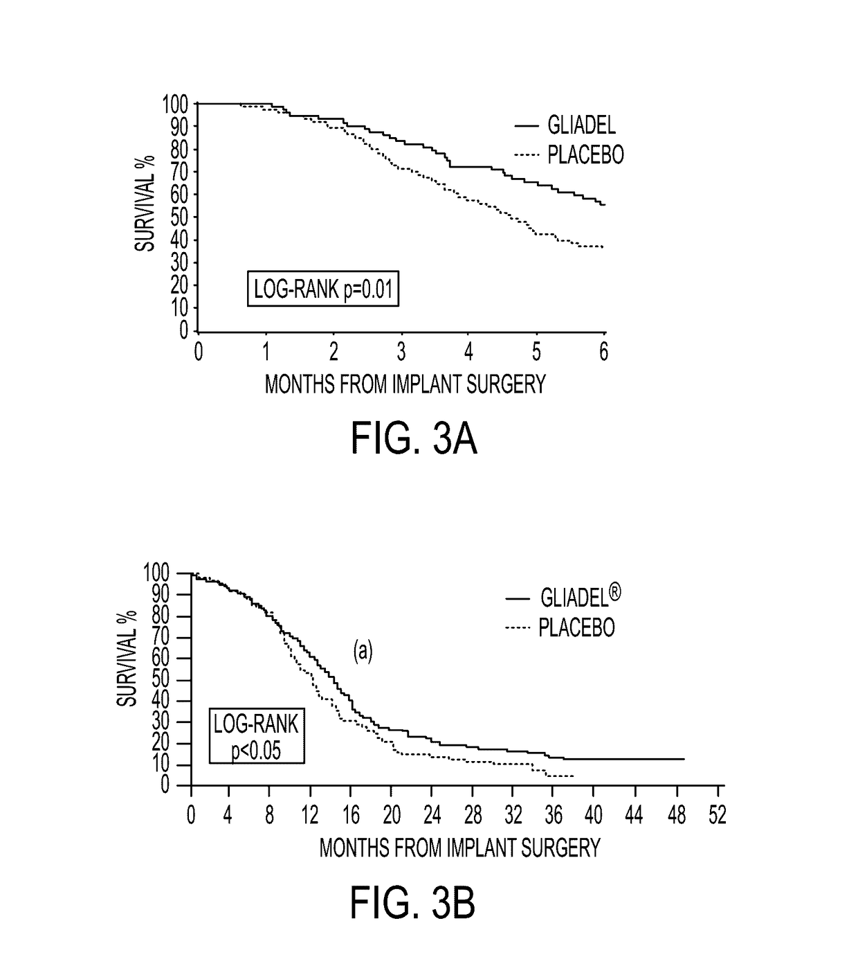
Examples
Embodiment Construction
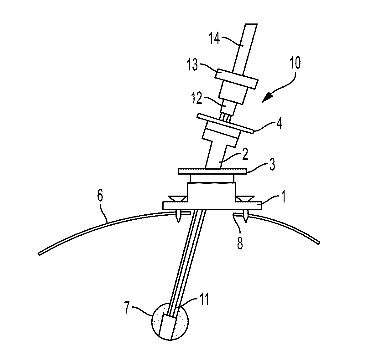
[0055]In frameless stereotaxis, the patient's head is immobilized and fiducials or markers which are visible to an infrared image guidance system are placed at reference points on the patient's skull and on the instruments entering the brain. Thus the guidance system can track the position of the instruments relative to the immobilized head. Surgeons can then visualize this information as it is coupled with preoperative MRI or CAT scan images of the brain and displayed on a monitor, as shown in FIG. 4(A)-(B). FIG. 4(A)-(B) are photographic depiction of screenshots from the StealthStation® Treatment Guidance System by Medtronic. In FIG. 4(A), reference points on the skull are visible. Also, the dot 16 on the skull shows the point of entry with the trajectory pointed directly towards the screen. Upper panes show MRI cross section images in planes which intersect to form the line containing the desired trajectory. FIG. 4(B) is a close up of the upper left pane from FIG. 4(A). The progr...
PUM
 Abstract
Description
Claims
Application Information